Prevalence of Fungal and Demodex Mite (Acari: Demodicidae) Skin Infections in A Tertiary Health Care Center in Garhwal Region of Uttarakhand ...
←
→
Page content transcription
If your browser does not render page correctly, please read the page content below
Section: Microbiology
Original Article                                                                            ISSN (O):2395-2822; ISSN (P):2395-2814

Prevalence of Fungal and Demodex Mite (Acari:
Demodicidae) Skin Infections in A Tertiary Health Care
Center in Garhwal Region of Uttarakhand.
Yogendra Pratap Mathuria1
1Associate   Professor, Department of Microbiology, Government Doon Medical College, Dehradun.

Received: April 2017
Accepted: April 2017

Copyright: © the author(s), publisher. It is an open-access article distributed under the terms of the Creative
Commons Attribution Non-Commercial License, which permits unrestricted non-commercial use, distribution, and
reproduction in any medium, provided the original work is properly cited.

                                                             ABSTRACT

Background: The prevalence of cutaneous mycoses has been increasing with constantly changing pattern. The likelihood
of fungal infections & demodicosis in the population of Garhwal region might be increased due to their poor personal
hygiene, agriculture work and close contact with soil and animals. Aims: To find out the prevalence of various mycotic &
demodex mite skin infections in Garhwal region of Uttarakhand and to compare and correlate it with site, gender and age
group of residents of Garhwal region of Uttarakhand. Methods: The patients with suspected superficial fungal infection
were studied in relation to age and sites involved. Skin scrapings were collected in a sterile black paper and KOH mount
were made. The fungal infections and demodicosis were classified on the basis of morphology, colour, thickness and
branching pattern of hyphae. Results: Out of total 2534 patients, maximum number of patients of superficial skin infection
were observed in month of September 286 (11.29%).Total 1340(52.88%) patients had fungal infection, while among face
infections 11(11.57%) had demodicosis. Maximum patients 817(32.24%) were in the age group of 21 – 30 years of age.
Dermatophytes (69.82%) were the commonest cause of fungal infection, followed by dematatious fungi, Candida species,
whereas minimum number of cases was found of Demodex mite. All the demodex mites were isolated from infections of
face. Conclusion: Dermatophytes were most common cause of mycotic skin infection and Demodex mite was the
commonest parasitic infection of face.

Keywords: Dermatophyte, Mycotic skin infection, Demodicosis, Demodex mite.

                   INTRODUCTION                                           Demodex is an ecto-parasite of pilo-sebaceous
                                                                          follicle and sebaceous gland, typically found on the
Cutaneous mycosis is one of the most common                               face including cheeks, nose, chin, forehead, temples,
infectious diseases worldwide and affects around 20                       eye lashes, brows, and also on the balding scalp,
– 25% of world’s population and the prevalence of                         neck and ears.[8,9] Apart from common factors like
cutaneous mycoses is still increasing with constantly                     temperature, humidity, rainfall, environmental light,
changing pattern.[1-3] Infestation with Demodex is                        climate, chemical composition and pH; other factors
ubiquitous in humans; with more prevalence in                             like human and/or animal presence in the vicinity are
healthy adults varying between 23-100%, whereas                           also of importance in amount and diversity of flora
Demodicosis is uncommon.[4,5] Two common                                  growing there.[10-14]
species of Demodex are D. folliculorum and D.
                                                                          Name & Address of Corresponding Author
brevis which are cosmopolitan.[6] The size of adult
                                                                          Dr. Yogendra Pratap Mathuria
mites is mostly of 0.1 mm to 0.4 mm long, with D.                         Associate professor,
brevis slightly shorter than D. folliculorum. They                        Department of Microbiology,
have a semi-transparent elongated body that consists                      Government Doon Medical College, Dehradun.
of two fused segments. Eight short segmented legs
attached to the first body segment. Females are                           In Garhwal region of Uttarakhand alpine conditions
somewhat shorter and rounder than males.[7]                               predominate with mild summers, humid monsoon
Dermatophytes, yeasts and non dermatophytic molds                         and cold winters but the city of Srinagar being a
can involve skin, hair and nails and are important                        valley has warm summers and humid monsoons.
microorganisms of soil. The infection is generally                        The major part of population is engaged in
restricted to non – living cornified layers of skin.                      agriculture, livestock rearing and manual labour and
Various clinical manifestations are seen varying                          is in close contact with soil and animals. Poor
from mere pruritis to favus. In majority of cases                         personal hygiene and inadequate environmental
however the infection presents as scaly lesion, spots                     sanitation increases the risk of contracting fungal
or blisters.                                                              infection and Demodicosis.[15, 16]

Annals of International Medical and Dental Research, Vol (3), Issue (4)                                                  Page 1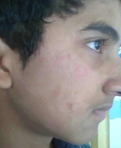
Mathuria; Prevalence of Fungal and Demodex Mite Skin Infections

                                                                                                                    Section: Microbiology
Though the infections are not serious in terms of
mortality but lesions are not self - curative and may
harbour       secondary       bacterial      infections.
Disfigurement caused due to infection affects the
self – esteem of patient and decreases the quality of
life. The infected individual acts as reservoir of
disease and can transfer it by direct or indirect
contact.[17]
Veer Chander Singh Garhwali Government Medical
Science and Research Institute (VCSGGMS&RI),               Figure 1: Distribution of samples according to the age
Srikot, Srinagar is one of the referral centres for        of patients.
Garhwal region. There are seven districts under
Garhwal division: Dehradun, Uttarkashi, Haridwar,          Out of total 2534 samples 1340 (52.88%) samples
Chamoli,      Pauri,    Tehri     and     Rudraprayag.     were found positive by direct microscopy for fungus
VCSGGMS&RI, Srinagar is closest referral center            [Figure 2] & 11(11.57%) cases of face infection
from Chamoli, Tehri, Rudraprayag and Pauri                 were found positive for Demodex mite [Figure 3].
districts so most of patients of these hilly are usually
referred to this medical college. This study was
undertaken to study the prevalence and pattern of
fungal and demodex skin infections in Garhwal
region of Uttarakhand.

     MATERIALS AND METHODS
This study was carried out in department of
Microbiology, VCSGGMS&RI, Srinagar, Garhwal.
                                                           Figure 2: Hyaline, septate, branching hyphae of
A total of 2534 clinically diagnosed patients of
                                                           Dermatophytes [40x].
superficial fungal skin infection reported in
outpatient departments for one complete year were
randomly selected and enrolled for the study.
History was taken in relation to name, age, and sites
involved. Patients under antifungal treatment were
excluded from the study group.
The affected area was cleansed with 70% ethyl
alcohol and skin scraping was taken from inflamed
border of active lesion using surgical blade (No. 23).
Skin scrapings were collected in a sterile black paper
and were used for KOH mount, 10% KOH was used
for skin scrapings, 20% for scrapings from palm and        Figure 3: Demodex brevis isolated from a patient
sole and 40% for nail clippings. Fungi were                [100x].
identified on the basis of morphology, pigmentation,
other microscopic characteristics and Demodex mite
on the basis of their unique morphology.

                    RESULTS
A total of 2534 patients with clinical suspicion of
fungal and demodex infection were included in the
study for the period of one year i.e. from January
2015 to December 2015 and samples were collected
                                                           Figure 4: Monthwise distribution of total & positive
from site of infection. Male: Female ratio of samples
                                                           samples.
was 2.3:1, percentage of positive samples for males
was 54.35% (962 positive out of 1770 samples) and          Maximum number of sample 286(11.28%) were
49.48% (378 positive out of 764 samples) for               collected in the month of September and minimum
females [Figure 1]. Demodex mites were isolated in         number of sample 165 (6.51%) were collected in
11(11.57%) positive samples from face.                     December. Maximum percentage of positive cases
                                                           73.90% (167 out of 256 samples) and minimum
                                                           44.50% (81 out of 182 samples) were seen in month
                                                           of July and January respectively [Figure 4].

Annals of International Medical and Dental Research, Vol (3), Issue (4)                                   Page 2
Mathuria; Prevalence of Fungal and Demodex Mite Skin Infections
Section: Microbiology
Figure 5: Distribution of total & positive samples
according to sites.
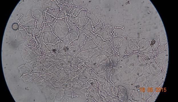
Figure 7: Fungal lesion on neck.
Maximum number of samples (763, 30.11%) were
collected from groin, thigh and buttock region,
followed by nails, (512, 20.21%) and minimum
samples were collected from face (126, 4.97%).
Maximum percentage of positive samples 79.03%
(603 out of 763 samples) was from groin, thighs &
buttock and minimum 25.20% (96 positive in total
381 samples) from foot and sole [Figure 5].
Figure 8: Demodex mite infection on cheek.
On microscopy examination, maximum fungal
isolates 1098(82%) were hyaline septate branching
hyphae, suggestive of Dermatophytes, followed by
Figure 6: Fungal lesion on nail. yeasts 123(9%) and Dematiaceous fungi, 119 (9%).
Table 1: Distribution of different isolates in relation to sites.
Site Total Samples (%) Positive samples Dermatophytes: Yeasts (%) Dematiaceous fungi
(%) hyaline, septate hyphae (%)
(%)
Hand 248(9.79%) 73(29.44%) 64(87.67%) 3(4.11%) 6(8.22%)
Trunk 231(9.11%) 107(46.32%) 92(85.98%) 2(1.87%) 13(12.15%)
Head & 273(10.77%) 163(90.01%) 7(3.87%)
Neck 181(66.3%) 11(6.01%)
Nails 512(20.21%) 196(38.28%) 79(40.31%) 70(35.71%) 47(23.98%)
Groin, 763(30.11%) 591(98.01%) 00(0%)
thighs, &
buttock 603(79.03%) 12(1.99%)
Foot & sole 381(15.03%) 96(25.20%) 38(39.58%) 12(17.39%) 46(47.92%)
Face 126(4.97%) 84(66.67%) 71(84.52%) 13(15.48%) 00(0%)
Total 2534(100) 1340(52.88%) 1098(43.33%) 123(4.85%) 119(4.70%)
Table 2: Agewise distribution of isolates from face.
Positive Dermetophytes+ Samples Positive Dermetophytes+
samples from (yeasts/Malassezia) + from Female (yeasts/Malassezia) + Total
Age/ Samples males [demodex mite] patients [demodex mite] Samples
0-1yr 3 1+(1) +[0] 2 0+(0)+[0] 5
1-10yrs 8 2+(3) +[0] 9 2+(4)+[0] 17
11-20yrs 16 10+(1)+[2] 19 12+(2)+[1] 35
21-30yrs 16 12+(0)+[3] 21 13+(0)+[3] 37
31-40yrs 8 6+(0)+[0] 7 6+(0)+[1] 15
41-50yrs 3 2+(0)+[0] 4 3+(0)+[1] 7
51-60yrs 4 2+(0)+[0] 3 2+(0)+[0] 7
>60yrs 1 0+(0)+[0] 2 1+(0)+[0] 3
59 35+(4)+[5] 67 39+(6)+[6] 126
Maximum percentage of Dermatophytes (98.01% groin, thigh & buttock region, followed by Head &
(591 out of total 603 isolates) were isolated from neck 90.01% (163 out of total181 isolates) and
Annals of International Medical and Dental Research, Vol (3), Issue (4) Page 3Mathuria; Prevalence of Fungal and Demodex Mite Skin Infections
Section: Microbiology
minimum percentage 39.58%(38 out of total 96 The surrounding area near groin, thigh and buttocks
isolates) were from foot & sole. area were most common affected sites [Table 1]
Maximum percentage of dematiaceous fungi followed by nails, head & neck. Less frequent
(47.92% ,46 out of total 196 isolates) were isolated changing of undergarments and poor personal
from foot & sole region, followed by nails 23.98% hygiene along with involvement in physically
(47 out of total196 isolates) and there were no cases strenuous work leading to heavy sweating may be
from groin, thigh & buttock region and face . responsible for more frequent involvement of groin
Maximum percentage of yeast (35.71%, 70 out of and surrounding area.
total 196 isolates) were from nails, followed by foot Dermatophytes were most common isolates
& sole 15.48% (13 out of total 84 isolates) and followed by Dematiaceous fungi and yeasts [Table
minimum percentage from trunk 1.87% (2 out of 2].
total 107 isolates) [Figure 6,7,8] [Table 1]. Yeast C. albicans was next most isolated in all
Dermatophytes 74 (77.89%) were the most common samples, while Malassezia spp. was most commonly
isolates from face, followed Demodex mites isolated from face in the age-group of 1-10years.
11(11.56%) and Malassezia yeast 10 (11.53%). Demodex mites were also isolated from patients (11,
[Table 2]. 11.57%) suffering from superficial cutaneous
mycosis of face, mainly from the 21-30 years age-
group as seen in other studies. Demodicosis was
more common in females ( Female :Male::1.2:1) as
seen in other studies,[29,30] but reverse female: male
ratio was noted by Roihu in Finland.[31] As Females
have more sebaceous glands therefore more chances
of Demodicosis infection in females.[32]
CONCLUSION
Figure 9: Isolates from face of male & female patients.
Superficial mycotic infections are very common in
Dermatophyte (25, 33.78% of total Dermatophyte Garhwal region of Uttarakhand whereas
isolates from face) were most commonly isolated Demodicosis are less common in face infections
from 21- 30 year age-group, followed by 22 despite the relatively cold climate conditions. This
(29.72%) from 11- 20 year age-group. Malassezia may be due to warm and humid days despite cold
yeast 7(70%) were commonly isolated from 01-10 nights due to proximity with river. Poor hygiene,
year age-group. Six (54.54%) demodex mites were occlusive clothing and involvement in agriculture
isolated from 21- 30 year age-group, which was and related activities increase the risk of fungal
maximum for any age-group. [Table 2 & Figure 9] infection. Dermatophytes are most common cause of
fungal infection but yeasts like C. albicans are also
quite common. Dematiaceous fungi are common
DISCUSSION
cause of superficial mycosis foot & sole and nails.
Further research on pattern of fungi found in soil at
In this study 2534 clinically suspected cases of
different regions, therapeutic efficacy of different
fungal skin infection attending Dermatology and
drugs, presence of fungal infection in farmers and
Venereal disease outpatient department of VCSG
patients not reporting to OPD will help make the
Government Medical Science and Research Institute,
results more applicable to general population.
Srikot, Srinagar were studied.
Research on hygiene habits of different age groups
Male predominance has been observed in the study
and occupation in relation to fungal infection will
but Male: Female ratio of 2.32:1[Figure 1] is greater
help better direct awareness programmes.
than most other studies,[15,17-23] but is comparable to
other studies carried out in Shimla, Central India and
Rajasthan.[24-28] This high Male: Female ratio may be REFERENCES
because females in rural area avoid visiting health
1. Pires CAA, Cruz NFS, Lobato AM, Sousa PO, Carneiro FRO,
facilities until their condition begins affecting their
Mendes AMD. Clinical, epidemiological, and therapeutic
work and home made remedies have failed to profile of dermatophytosis. An Bras dermatol.
provide relief. 2013;88(2):259-264.
Age range of patients varied from 3 months to 81 2. Havlickova B, Czaika VA, Friedrich M. Epidemiological
years. Maximum number of patients was in the age trends in skin mycoses Worldwide. Mycoses. 2008;51: 2–15.
group of 21 – 30 years, followed by 11-20 years & 3. Marques SA, Robles AM, Tortorano AM, Tuculet MA,
Negroni R, Mendes RP. Mycoses associated with AIDS in the
31-40years [Figure 1] which has also been third world. Med Mycol. 2000;38 Suppl 1:269–279
observed in most of the researches.[23,24,26,27] The 4. Norn MS. Demodex folliculorum. Incidence, regional
reason of high prevalence in this group may be their distribution, pathogenicity. Dan Med Bull. 1971;18:14–7.
more active life style and involvement in outdoor
activities.
Annals of International Medical and Dental Research, Vol (3), Issue (4) Page 4Mathuria; Prevalence of Fungal and Demodex Mite Skin Infections
Section: Microbiology
5. Rufli T, Mumcuoglu Y. The hair follicle mites Demodex India. Springerplus. 2014;3: 134. Doi:10.1186/2193-1801-3-
folliculorum and Demodex brevis: Biology and medical 134.
importance. A review. Dermatologica. 1981;162:1–11 28. Sarma S, Borthakur A K. A clinico-epidemiological study of
6. Woolley Tyler A. Acarology: mites and human welfare. 1988. dermatophytoses in northeast india. Indian J dermatol venereol
7. Rufli T, Mumcuoglu Y. The hair follicle mites Demodex leprol 2007;73: 427-428.
folliculorum and Demodex brevis: biology and medical 29. Youssefi MR, Pour RT, Rahimi MT. Prevalence of Demodex
importance. A review. Dermatologica. 1981;162(1):1-11. Mites (Acari:Demodicidae) parasitzing Human in Babool,
8. Aylesworth R, Vance C. Demodex folliculorum and Demodex North of Iran.Acad.J. Entomology,2012, 5(1),62-64.
brevis in cutaneous biopsies. J Am Acad Dermatol. 30. Ghazaei, M.R. and A. Farhad Pour, 2009. The reporton a Case
1982;7:583–9. of Demodcosis. The Scientific and Research Journal of Arak
9. Basta- Juzbasic A, Subic JS, Ljubojevic S. Demodex Medical SciencesUniversity. 1(46): 107-110
folliculorum in development of dermatitis rosaceiformis 31. Roihu, T.and A.L. Kariniemi, 1998. Demodex mites in acne
steroidica and rosacea-related diseases. Clin Dermatol. rosacea. J. Cutan Pathol., 25(10): 550-2.
2002;20:135–40 32. Eston, M.D. and M. Dirk, 2010. Demodex mites: facts and
10. Singh BS, Singh DV, Singh BG, Singh RJ. Incidence of controversy Clinics Dermatol., 2: 502-504.
keratinophilic fungi and related dermatophytes in Agra soil.
Adv Pl Sci. 1990;3(1):8–15.
11. Jain N, Bhadauria S, Sharma M, Kumar P. Keratinophilic and How to cite this article: Mathuria YP. Prevalence of
related fungal flora of Jaipur II. J Phyto Res. 1999;12(1– Fungal and Demodex Mite (Acari: Demodicidae) Skin
2):105–106. Infections in A Tertiary Health Care Center in Garhwal
12. Anbu Pariasamy, Hilda A, Gopinath S. Keratinophilic fungi of Region of Uttarakhand. Ann. Int. Med. Den. Res. 2017; 3(4):
poultry farm and feather dumping soil in Tamilnadu, India. MB01-MB05.
Mycopathologia. 2004;158(3):303–309
13. Deshmukh SK. Keratinophilic fungi on feathers of pigeon in Source of Support: Nil, Conflict of Interest: None declared
Maharashtra, India. Mycoses. 2004;47(5–6):213–215.
14. Deshmukh SK, Agrawal SC, Jain PC. Isolation of
dermatophytes and other Keratinophilic fungi from soils of
Mysore (India) Mycoses. 2000;43:55–57.
15. Padhya AA, Thirumalachar MJ. Dermatophytosis in Poona,
India. Observation on incidence, clinical features,
environmental factors and causal agents studied during 1958
to 1963 at Sasson hospitals. Mycopath Mycol Appl 1970;
40:225-236.
16. Das Gupta SN, Shome SK. Studies in medical mycology - on
the occurrence of mycotic diseases in Lucknow. Mycopath
Mycol Appl 1959; 10:177-186.
17. Erbaqciz, Tuncela, Zer Y et al. A prospective epidemiological
survey on the prevalence of onychomycosis and
dermatophytosis in male boarding school
residents.Mycopathologica 2005;159:347-352.
18. Sharma M, Sharma R. Profile of dermatophytic and other
fungal infections in Jaipur. Indian J Microbiol. 2012;52:270–
274
19. Singh S, Beena PM. Profile of dermatophyte infections in
baroda. Indian JDermatol venereol leprol. 2003;69:281–283.
20. . Patwardhan N, Dave R. Dermatomycosis in and around
Aurangabad. Indian J Pathol microbiol 1999; 42:455-462.
21. Ranganathan S, Menon T, Sentamil G S. Effect of socio-
economic status on the prevalence of dermatophytosis in
Madras. Indian j dermatol venereal Leprol 1995;61:16-18
22. Peerapur B V, Inamdar A C, Pushpa P V, Srikant B.
Clinicomycological study of dermatophytosis in bijapur.
Indian j med microbiol 2004;22:273-274
23. Hanumanthappa, Sarigini k, Shilpashree P.
Clinicomycological. Study of 150 Cases of dermatophytosis
in a tertiary care hospital in south India. Indian J Dermatol
2012; Jul-Aug; 57 (4): 322–323.
24. Raghavendra K R, Yadav D, Kumar A, Sharma M, Bhuria J,
Chand AE. The nondermatophyte molds: Emerging as leading
cause of onychomycosis in south-east Rajasthan. Indian
Dermatol online J 2015; 6:92-97
25. Garg A, Venkatesh V, Singh M, Pathak KP, Kaushal GP,
Agrawal SK. Onychomycosis in central india: a
clinicoetiologic correlation. Int J Dermatol. 2004;43:498–502.
Doi: 10.1111/j.1365-4632.2004.02125.x.
26. Bhagra S, Ganju SA, Kanga A, Sharma NL, Guleria RC.
Mycological pattern of dermatophytosis in and around shimla
hills. Indian J Dermatol 2014;59:268-270.
27. Bhatia VK, Sharma PC. Epidemiological studies on
dermatophytosis in human patients in Himachal pradesh,
Annals of International Medical and Dental Research, Vol (3), Issue (4) Page 5You can also read