Urinalysis and Urinary GGT-to-Urinary Creatinine Ratio in Dogs with Acute Pancreatitis
←
→
Page content transcription
If your browser does not render page correctly, please read the page content below
veterinary
           sciences
Article
Urinalysis and Urinary GGT-to-Urinary Creatinine
Ratio in Dogs with Acute Pancreatitis
Eleonora Gori * , Alessio Pierini, Ilaria Lippi, Noemi Boffa, Francesca Perondi and
Veronica Marchetti
 Department of Veterinary Sciences, University of Pisa, Via Livornese Lato Monte, 56121 Pisa, Italy;
 pierini.alessio2004@gmail.com (A.P.); ilariausa@gmail.com (I.L.); noe.boffa@gmail.com (N.B.);
 f.perondi87@gmail.com (F.P.); veronica.marchetti@unipi.it (V.M.)
 * Correspondence: eleonora.gori@vet.unipi.it; Tel.: +39-0502210100

 Received: 4 December 2018; Accepted: 12 March 2019; Published: 13 March 2019

 Abstract: In acute pancreatitis (AP), kidney injury (KI) can occur. Urinalysis and some urinary
 biomarkers have been proposed as prognostic tools in human AP. The aim of the study was to evaluate
 urinalysis and urinary GGT-to-urinary creatinine (uGGT/uCr) in canine AP and their association
 with possible outcomes. AP diagnosis was based on clinical and laboratory parameters, abnormal
 SNAP® cPL™ test and compatible imaging. Urinary KI (uKI) was defined if dogs had urinary casts
 and/or proteinuria. Dogs (n = 70) were divided in survivors and non-survivors according to the
 15-day outcome. Data were analyzed using statistical software. Seventy dogs were retrospectively
 included, of which 24 dogs (34%) died. uKI was detected in 36 dogs (37%) which was associated with
 mortality (p = 0.01, Odds ratio (OR) 3.9, 95% CI 1.3–11.56). Non-survivors showed higher dipstick
 bilirubin levels than survivors (p = 0.0022). By excluding active sediments, urine protein-to-creatinine
 ratio (UPC) ≥2 was associated with mortality (p = 0.001, OR 47.5, 95% CI 4–571.9). The uGGT/uCr
 was available in 40 dogs, although no association of this factor with any outcome was found. The UPC
 ≥2 can be a negative prognostic factor in canine AP and further studies on uGGT/uCr are warranted.

 Keywords: urinalysis; dog; acute pancreatitis; kidney injury; urinary GGT-to-creatinine ratio;
 prognosis

1. Introduction
     Acute pancreatitis (AP) is the most common disease of the exocrine pancreas in dogs, with a very
variable clinical presentation [1]. Fortunately, most dogs show mild to moderate clinical signs that are
characterized by various degrees of vomiting, lethargy, inappetence, abdominal pain, diarrhea [1–3].
More severe forms of AP are characterized by the development of a systemic inflammatory response
syndrome (SIRS) [4].
     During AP-induced SIRS, multiple organ impairment (MODS) can occur as the result of a
“cytokine storm” that can damage more sensible organs and apparatus, especially kidneys and
lungs [4–6]. The AP diagnosis can be made based upon clinical presentation, hematobiochemical
profile (acute inflammation parameters), diagnostic imaging (abdominal ultrasound) and specific tests
(canine pancreas-specific lipase; cPL) [1,6,7]. To evaluate the renal impairment during AP in dogs, as
well as the evaluation of serum urea and creatinine [6], urinalysis can help the clinician in the overview
of renal function.
     During AP, kidney injury (KI) can occur via hypovolemia, cytokine-induced ischemia,
inflammation and oxidative stress [5]. In people, urinalysis and some urinary biomarkers have been
proposed as useful prognostic tools in AP [8]. Urinary GGT-to-urinary creatinine ratio (uGGT/uCr)

Vet. Sci. 2019, 6, 27; doi:10.3390/vetsci6010027                                   www.mdpi.com/journal/vetsci
Vet. Sci. 2019, 6, 27 2 of 7
has been evaluated in various studies as a urinary KI (uKI) marker [9,10] as it seems to be a good
marker for the detection of acute kidney injury (AKI), especially as a tubular damage marker [11].
To the best of the authors’ knowledge, there have been no previous studies that evaluated
urinalysis in canine AP. The aim of the study was to evaluate urinalysis parameters and urinary
GGT-to-urinary creatinine ratio (uGGT/uCr) in canine AP and to determine their association with
possible outcomes.
2. Materials and Methods
Dogs with AP who had been hospitalized at the Veterinary Teaching Hospital between September
2016 and January 2018 were identified from the hospital management system. Since each owner signs
an informed consent to allow to use of dogs’ medical records in our veterinary teaching hospital
and this study involved a retrospective research in medical records, a formal ethical approval was
not necessary.
The diagnosis of AP was based on (1) the acute onset of two or more of the following clinical
signs: abdominal pain, diarrhea, vomiting or anorexia/hyporexia; (2) abdominal ultrasound (Xario
XG, Toshiba, Tokyo, Japan) suggestive of AP without other identifiable extra-pancreatic diseases; and
(3) abnormal SNAP®cPL test result (Idexx Laboratories, Milan, Italy).
Abdominal ultrasound was considered to be compatible with AP diagnosis if there were the
following ultrasonographic changes: hypoechoic areas within the pancreatic parenchyma, hyperechoic
mesenteric areas surrounding the pancreas, various degrees of pancreas enlargement and abdominal
effusion [1]. Dogs with clinical signs and clinicopathological features compatible with AP but without
a positive abdominal ultrasound at hospital admission were rechecked every 24 h and included if they
developed ultrasonographic findings compatible with AP, within 48 h from their admission.
Dogs with a history of renal diseases (clinical records/history, bloodwork and diagnostic imaging),
urinary tract infection and/or on hemodialysis treatment were excluded, along with dogs with
acute abdomen of non-pancreatic origin, and dogs that had received known nephrotoxic drugs
(e.g., non-steroidal anti-inflammatory drugs, aminoglycosides).
In the veterinary teaching hospital, urine samples are collected and analyzed within 12 h
of the collection (IDEXX VetLab UA Analyzer and Idexx UA Strips, Idexx, Milan, Italy). Urine
protein-to-creatinine ratio (UPC) and uGGT/uCr are routinely performed in the hospital using a
biochemistry analyzer (Liasys, Assel SRL, Rome, Italy). For uGGT/uCr, a cut-off value of 105 U/g was
used [11]. uKI was defined if dogs had urinary casts and/or proteinuria. Sediments were classified as
active if there were one or more of the following findings: bacteriuria and >5 RBCs, WBCs, or epithelial
cells/hpf. Dogs were divided into two groups (survivors and non-survivors) according to 15-day
outcome from the hospital admission. Normal distribution was assessed using D’Agostino–Pearson
test. Urine specific gravity (USG), UPC and uGGT/uCr were evaluated in association with the outcome
using the Mann–Whitney U-test. pH was compared between outcome groups using an unpaired t-test.
A chi square test was used to evaluate dipstick parameters in association with the outcome. Fisher’s
exact test was used to compare the severity of UPC (≥2 orVet. Sci. 2019, 6, 27 3 of 7
Shepherd, German Pinscher, Siberian Husky, Golden Retriever, Jack Russell Terrier, Yorkshire Terrier.
Thirty-three dogs (47%) were females, eight of them intact, and the remaining 37 dogs (53%) were
males, six of them neutered.
Twenty-four dogs (34%) died. Seven out of 24 dogs were euthanized due to a worsening in
clinical condition, or to a progression of the disease. Urine samples were collected by free catch
(n = 43), cystocentesis (n = 19) or catheterization (n = 8). Forty dogs showed active sediment (57%).
uKI was detected in thirty-six dogs (37%) and was associated with mortality (p = 0.01, OR 3.9, 95% CI
1.3–11.56). Non-survivor dogs showed higher dipstick bilirubin levels compared to the surviving
dogs (p = 0.005). SG, pH and the other dipstick parameters were similar between groups (Table 1).
Non-survivor dogs showed higher values of UPC compared to survivor dogs (p = 0.0087). By excluding
dogs with active sediment (n = 40), UPC and its severity (UPC ≥2) were associated with mortality
(p = 0.03 and p = 0.001, OR 47.5, 95% CI 4–571.9, respectively; Figure 1). uGGT/uCr was available in
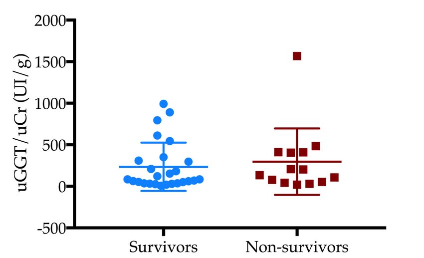
40 dogs (57%). Twenty-one dogs (53%) had uGGT/uCr over the cut-off level which was not associated
with the outcome. No statistical differences were found in uGGT/uCr values between survivor and
non-survivor dogs (Figure 2).
Table 1. Evaluation of dipstick parameters between survivors and non-survivors.
Parameter Survivors (n = 46) Non-Survivors (n = 24) p-Values
pH * 6.5 ± 1 6.3 ± 0.9 0.35
SG § 1018 (1007–1050) 1017 (1004–1048) 0.42
PRO
neg 30 (43%) 10 (14%)
TR 0 (0%) 0 (0%)
1+ 8 (11%) 6 (9%) 0.09
2+ 5 (7%) 2 (3%)
3+ 3 (4%) 6 (9%)
GLU
neg 39 (56%) 24 (34%)
1+ 2 (3%) 0 (0%)
2+ 1 (1%) 0 (0%) 0.24
3+ 0 (0%) 0 (0%)
4+ 4 (6%) 0 (0%)
KET
neg 38 (54%) 20 (29%)
1+ 5 (7%) 4 (6%)
0.3
2+ 3 (4%) 0 (0%)
3+ 0 (0%) 0 (0%)
UBG
norm 16 (24%) 6 (9%)
1+ 24 (35%) 11 (15%)
2+ 5 (7%) 3 (4%) 0.23
3+ 1 (1%) 3 (4%)
4+ 0 (0%) 1 (1%)
BIL
neg 34 (49%) 15 (22%)
1+ 3 (4%) 1 (1%)
0.022
2+ 8 (11%) 2 (3%)
3+ 1 (1%) 6 (9%)
BLD/HGB
neg 23 (33%) 7 (10%)
1+ 3 (4%) 0 (0%)
2+ 6 (9%) 4 (6%) 0.17
3+ 4 (6%) 2 (3%)
4+ 10 (14%) 11 (15%)
SG, specific gravity; PRO, protein; GLU, glucose; KET, ketones; UBG, urobilinogen; BIL, bilirubin; BLD/HGB,
blood/hemoglobin. * p-value obtained with unpaired t-test. § p-value obtained using a Mann–Whitney U-test.
All the other p-values were obtained using a chi square test.110 SG, specific 4+ 10 (14%) GLU, glucose; KET,
gravity; PRO, protein; 11 (15%)
ketones; UBG, urobilinogen; BIL,
110
111 SG, specific
bilirubin; gravity; PRO,
BLD/HGB, protein; GLU,*glucose;
blood/hemoglobin. KET, ketones;
p-value obtained UBG, urobilinogen;
with unpaired BIL,
t-test. § p-value
111
112 bilirubin; BLD/HGB, blood/hemoglobin. * p-value obtained with unpaired t-test. §
obtained using a Mann–Whitney U-test. All the other p-values were obtained using a chi p-value
112
113 obtained
square test.using a Mann–Whitney U-test. All the other p-values were obtained using a chi
113 Vet. Sci. 2019,
square6, 27test. 4 of 7
114
114
115 Figure 1. Evaluation of urine protein-to-creatinine ratio (UPC) severity in the outcome
115
116 Figure
Figure
groups Evaluation
1. 1. of urine
Evaluation
(blue pattern: protein-to-creatinine
of urine
survivors; ratio (UPC)
protein-to-creatinine severity
ratio (UPC)
red pattern: non-survivors) in the outcome
severity
(p = 0.001). groups
in the outcome
116 (blue pattern:
groups survivors;
(blue pattern:red pattern: non-survivors)
survivors; (p = 0.001).
red pattern: non-survivors) (p = 0.001).
117
117
118 Figure 2. Urinary
Figure GGT-to-urinary
2. Urinary creatinine
GGT-to-urinary (uGGT/Cr)
creatinine values between
(uGGT/Cr) survivors survivors
values between and non-survivors
and non-
118
119 (p Figure
> 0.05). 2. Urinary
survivors GGT-to-urinary creatinine (uGGT/Cr) values between survivors and non-
(p > 0.05).
119 survivors (p > 0.05).
4. Discussion
120 4. Discussion
120 As far as we are aware, during AP, various classes of cytokines (“cytokine storm”), especially
4. Discussion
121 As far as we are aware, during AP, various classes of cytokines (“cytokine storm”), especially
interleukin-1, -2, -6 and tumor necrosis factor alfa, and reactive oxygen species can cause multiple
121
122 As far as -2,
interleukin-1, we -6areand
aware, during
tumor AP,factor
necrosis various classes
alfa, of cytokines
and reactive (“cytokine
oxygen species storm”),
can causeespecially
multiple
organ impairment, especially in the kidneys and lungs [4].
122
123 interleukin-1, -2, -6 especially
organ impairment, and tumorinnecrosis factor
the kidneys alfa,
and and[4].
lungs reactive oxygen species can cause multiple
In both human and veterinary medicine, there is constant research conducted on early biomarkers
123
124 organInimpairment,
both human especially in the kidneys
and veterinary and lungs
medicine, there[4].is constant research conducted on early
that can predict renal impairment and acute kidney injury (AKI). In humans, the development of AKI
124
125 In boththat
biomarkers human and veterinary
can predict medicine, and
renal impairment thereacute
is constant research
kidney injury conducted
(AKI). on early
In humans, the
is responsible for 70–80% of early deaths in patients with AP [5,12–14]. For this reason, predicting
125
126 biomarkers that can predict renal impairment and acute kidney injury (AKI).
development of AKI is responsible for 70–80% of early deaths in patients with AP [5,12–14]. For this In humans, the
the severity and understanding the prognosis of these patients is of crucial importance. In dogs with
126
127 development of AKIthe
reason, predicting is responsible
severity andforunderstanding
70–80% of earlythe deaths in patients
prognosis with AP
of these [5,12–14].
patients is ofFor this
crucial
AP, serum creatinine has been proposed as a negative prognostic marker, as well as the presence of
127
128 reason, predicting
importance. In dogsthe withseverity and creatinine
AP, serum understanding
has beenthe proposed
prognosisasofa negative
these patients is of marker,
prognostic crucial
AKI [6,15]. Unfortunately, serum creatinine levels have limited value as an early marker of renal
128
129 importance.
as well as theInpresence
dogs with AP, serum
of AKI creatinine has been
[6,15]. Unfortunately, serumproposed as levels
creatinine a negative
haveprognostic
limited valuemarker,
as an
impairment, as it only changes when the majority of the renal mass is damaged [16].
129
130 as wellmarker
early as the of
presence of AKI [6,15].
renal impairment, as Unfortunately,
it only changes serum
when the creatinine
majoritylevels have
of the limited
renal value
mass is as an
damaged
To date, this is the first study of an evaluation of urinalysis abnormalities and uGGT/uCr in dogs
130
131 early
[16]. marker of renal impairment, as it only changes when the majority of the renal mass is damaged
with AP. In this study, more than the half of the population showed an active sediment. This finding
131 [16].
could be due to the high prevalence of free catches in our sampling method, compared to cystocentesis.
In fact, voided samples of urine may be contaminated with cells and bacteria located in the distal
urethra, genital tract, and/or on the skin and hair [17].Vet. Sci. 2019, 6, 27 5 of 7
High urinary bilirubin concentrations were associated with mortality. Increased bilirubinuria
may be present in human patients with AP [8]. The renal threshold for bilirubin is very low in dogs,
therefore small amounts of bilirubin may be present in the urine of healthy dogs, even when serum
bilirubin is normal [18]. During AP, bilirubinuria could be caused by some subclinical hemolytic
processes or possible disseminated intravascular coagulation due to systemic inflammation and also as
a result of an impairment of a bile duct outflow [1,8]. On the other hand, increased urine bilirubin may
be observed during AP-induced hepatic injury [1,8]. However, only three non-survivor dogs showed
mild hyperbilirubinemia at the biochemical panel.
In the present study, non-survivors showed higher values of UPC than survivors. We evaluated
the UPC excluding active sediment in order to avoid bias in the interpretation of this result. In addition,
a study on the comparison of UPC using cystocentesis versus free catch in dogs showed how the
UPC was minimally affected by free catch, thus allowing the correct grading of proteinuria with this
method [19]. A human paper on urinalysis in humans demonstrated how high urinary protein and
low specific gravity were associated with more severe acute disease and with the development of
AKI [8]. To the best of our knowledge, there is only one veterinary study evaluating renal impairment
in dogs with AP, which showed how high UPC was associated with this outcome [15]. Urinary kidney
injury (uKI) was defined as the presence of casts and/or proteinuria. Casts originate in the kidney, and
their presence supports a diagnosis of renal impairment, as well as the presence of proteinuria [18].
We failed to find an association between uGGT/uCr and the outcome. Urinary GGT is located in
the metabolically active portion of the proximal tubule. It can be found in the urine as a consequence
of acute tubular injury [9,10]. Recently, Lippi et al. [11] studied uGGT/uCr in healthy dogs and in dogs
with various renal diseases (AKI, stable chronic kidney disease (CKD), lower urinary tract infections).
In the latter study, the authors showed how GGT/uCr could be a potentially good marker for the
diagnosis of AKI because dogs with AKI showed significantly higher levels of uGGT/uCr as compared
to healthy dogs [11]. In azotemic dogs, uGGT/uCr may increase as a consequence of a reduced renal
creatinine clearance. The healthy dogs showed a maximum value of uGGT/uCr of 105.6 UI/g and for
this reason we chose this value as the threshold for our dogs. In our study, more than half of the dogs
had a uGGT/uCr value over the chosen cut-off level (>105 U/g), proving that, even if uGGT/uCr was
not associated with the outcome, a mild-to-moderate non-fatal tubular damage during AP could occur.
This study has several limitations. First of all, there was not a standardization in the urine
sampling method. Cystocentesis may be the best way to prevent urine contamination with bacteria,
cells, etc. from the lower urogenital tract [17]. Secondly, dogs may have had other diseases besides
AP that could have affected urinalysis, such as endocrinopathies and hepatopathies. The authors
could not rule out CKD stage 1 dogs, due to the lack of glomerular filtration rate (GFR) or Symmetric
dimethylarginine (SDMA) evaluation. We were only able to collect and analyze 40 urine samples
for the evaluation of uGGT/uCr. Increasing the number of samples should provide more interesting
results, especially including a basal value and a follow-up level in the urine. Although histopathology
is still considered the gold standard for the differentiation of acute from chronic pancreatitis, this
investigation is not feasible in clinical practice. To date, a lack of histopathology in AP diagnosis has
been replaced by using multiple diagnostic investigations as specific tests for pancreatic specific lipase
such as through a hematobiochemical profile and diagnostic imaging exams.
Urinalysis is a cheap and feasible exam that could help the clinician to assess renal impairment.
In dogs with AP kidney injury it is fairly common and seems to be a negative prognostic factor.
Proteinuria, during AP, resulted in a greater negative prognostic marker in urinalysis. Further
research needs to include large-scale prospective studies of the significance and trends associated with
uGGT/uCr in canine AP.Vet. Sci. 2019, 6, 27 6 of 7
Author Contributions: Conception, E.G., V.M., N.B., I.L.; writing—original draft preparation, E.G.;
writing—review and editing, E.G., A.P., I.L., N.B, F.P., V.M.; statistical analysis, A.P., E.G.; supervision, V.M.;
project administration, E.G., V.M.
Funding: This research received no external funding.
Conflicts of Interest: The authors declare no conflict of interest.
References
1. Xenoulis, P.G. Diagnosis of pancreatitis in dogs and cats. J. Small Anim. Pract. 2015, 56, 13–26. [CrossRef]
[PubMed]
2. Hess, R.S.; Saunders, H.M.; Van Winkle, T.J.; Shofer, F.S.; Washabau, R.J. Clinical, clinicopathologic,
radiographic, and ultrasonographic abnormalities in dogs with fatal acute pancreatitis: 70 cases (1986–1995).
J. Am. Vet. Med. Assoc. 1998, 213, 665–670. [PubMed]
3. Pápa, K.; Máthé, A.; Abonyi-Tóth, Z.; Sterczer, A.; Psáder, R.; Hetyey, C.; Vajdovich, P.; Vörös, K. Occurrence,
clinical features and outcome of canine pancreatitis (80 cases). Acta Vet. Hung. 2011, 59, 37–52. [CrossRef]
[PubMed]
4. Mansfield, C. Pathophysiology of acute pancreatitis: Potential application from experimental models and
human medicine to dogs. J. Vet. Intern. Med. 2012, 26, 875–887. [CrossRef] [PubMed]
5. Petejova, N.; Martinek, A. Acute kidney injury following acute pancreatitis: A review. Biomed. Pap. Med. Fac.
Univ. Palacky Olomouc. Czech Repub. 2013, 157, 105–113. [CrossRef] [PubMed]
6. Marchetti, V.; Gori, E.; Lippi, I.; Luchetti, E.; Manca, M.L.; Pierini, A. Elevated serum creatinine and
hyponatraemia as prognostic factors in canine acute pancreatitis. Aust. Vet. J. 2017, 95, 444–447. [CrossRef]
[PubMed]
7. Tvarijonaviciute, A.; García-Martínez, J.D.; Caldin, M.; Martínez-Subiela, S.; Tecles, F.; Pastor, J.; Ceron, J.J.
Serum paraoxonase 1 (PON1) activity in acute pancreatitis of dogs. J. Small Anim. Pract. 2015, 56, 67–71.
[CrossRef] [PubMed]
8. Rybak, K.; Sporek, M.; Gala-Bładzi ˛ ńska, A.; Mazur-Laskowska, M.; Dumnicka, P.; Walocha, J.; Drożdż, R.;
Kuźniewski, M.; Ceranowicz, P.; Kuśnierz-Cabala, B. [Urinalysis in patients at the early stage of acute
pancreatitis]. Prz. Lek. 2016, 73, 88–92. [PubMed]
9. Clemo, F.A. Urinary enzyme evaluation of nephrotoxicity in the dog. Toxicol. Pathol. 1998, 26, 29–32.
[CrossRef] [PubMed]
10. Cobrin, A.R.; Blois, S.L.; Kruth, S.A.; Abrams-Ogg, A.C.G.; Dewey, C. Biomarkers in the assessment of acute
and chronic kidney diseases in the dog and cat. J. Small Anim. Pract. 2013, 54, 647–655. [CrossRef] [PubMed]
11. Lippi, I.; Perondi, F.; Meucci, V.; Bruno, B.; Gazzano, V.; Guidi, G. Clinical utility of urine kidney injury
molecule-1 (KIM-1) and gamma-glutamyl transferase (GGT) in the diagnosis of canine acute kidney injury.
Vet. Res. Commun. 2018, 225, 32–36. [CrossRef] [PubMed]
12. Zhou, J.; Li, Y.; Tang, Y.; Liu, F.; Yu, S.; Zhang, L.; Zeng, X.; Zhao, Y.; Fu, P. Effect of acute kidney injury on
mortality and hospital stay in patient with severe acute pancreatitis. Nephrology 2015, 20, 485–491. [CrossRef]
[PubMed]
13. Kumar, R.; Pahwa, N.; Jain, N. Acute kidney injury in severe acute pancreatitis: An experience from a tertiary
care center. Saudi J. Kidney Dis. Transpl. 2015, 26, 56–60. [PubMed]
14. Naqvi, R. Acute Kidney Injury in association with Acute Pancreatitis. Pak. J. Med. Sci. 2018, 34, 606–609.
[CrossRef] [PubMed]
15. Gori, E.; Lippi, I.; Guidi, G.; Perondi, F.; Pierini, A.; Marchetti, V. Acute pancreatitis and acute kidney injury
in dogs. Vet. J. 2019, 245, 77–81. [CrossRef] [PubMed]
16. Finco, D.R.; Brown, S.A.; Vaden, S.L.; Ferguson, D.C. Relationship between plasma creatinine concentration
and glomerular filtration rate in dogs. J. Vet. Pharmacol. Ther. 1995, 18, 418–421. [CrossRef] [PubMed]
17. Osborne, C.A.; Lulich, J.P.; Albasan, H. The ins and outs of urine collection. In Nephrology and Urology of
Small Animals, 1st ed.; Bartges, J., Polzin, D.J., Eds.; Blackwell Publishing Ltd.: Oxford, UK, 2014; pp. 28–42.
ISBN 978-0-8138-1717-0.Vet. Sci. 2019, 6, 27 7 of 7
18. Barsanti, J.A. Urinary disorders. In Small Animal Clinical Diagnosis by Laboratory Methods, 5th ed.;
Willard, M.D., Tveden, H., Eds.; Elsevier Saunders: St. Louis, MO, USA, 2012; pp. 126–155,
ISBN 978-1437706574.
19. Beatrice, L.; Nizi, F.; Callegari, D.; Paltrinieri, S.; Zini, E.; D’Ippolito, P.; Zatelli, A. Comparison of urine
protein-to-creatinine ratio in urine samples collected by cystocentesis versus free catch in dogs. J. Am. Vet.
Med. Assoc. 2010, 236, 1221–1224. [CrossRef] [PubMed]
© 2019 by the authors. Licensee MDPI, Basel, Switzerland. This article is an open access
article distributed under the terms and conditions of the Creative Commons Attribution
(CC BY) license (http://creativecommons.org/licenses/by/4.0/).You can also read