Primary malignant teratoma of the kidney: a rare case report and literature review - Translational ...
←
→
Page content transcription
If your browser does not render page correctly, please read the page content below
Case Report

Primary malignant teratoma of the kidney: a rare case report and
literature review
Jun Dai1#^, Hong-Chao He1#, Xiao-Qun Yang2, Xin Huang1, Chen Fang1, Wei He1, Ju-Ping Zhao1,
Fu-Kang Sun1
1
Department of Urology, Ruijin Hospital, Shanghai Jiao Tong University, School of Medicine, Shanghai, China; 2Department of Pathology, Ruijin
Hospital, Shanghai Jiao Tong University, School of Medicine, Shanghai, China
#
These authors contribute equally to this work.
Correspondence to: Fu-Kang Sun, MD; Ju-Ping Zhao, MD. Department of Urology, Ruijin Hospital, Shanghai Jiaotong University, School of
Medicine, No.197, Ruijin Er Road, Shanghai 200025, China. Email: sunfukang6@126.com; zhaojp01@126.com.

                 Abstract: Teratomas originate from pluripotent cells and can differentiate along one or more embryonic
                 germ lines. Renal teratoma is infrequent and malignant renal teratoma is even rarer. Experience in the
                 diagnosis and treatment of this uncommon malignancy is seriously limited. In this report, we described
                 the case of a 64-year-old female who complained of right flank pain for 4 months. Computed tomography
                 (CT) revealed a hypodense mass (50 mm in maximum diameter) with slow contrast enhancement and
                 obscure boundary located in the lower pole of the right kidney. CT also showed multiple retroperitoneal
                 lymphadenectasis. Retroperitoneal laparoscopic right radical nephrectomy along with regional
                 lymphadenectomy was successfully performed, and postoperative pathological examination confirmed
                 malignant teratoma of the kidney. After surgery, the patient received adjuvant chemotherapy with BEP
                 (bleomycin, etoposide, and cisplatin) protocol. At the 6-month follow-up, pulmonary and liver metastases
                 were discovered by CT and the patient refused any further treatment. Unfortunately, she died at 16 months
                 postoperatively. Although primary renal malignant teratoma is extremely rare, this kind of tumor should
                 be taken into consideration. Currently, there is no therapeutic standard consensus for this disease and
                 the prognosis remains unclear. Early detection and surgical intervention is critical, and more research on
                 postoperative adjuvant therapy should be performed.

                 Keywords: Case report; malignant teratoma; kidney; prognosis; pathology

                 Submitted Dec 22, 2020. Accepted for publication Mar 05, 2021.
                 doi: 10.21037/tau-21-97
                 View this article at: http://dx.doi.org/10.21037/tau-21-97

Introduction                                                                  regions. Renal teratoma is infrequent, and malignant
                                                                              mass is even rarer. Teratomas are divided into benign
Teratomas are neoplasms composed of one or more
embryonic germ layers: the ectoderm, endoderm, and                            teratoma and malignant teratoma. Skin with dermal
mesoderm. Teratomas are the most common germ                                  appendages, bronchial structures, neuroglial tissue and
cell tumors, which commonly arise in the gonads and                           teeth are commonly seen in teratoma. Malignant teratoma
often occur in infancy and childhood (1). Extragonadal                        is composed of embryonic tissue, mainly neural tissue
teratomas are seldom seen and mainly occur in the anterior                    or contains a malignant component of a type typically
mediastinum, retroperitoneum, and sacrococcygeal                              encountered in other organs and tissues, e.g., sarcomas and

^ ORCID: 0000-0002-9175-6238.

© Translational Andrology and Urology. All rights reserved.             Transl Androl Urol 2021;10(4):1807-1812 | http://dx.doi.org/10.21037/tau-21-97
1808                                                                                   Dai et al. Primary malignant teratoma of the kidney

                                                                        org/10.21037/tau-21-97).

                                                                        Case presentation

                                                                        The patient was a 64-year-old female. She complained of
                                                                        right flank pain for 4 months; the pain was intermittent and
                                                                        spontaneous recurrent without obvious cause. She denied
                                                                        gross hematuria, palpable mass, and emaciation. She had no
                                                                        symptoms of carcinoid syndrome. Aside from hypertension,
                                                                        she had no other previous medical history. General physical
                                                                        examination revealed a slight percussion pain in the right
Figure 1 CT features of a location in the right kidney. CT scan         renal region and there was no palpable mass in the abdominal
shows a hypodense mass, 50 mm in diameter, with slow contrast           region. The biochemical variables and the routine blood
enhancement located in the lower pole of the right kidney and           were normal. Computed tomography (CT) revealed a
lymphadenopathy in the retroperitoneum.                                 hypodense mass (50 mm in maximum diameter) with slow
                                                                        contrast enhancement and obscure boundary located in the
                                                                        lower pole of the right kidney. CT also displayed multiple
                                                                        retroperitoneal lymphadenectasis (Figure 1). An abnormal
                                                                        uptake of fluorodeoxyglucose (FDG) was observed in
                                                                        the low pole of the right kidney by positron emission
                                                                        tomography (PET). Consequently, malignant renal mass
                                                                        was diagnosed, with multiple para-aortic lymph node
                                                                        metastases. All procedures performed in studies involving
                                                                        human participants were in accordance with the ethical
                                                                        standards of the institutional and/or national research
                                                                        committee(s) and with the Helsinki Declaration (as revised
                                                                        in 2013). Written informed consent was obtained from the
                                                                        family members of the patient for publication of this report
                                                                        and any accompanying images.
                                                                           Retroperitoneal laparoscopic right radical nephrectomy
                                                                        along with regional lymphadenectomy was successfully
                                                                        performed under general anesthesia. During the operation,
                                                                        multiple enlarged lymph nodes surrounding renal vessel
                                                                        were also noted and resected.
Figure 2 Tumor specimen. Resected tumor (50 mm × 50 mm ×                   Macroscopically, the mass was located in the mid and
30 mm) located in the mid and lower poles of the left kidney. The       lower poles of the right kidney, and measured approximately
cut surface showed an off-white color with moderate hardness.           50 mm × 50 mm × 30 mm (Figure 2). The cut surface
                                                                        showed an off-white color with moderate hardness. The
                                                                        renal mass with obscure boundary seemed to have an
carcinomas. According to literature reviews, there is no                aggressive ability.
more than 30 cases of renal teratomas and less than 10 renal               Microscopically, pathological examination showed
malignant teratomas have been sporadically reported to                  that the tumor was mainly composed of neuroglial tissue
date. Experience in the diagnosis and treatment of this rare            elements and carcinoma components. Immunohistochemical
malignancy is seriously limited. Therefore, we present a                analysis demonstrated that the tumor cells were positive for
case of renal malignant teratoma in a 64-year-old female                SALL-4, AE1/AE3 (epithelial components), EMA (epithelial
patient and review the related literature.                              components), SYN (glial components and ganglion cells),
   We present the following article in accordance with                  CD56 (colloid components and ganglion cells), S-100 (glial
the CARE reporting checklist (available at http://dx.doi.               components and ganglion cells), GFAP (glial components),

© Translational Andrology and Urology. All rights reserved.         Transl Androl Urol 2021;10(4):1807-1812 | http://dx.doi.org/10.21037/tau-21-97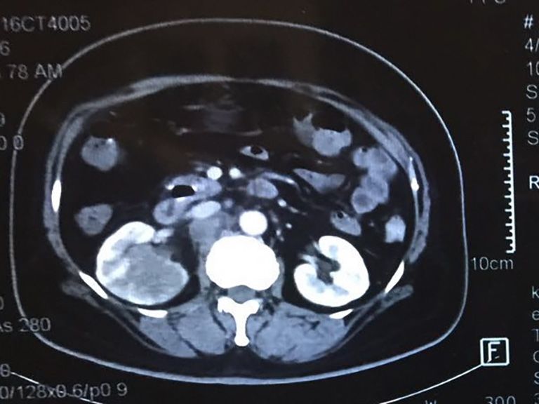
Translational Andrology and Urology, Vol 10, No 4 April 2021                                                                                  1809

    A                                                 B                                         C

   D                                                  E                                          F

Figure 3 IHC findings of primary malignant teratoma of the kidney. (A) AE1/AE3 staining was positive for epithelial components (original
magnification, ×200); (B) synaptophysin staining was positive in the colloid components and ganglion cells (original magnification, ×200);
(C) CD56 staining was positive in the colloid components and ganglion cells (original magnification, ×200); (D) S-100 staining was positive
in the colloid components and ganglion cells (original magnification, ×200); (E) GFAP staining was positive in the colloid components
(original magnification, ×200); (F) Ki67 staining was focally positive in the teratoma component (original magnification, ×200).

Desmin (mesenchymal cell), and Vimentin (mesenchymal                      the patient died at 16 months postoperatively.
and nerve tissue); focally positive for Ki67 (70%); but were
negative for SMA, CD117, D2-40, PLAP, Oct3/4, CD30,
                                                                          Discussion
WT-1, CgA, Bcl-2, Calponin, CD34, AFP, HCG and NUT
(Figure 3). Thus, renal malignant teratoma was diagnosed                  Teratomas are rare neoplasms that most commonly occur
on the basis of above pathological findings.                              in the gonads (ovaries and testes). They are also typically
   Postoperative recovery of the patient was uneventful                   found in the anterior mediastinum, retroperitoneum, and
and the patient was discharged 5 days after surgery. Due                  sacrococcygeal regions, as well as in the central nervous
to the extreme rarity of renal malignant teratoma, there                  system, but have relatively rare (incidence 5%) in other
is no uniform consensus regarding adjuvant treatment. In                  systems, such as abdominal organs (2,3). The kidney is one of
this case, given the multiple lymph node metastases, post-                the least common sites of teratomas. This peculiar distribution
operative adjuvant chemotherapy was administered with                     is probably due to the arrest of primitive germ cells during
BEP (bleomycin, etoposide, and cisplatin) protocol, which                 their migration from the yolk sac to the genital ridge. The
was extrapolated from the guidelines for ovarian malignant                proximity of the genital ridge to the nephrogenic anlage may
germ cells tumors. The patient received three courses of                  explain how germ cells could be displaced into the kidney (2).
chemotherapy with an interval of 2 weeks. No serious                         Primary renal teratoma was first reported by McCurdy
adverse event was observed and routine laboratory tests were              in 1934 (4) as an extremely rare tumor. At present, less
normal. However, abdominal wall metastatic nodules and                    than 30 cases can be found in a MEDLINE search, and
retroperitoneal lymph nodes were detected by abdominal                    there are very few reports regarding malignant teratomas
CT at 3 months postoperatively. At the 6-month follow-up,                 of the kidney (5). The clinical characteristics, pathological
pulmonary and liver metastases were revealed by CT and                    features, and relevant information of most renal malignant
the patient refused any further treatment. Unfortunately,                 teratomas are listed in Table 1. As shown in Table 1, only one

© Translational Andrology and Urology. All rights reserved.           Transl Androl Urol 2021;10(4):1807-1812 | http://dx.doi.org/10.21037/tau-21-97
Table 1 Clinical characteristics, pathologic features, and relevant information regarding renal malignant teratomas (since 2000)
Adjacent invasions 1810
Year of Adjuvant Outcome
Case Journal Gender Age Side Clinical presentation Components of teratoma or lymph node
publication therapy (months/years)
metastasis
Chemotherapy
Malignant epithelioid
Aching pain in the left (bleomycin, 18 months
1 2018 Medicine Male 47 years L components, with a small +
waist etoposide, (no recurrence)
amount of brain tissue
cisplatin)
Keratinizing stratified
squamous epithelium with
skin adnexae, cartilage,
mucinous columnar
Diagnostic Abdominal distension
2 2013 Female 6 months L epithelium, bone, melanin − − N/A
Pathology and pain
containing cells and
neuroglial cells with
occasional foci of immature
© Translational Andrology and Urology. All rights reserved.
neuroectodermal tissue
Pediatric A variety of tissue derived
A left sided abdominal
3 2010 Blood & Male 6 months L from all the 3 germ cell − − N/A
mass
Cancer layers.
Constipation and a Mature teratoma with rare 11 years
4 2001 Urology Male 2 months L N/A −
palpable left flank mass foci of immature elements (no recurrence)
Chemotherapy
The Journal Poor appetite and poor Yolk sac tumor and (etoposide, 7 months
5 2000 Female 2 years L +
of Urology activity immature teratoma vinblastine, BP- (no recurrence)
16, bleomycin)
N/A, not applicable.
Transl Androl Urol 2021;10(4):1807-1812 | http://dx.doi.org/10.21037/tau-21-97
Dai et al. Primary malignant teratoma of the kidneyTranslational Andrology and Urology, Vol 10, No 4 April 2021 1811
case of an adult patient was reported, and our patient should chemotherapy with BEP regimen is controversial. In fact,
be the oldest one. Some reports focused on the surgery there was no improved survival benefit with respect to adjuvant
and pathologic features, and few reports concerned the chemotherapy in our case. Therefore, further investigations
postoperative adjuvant treatment protocol. regarding treatment regimes are needed. Prognosis
It is difficult to make a definitive diagnosis before surgery. in this rare entity is uncertain till now. Among 3 cases
There are neither specific symptoms and signs, nor special with survival reported in table 1, adjacent invasions or
presentations in imaging and biochemical tests for the lymph node metastases occurred in 2 patients who received
diagnosis of primary renal malignant teratoma. Clinical adjuvant chemotherapy postoperatively. Though there was
symptoms could include an abdominal mass, abdominal pain, no recurrence or metastasis in these 2 patients, follow-up
abdominal discomfort, hematuria, anorexia, vomiting, and was only 18 and 7 months respectively. As for the other case
constipation (6). These symptoms and signs are not helpful whose survival time arrived up to 11 years, the tumor was
for differential diagnosis from other renal cell carcinomas. mature teratoma with rare foci of immature elements and
On radiological evaluation, such as ultrasonography, CT, and without lymph node metastasis. We should be aware that renal
magnetic resonance imaging (MRI), the features of primary malignant teratoma is a rare malignancy with an aggressive
renal malignant teratoma are also similar to other renal ability and poor prognosis, and early detection and surgical
cell carcinomas. In the literature, heterogeneous masses, intervention is critical.
sometimes with cystic areas and coarse foci of calcifications or
necrosis can be observed in imaging, however none of these
Conclusions
has diagnostic significance. Hence, the diagnosis of primary
renal malignant teratoma is mainly based on pathological Although primary renal malignant teratoma is extremely
tests combined with immunohistochemistry (IHC). rare, this kind of tumor should be taken into consideration.
Pathologically, there are several characteristics of Such rare and challenging cases should be referred
malignant renal teratomas. Beckwith (7) has reported two to an experienced pathologist for confirmation of
criteria for this tumor: (I) the primary tumor should be histopathological diagnosis. In addition, there is currently
unequivocally of intrarenal origin; the entire lesion should no therapeutic standard consensus for this disease and the
be contained within the renal capsule and there should be prognosis remains unclear. Early detection and surgical
no teratomas in remote sites that might have metastasized intervention are critical, and more research regarding
to the kidney; and (II) the tumor should exhibit unequivocal postoperative adjuvant therapy should be conducted.
heterotopic organogenesis. When a teratoma contains a
type of malignant component typically encountered in
Acknowledgments
other organs and tissues, such as sarcomas or carcinomas, it
is known as a teratoma with somatic type malignancies (8). Funding: This research was funded by the National Natural
In our case, the tumor had neuroglial tissue elements Science Foundation of China (Grant number 81972494) and
and carcinoma elements, and satisfied Beckwith’s criteria. the Practice Project on the Production, Study and Research
Thus, the diagnosis of renal malignant teratoma is for University Teachers in Shanghai (RC20200042)
accurate. Malignant teratomas have a strong resemblance
to small, blue, round cell tumors, which commonly
Footnote
include Wilm’s tumor, metanephric adenoma, lymphoma,
peripheral neuroectodermal tumor, rhabdomyosarcoma, Reporting Checklist: The authors have completed the CARE
and rare metastatic small cell tumors from the lung (9,10). reporting checklist. Available at http://dx.doi.org/10.21037/
Therefore, the diagnosis and differential diagnosis of tau-21-97
malignant teratoma is especially difficult when the tumor
contains various heterogeneous elements, which is also the Conflicts of Interest: All authors have completed the ICMJE
main reason for its misdiagnosis. uniform disclosure form (available at http://dx.doi.
Due to the rare incidence of renal malignant teratoma, org/10.21037/tau-21-97). The authors have no conflicts of
there remains a lack of treatment consensus and standardized interest to declare.
protocol. Radical nephrectomy is considered the first choice
for treatment, however the clinical benefit of adjuvant Ethical Statement: The authors are accountable for all
© Translational Andrology and Urology. All rights reserved. Transl Androl Urol 2021;10(4):1807-1812 | http://dx.doi.org/10.21037/tau-21-971812 Dai et al. Primary malignant teratoma of the kidney
aspects of the work in ensuring that questions related hepatoblastoma and teratoma of the liver in a 3-year-old
to the accuracy or integrity of any part of the work are child: a unique combination and clinical challenge. Diagn
appropriately investigated and resolved. All procedures Pathol 2009;4:37.
performed in studies involving human participants were in 4. Liu YC, Wang JS, Chen CJ, et al. Intrarenal mixed germ
accordance with the ethical standards of the institutional cell tumor. J Urol 2000;164:2020-1.
and/or national research committee(s) and with the Helsinki 5. Govender D, Nteene LM, Chetty R, et al. Mature renal
Declaration (as revised in 2013). Written informed consent teratoma and a synchronous malignant neuroepithelial
was obtained from the family members of the patient for tumour of the ipsilateral adrenal gland. J Clin Pathol
publication of this report and any accompanying images. 2001;54:253-4.
6. Mochizuki K, Ohno Y, Tokai Y, et al. Congenital intrarenal
Open Access Statement: This is an Open Access article teratoma arising from a horseshoe kidney. J Pediatr Surg
distributed in accordance with the Creative Commons 2006;41:1313-5.
Attribution-NonCommercial-NoDerivs 4.0 International 7. Yaqoob N, Ahmed Z, Jafri N, et al. Renal teratoma: a rare
License (CC BY-NC-ND 4.0), which permits the non- entity. J Pak Med Assoc 2003;53:492-3.
commercial replication and distribution of the article with 8. Epstein JI, Egevad L, Amin MB, et al. The 2014
the strict proviso that no changes or edits are made and the International Society of Urological Pathology (ISUP)
original work is properly cited (including links to both the Consensus Conference on Gleason Grading of Prostatic
formal publication through the relevant DOI and the license). Carcinoma: Definition of Grading Patterns and
See: https://creativecommons.org/licenses/by-nc-nd/4.0/. Proposal for a New Grading System. Am J Surg Pathol
2016;40:244-52.
9. Geethamani V, Kusuma V, Gowda KM, et al. Adult Wilms'
References
tumour: a case report with review of literature. Diagn
1. Nirmal TJ, Krishnamoorthy S, Korula A. Primary Pathol 2006;1:46.
intrarenal teratoma in an adult: A case report and review of 10. Idrissi-Serhrouchni K, El-Fatemi H, El madi A, et al.
literature. Indian J Urol 2009;25:404-6. Primary renal teratoma: a rare entity. Diagn Pathol
2. Jones NM, Kiely EM. Retroperitoneal teratomas-- 2013;8:107.
potential for surgical misadventure. J Pediatr Surg
2008;43:184-6; discussion 187. (English Language Editor: A. Kassem)
3. Moll A, Krenauer A, Bierbach U, et al. Mixed
Cite this article as: Dai J, He HC, Yang XQ, Huang X, Fang
C, He W, Zhao JP, Sun FK. Primary malignant teratoma of the
kidney: a rare case report and literature review. Transl Androl
Urol 2021;10(4):1807-1812. doi: 10.21037/tau-21-97
© Translational Andrology and Urology. All rights reserved. Transl Androl Urol 2021;10(4):1807-1812 | http://dx.doi.org/10.21037/tau-21-97You can also read