A Fully Automated, End-to-End Prostate MRI Workflow Solution Incorporating Dot, Ultrashort Biparametric Imaging and Deep-Learning-based ...
←
→
Page content transcription
If your browser does not render page correctly, please read the page content below
Oncological Imaging                                                                                     MAGNETOM Flash (76) 1/2020

A Fully Automated, End-to-End Prostate
MRI Workflow Solution Incorporating Dot,
Ultrashort Biparametric Imaging and Deep-
Learning-based Detection, Classification,
and Reporting
David J. Winkel, M.D.1; Robert Grimm, Ph.D.2; Thomas Benkert, Ph.D.2; Berthold Kiefer, Ph.D.2; Daniel T. Boll, M.D.1

1
    Department of Radiology, University Hospital Basel, Switzerland
2
    MR Applications Predevelopment, Siemens Healthineers, Erlangen, Germany

Introduction
For more than a decade, magnetic resonance imaging               based on the expertise level. However, even among expert
(MRI) has been established as a powerful tool for prostate       radiologists, agreement on prostate cancer classification
cancer diagnosis. The PROMIS study has demonstrated              based on established guidelines is imperfect [7, 8].
that prostate MRI is a suitable triage tool for biopsy-naïve
men, reducing the number of unnecessary biopsies by              This all points to a clear need for
a quarter while improving the detection of clinically signifi-   1. Efficient, reproducible, and robust data
cant cancer [1]. The PRECISION study randomized patients             acquisition workflow
to either systematic biopsies or MRI; with no biopsy if          2. Optimized and fast sequence design
MRI was negative, and targeted biopsy if MRI was positive.       3. Automated detection, classification, and reporting
Targeted biopsies guided by MRI detected significantly               workflows in prostate MRI examinations
more clinically significant cancers while reducing the
number of clinically insignificant cancers [2]. Because
of these findings, MRI for prostate cancer diagnosis has
been integrated into established guidelines [3].
     Increasing demand for prostate MRI examinations can
be expected, as the incidence of prostate cancer increases
with age and life-expectancy in developed countries is
rising. Furthermore, prostate MRI has been discussed in
the literature as a screening tool, similar to breast cancer
screening [4]. However, several limitations need to be
addressed in order to prepare for this increasing prostate
MRI workload. Variation in MRI data acquisitions could be
reduced [5]. Another limitation is the relatively long acqui-
sition time of multiparametric MRI examinations (mpMRI)
employing T2-weighted (T2w), diffusion-weighted imaging
(DWI) and dynamic-contrast enhanced (DCE) MRI. Several
studies have shown that an approach without DCE MRI,
called biparametric MRI (bpMRI), yields comparable results       1  Image acquisition using the Prostate Dot Engine1 including
to mpMRI of the prostate [6]. Potentially even more                  automated prostate contour detection, prostate centering, field
important topic is the varying interpretation performance            of view adaption and three-dimensional correction of spatial axes.

78       siemens.com/magnetom-world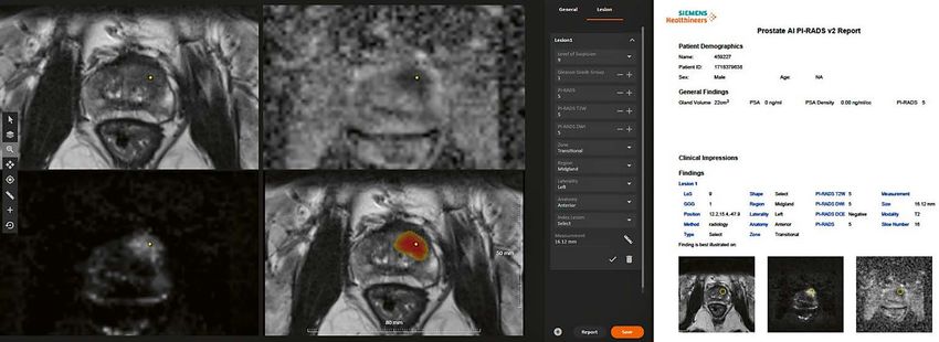
MAGNETOM Flash (76) 1/2020 Oncological Imaging
This is a chain of independent, yet highly interlinked Sequence specifications
stages. Well-registered and reformatted images with The biparametric protocol consists of a T2-weighted turbo
reproducible high image quality are a key prerequisite spin-echo (TSE) pulse sequence in axial, sagittal and
for optimal and reproducible artificial intelligence- coronal orientations and an improved single shot DWI EPI
based analyses. sequence (ZOOMitPRO, Siemens Healthcare, Erlangen,
In this article, we outline an end-to-end solution Germany) with consecutive computation of the apparent
that addresses all the limitations above, incorporating diffusion coefficient. Unlike other DWI techniques,
day optimizing throughput (Dot), ultrashort bpMRI ZOOMitPRO magnifies the prostate (in the phase-encoding
and deep-learning-based lesion detection, classification direction) and is free of infolding artifacts. Either a smaller
and reporting. We present two example cases using quadratic FOV or only a reduced FOV in the phase-encod-
the proposed workflow in order to illustrate its feasibility. ing direction (‘stripe’) is excited (see Figure 2A). As there
is no signal from the non-excited regions, only the small
stripe needs to be encoded (see Figures 2B, C). That
Material and methods
means the encoding time can be decreased while main-
Prostate Dot Engine taining spatial resolution, or the spatial resolution can
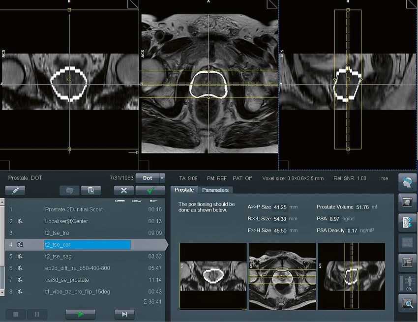
The Prostate Dot Engine1 is a prototype software tool be increased, or a combination of the two. Furthermore,
designed to provide a fast, robust, and standardized image decreased encoding time reduces spatial distortion.
acquisition workflow. After acquiring the Turbo-Spin Echo
(TSE) scout, the Prostate Dot automatically centers the Prostate AI
prostate in the field of view, adapts the size of the field The output of the Prostate Dot Engine goes into the AI
of view and performs a three-dimensional correction of prototype (Prostate AI1, Siemens Healthcare, Erlangen,
spatial axes. Slices can be aligned either strictly orthogonal Germany) for fully automatic prostate lesion detection,
or automatically defined by the orientation of the urethra, classification and reporting.
i.e., perpendicular to the urethra for the axial planes.
Furthermore, the prostate is segmented for standardized As illustrated in Figure 3, Prostate AI contains two parts:
volume assessment. After coil placement, the Dot work- 1. A preprocessing pipeline
flow does not require further adaptations by technicians, 2. A component for lesion detection and classification,
and it allows interruptions and corrections of the scan based on deep learning
process at any time. A screenshot of the Prostate Dot The preprocessing pipeline takes the acquired bpMRI
Engine can be found in Figure 1. sequences and generates the required well-formatted
and transformed data volumes. From the DWI series, a
Work in progress: the application is currently under development and is not for
1 logarithmic extrapolation method is adopted to compute
sale in the U.S. and in other countries. Its future availability cannot be ensured. a new DWI volume with b-value of 2000 s/mm2. This step
2A 2B 2C
2 Single-shot DWI EPI sequence (ZOOMitPRO) with image examples from one study object: (2A) reduced FOV in phase-encoding direction
(blue stripe); (2B) resulting image in comparison to (2C) the conventional RESOLVE technique.
siemens.com/magnetom-world 79
Oncological Imaging                                                                                                              MAGNETOM Flash (76) 1/2020

                                                                                                                                                   Prostate
                                                                                                                                                  Centering

                                                              Automated Dot                              Axial Scout                                  FOV
                                                                  Engine                                  Images                                     Sizing

                                                                                                                                               3D Correction
                                                                                                                                               of Spatial Axes

                                                                                                T2-weighted Turbo-Spin
                                                                  TA = 3:37
                                                                                                 Echo in axial, sagittal
                                                                    min
                                                                                                and coronal orientations
                                                                                                                                                 Biparametric
                                                                                                                                                   Imaging
                                                                                                   Diffusion-Weighted
                                                                  TA = 2:43
                                                                                                      Imaging using
                                                                    min
                                                                                                         ZOOMit

                                                             DWI-2000 & ADC
                                                              Computation

                                                                                                                                            Volume Centering,
                       Volume Parsing                             ADC
                                                                                                       Registration                            Cropping &
                         & Loading                             Computation
                                                                                                                                             Transformation

  Raw DICOM                                                       T2WI
  Sequences                                                    Segmentation

     PI-RADS 3
                          PI-RADS                      FP Reduction                                                    Localization
     PI-RADS 4                                                                              Crop
                        Scoring Net                        Net                                                             Net
     PI-RADS 5

      bpMRI                           PI-RADS 1&2 vs.                  Subvolumes                    PI-RADS 1&2 vs.
                                        PI-RADS ≥ 3                                                    PI-RADS ≥ 3

                             Data                     Review, Edit &                      Report
                         Visualization                Finalize Report                   Generation

 3  Image acquisition workflow using the automated Prostate Dot Engine and biparametric imaging (orange); deep learning architecture with
     preprocessing pipeline (gray); deep learning-based lesion detection and classification component (blue).

Dot = day optimizing throughput, FOV = field of view, 3D = three-dimensional, TA = time of acquisition, DICOM = Digital Imaging and Communications in Medicine,
ADC = apparent diffusion coefficient, FP = false positive, PI-RADS = Prostate Imaging Reporting- and Data System

80       siemens.com/magnetom-world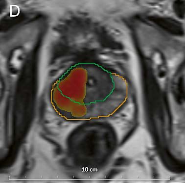
MAGNETOM Flash (76) 1/2020                                                                                                   Oncological Imaging

4  Data visualization platform with the T2w images, ADC map, and high b-value image as well as the T2w image overlaid with the AI-generated
    heatmap (in red and yellow). Prostate AI automatically detected the suspect lesion in the transition zone (TZ, yellow dot) and pre-populated all
    relevant information according to current PI-RADS guidelines. Next, a machine-readable report based on this information is generated.

can eliminate the b-value variances among the datasets                     Cases
and also improve lesion detection performance [10]. Also,
                                                                           Case 1
apparent diffusion coefficient (ADC) maps are computed.
                                                                           Figures 5A-D demonstrate a lesion in the right midgland
Next, whole-organ gland segmentation is performed on
                                                                           PZpl/PZa of a 62-year-old man, with a maximum diameter
the T2w volume using a learning-based method as
                                                                           of 30.2 mm and a mean ADC-value of 758 µm²/s.
presented in Yang et al. [11]. After segmentation, a rigid
                                                                           Prostate AI detected the lesion and assigned a PI-RADS 5
registration is conducted to align T2w and DWI images. The
                                                                           category. Biopsy results revealed a Gleason 4+3 = 7
preprocessing pipeline can eliminate both geometric and
                                                                           pattern.
intensity variances across sequences and patient studies.
     Prostate AI then automatically detects clinically rele-
                                                                           5A                                   5B
vant lesions and classifies each detected lesion according
to PI-RADS categories. This is achieved by a sequence of
coupled deep neural networks that are trained separately.
First, a fully convolutional localization net is able to gener-
ate a semantic lesion candidate heatmap (see Figures 5
and 6); then a sub-volume-based false positive reduction
net further improves detection accuracy by removing the
false positives; finally another sub-volume-based PI-RADS
scoring net stages the level of malignancy for each detec-
tion according to PI-RADS categories.                                       5C                                  5D
     In a last step, Prostate AI displays the detection and
classification results on a dedicated platform. As the ability
of the interpreting radiologist to accept or reject AI-based
findings has been identified as a prerequisite for adoption
of these techniques [12], these capabilities have been
implemented. The user is then able to create a machine-
readable report with all relevant information for the
referring physician (see Figure 4). This report can be sent
to the local RIS/PACS system.

                                                                                                           siemens.com/magnetom-world            81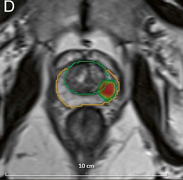
Oncological Imaging                                                                               MAGNETOM Flash (76) 1/2020

Case 2                                                         there is no study investigating workflow differences, such
Figures 6A-D demonstrate a lesion in the left apical PZpl      as time-saving metrics, between Dot-guided and conven-
of a 51-year-old man, with a maximum diameter of               tional, technician-guided workflows. Those studies are
10.2 mm and a mean ADC-value of 961 µm²/s. Prostate AI         currently planned, and their results will contribute to reveal
detected the lesion and assigned a PI-RADS 4 category.         the value of Dot engines in clinical routine.
Biopsy results revealed a Gleason 3+3 = 6 pattern.                  Concerning the use of abbreviated protocols consisting
                                                               of T2-weighted and DWI only – so-called biparametric
                                                               prostate MRI – several studies [6, 13, 14] have shown
6A                             6B                              comparable results as obtained with conventional, mpMRI
                                                               protocols including DCE-MRI. We added another compo-
                                                               nent to our suggested workflow, that is performing DWI
                                                               with the ZOOMitPRO. As shown in Figure 2, ZOOMitPRO uses
                                                               a reduced FOV in the phase-encoding direction compared
                                                               with either standard single shot DWI EPI or RESOLVE
                                                               (REadout Segmentation Of Long Variable Echo trains).
                                                               The resulting decreased acquisition time can be invested
                                                               in a superior spatial resolution. Future studies are needed
                                                               to systematically investigate differences between different
6C                             6D
                                                               types of DWI acquisition schemes compared to the
                                                               ZOOMitPRO technique.
                                                                    The last component in our workflow is the use of
                                                               AI-based lesion detection and classification. Schelb et al.
                                                               [15] used the input from T2w sequences and DWI to train
                                                               a deep learning algorithm (Unet) on the histopathological
                                                               outcome, serving as ground truth. They were able to
                                                               show that this algorithm achieved a similar performance
                                                               to human readers using the PI-RADS assessment score.
                                                               Cao et al. [16] used the input of mpMRI images to build a
                                                               convolutional neural network trained on histopathological
                                                               data and used this algorithm to detect suspicious lesions
Conclusion                                                     and to predict the Gleason score. The results were promis-
In this article, we outlined an end-to-end concept to allow    ing, with a high sensitivity for lesion detection – compara-
a standardized workflow with a reproducible and fast data      ble to expert human readers – and a high classification
acquisition with optimized imaging sequences and an            performance with regards to clinically significant cancer.
AI-empowered data analysis including automated detec-          However, the usefulness of these algorithms needs to be
tion, classification and reporting of suspicious lesions in    proven in larger multi-reader, multi-case (MRMC) studies,
biparametric prostate MRI examinations.                        systematically examining their influence on interpretation
     Reproducible and fast data acquisition concepts are       performance and speed, with and without those solutions.
not only contributing to a standardized reporting per-              We have identified a need to re-structure existing
formed by human readers but would also help artificial         prostate MRI workflows, as patient or – in case of screen-
intelligence-based solutions to reliably process input data.   ing approaches – participant throughput is expected
Preliminary results from a study conducted at the Universi-    to increase. In our vision, current workflows need more
ty of Innsbruck in Austria including 50 patients referred      reliable, reproducible and fast data acquisition steps.
for a prostate MRI examination, compared the tilting angle     Furthermore, recent research has shown that deep learn-
of the auto-alignment of the Prostate Dot Engine against       ing algorithms can compete with human intelligence in
axes determined manually by an experienced radiologist,        prostate MRI reporting. We outlined a possible end-to-end
serving as the reference-standard. The investigators were      solution and demonstrated its feasibility with two case
able to show a mean ± SD deviation of the tilting angle        examples. Future research will investigate what impact
of 5.5 ± 4.4 degrees (Ch. Kremser, W. Judmaier, Med.           the individual components or the combination of those
Uni Innsbruck, unpublished results). However, to date,         components will have on the future of prostate MRI.

82     siemens.com/magnetom-world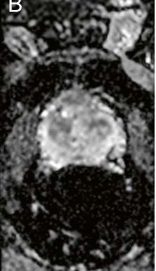
MAGNETOM Flash (76) 1/2020                                                                                                 Oncological Imaging

References

1 Ahmed HU, El-Shater Bosaily A, Brown LC, et al.                       11 Yang D, Xu D, Zhou SK, et al. Automatic Liver Segmentation Using
   Diagnostic accuracy of multi-parametric MRI and TRUS biopsy in          an Adversarial Image-to-Image Network. In: Descoteaux M,
   prostate cancer (PROMIS): a paired validating confirmatory study.       Maier-Hein L, Franz A, et al (eds) Medical Image Computing
   Lancet. 2017;389:815–822.                                               and Computer Assisted Intervention − MICCAI 2017. Springer
   https://doi.org/10.1016/S0140-6736(16)32401-1                           International Publishing, Cham. 2017; pp 507–515.
2 Kasivisvanathan V, Rannikko AS, Borghi M, et al.                      12 Padhani AR, Turkbey B. Detecting Prostate Cancer with Deep
   MRI-Targeted or Standard Biopsy for Prostate-Cancer Diagnosis.          Learning for MRI: A Small Step Forward. Radiology. 2019;192012.
   N Engl J Med. 2018;378:1767–1777.                                       https://doi.org/10.1148/radiol.2019192012
   https://doi.org/10.1056/NEJMoa1801993                                13 Kuhl CK, Bruhn R, Krämer N, et al. Abbreviated Biparametric Prostate
3 EAU Guidelines. Presented at the EAU Annual Congress Barcelona           MR Imaging in Men with Elevated Prostate-specific Antigen.
   2019. ISBN 978-94-92671-04-2.                                           Radiology. 2017;282:493–505.
4 Kim SJ, Vickers AJ, Hu JC. Challenges in Adopting Level 1 Evidence       https://doi.org/10.1148/radiol.2017170129
   for Multiparametric Magnetic Resonance Imaging as a Biomarker        14 Woo S, Suh CH, Kim SY, et al. Head-to-head comparison between
   for Prostate Cancer Screening.                                          biparametric and multiparametric MRI for the diagnosis of prostate
   JAMA Oncol. 2018;4:1663–1664.                                           cancer: A systematic review and meta-analysis. Am J Roentgenol.
   https://doi.org/10.1001/jamaoncol.2018.4160                             2018;211:W226–W241. https://doi.org/10.2214/AJR.18.19880
5 Padhani AR, Barentsz J, Villeirs G, et al. PI-RADS Steering Commit-   15 Schelb P, Kohl S, Radtke JP, et al. Classification of Cancer at Prostate
   tee: The PI-RADS Multiparametric MRI and MRI-directed Biopsy            MRI: Deep Learning versus Clinical PI-RADS Assessment. Radiology.
   Pathway. Radiology. 2019;292:464–474.                                   2019;190938. https://doi.org/10.1148/radiol.2019190938
   https://doi.org/10.1148/radiol.2019182946                            16 Cao R, Bajgiran AM, Mirak SA, et al. Joint Prostate Cancer
6 Weiss J, Martirosian P, Notohamiprodjo M, et al.                         Detection and Gleason Score Prediction in mp-MRI via FocalNet.
   Implementation of a 5-Minute Magnetic Resonance Imaging                 IEEE Trans Med Imaging. 2019;38:2496–2506.
   Screening Protocol for Prostate Cancer in Men with Elevated             https://doi.org/10.1109/TMI.2019.2901928
   Prostate-Specific Antigen before Biopsy.
   Invest Radiol. 2018;53:186–190.
   https://doi.org/10.1097/RLI.0000000000000427
7 Weinreb JC, Barentsz JO, Choyke PL, et al. PI-RADS Prostate
   Imaging - Reporting and Data System: 2015, Version 2. Eur Urol.
   2016;69:16–40. https://doi.org/10.1016/j.eururo.2015.08.052
8 Rosenkrantz AB, Ginocchio LA, Cornfeld D, et al.
   Interobserver Reproducibility of the PI-RADS Version 2 Lexicon:
   A Multicenter Study of Six Experienced Prostate Radiologists.
   Radiology. 2016;280:793–804.
                                                                                                      Contact
   https://doi.org/10.1148/radiol.2016152542                                                          David J. Winkel, M.D.
9 Maas MC, Fütterer JJ, Scheenen TW. Quantitative evaluation of                                       Department of Radiology
   computed high b value diffusion-weighted magnetic resonance                                        University Hospital Basel
                                                                                                      Spitalstrasse 21
   imaging of the prostate. Invest Radiol. 2013;48:779.
                                                                                                      4031 Basel
10 Rosenkrantz AB, Parikh N, Kierans AS, et al. Prostate Cancer
                                                                                                      Switzerland
   Detection Using Computed Very High b-value Diffusion-weighted                                      Tel.: +41 61 328 65 22
   Imaging: How High Should We Go? Acad Radiol. 2016;23:704–711.                                      davidjean.winkel@usb.ch
   https://doi.org/10.1016/J.ACRA.2016.02.003

                                                                                                         siemens.com/magnetom-world             83
You can also read