Correlation of hepcidin and serum ferritin levels in thalassemia patients at Chiang Mai University Hospital
←
→
Page content transcription
If your browser does not render page correctly, please read the page content below
Bioscience Reports (2021) 41 BSR20203352
https://doi.org/10.1042/BSR20203352

Research Article

Correlation of hepcidin and serum ferritin levels in
thalassemia patients at Chiang Mai University
Hospital
Adisak Tantiworawit1 , Sujaree Khemakapasiddhi1 , Thanawat Rattanathammethee1 , Sasinee Hantrakool1 ,
Chatree Chai-Adisaksopha1 , Ekarat Rattarittamrong1 , Lalita Norasetthada1 , Pimlak Charoenkwan2 ,

                                                                                                                                                                                           Downloaded from http://portlandpress.com/bioscirep/article-pdf/41/2/BSR20203352/904254/bsr-2020-3352.pdf by guest on 18 March 2021
  Somdet Srichairatanakool3 and Kanda Fanhchaksai2
1 Division
        of Hematology, Department of Internal Medicine, Faculty of Medicine, Chiang Mai University, Chiang Mai 50200, Thailand; 2 Division of Hematology and Oncology,
Department of Pediatrics, Faculty of Medicine, Chiang Mai University, Chiang Mai 50200, Thailand; 3 Department of Biochemistry, Faculty of Medicine, Chiang Mai University,
Chiang Mai 50200, Thailand
Correspondence: Kanda Fanhchaksai (kanda.f@cmu.ac.th)

                                           Hepcidin is a key iron-regulatory hormone, the production of which is controlled by iron
                                           stores, inflammation, hypoxia and erythropoiesis. The regulation of iron by hepcidin is of
                                           clinical importance in thalassemia patients in which anemia occurs along with iron over-
                                           load. The present study aimed to evaluate the correlation between serum hepcidin and fer-
                                           ritin levels in thalassemia patients. This cross-sectional study investigated 64 patients with
                                           thalassemia; 16 β-thalassemia major (BTM), 31 β-thalassemia/hemoglobin (Hb) E (BE), and
                                           17 Hb H + AE Bart’s disease (Hb H + AE Bart’s). The levels of serum hepcidin and ferritin,
                                           and Hb of the three groups were measured. The median values of serum ferritin and Hb were
                                           significantly different among the three groups, whereas serum hepcidin values were not ob-
                                           served to be significantly different. The correlation of the serum hepcidin and ferritin levels
                                           was not statistically significant in any of the three groups of thalassemia patients with BTM,
                                           BE, or Hb H + AE Bart’s (r = −0.141, 0.065 and −0.016, respectively). In conclusion, no sta-
                                           tistically significant correlations were observed between serum hepcidin with any variables
                                           including serum ferritin, Hb, age, labile plasma iron (LPI), and number of blood transfusion
                                           units among the three groups of thalassemia patients. Likely, the regulation of hepcidin in
                                           thalassemia patients is affected more by erythropoietic activity than iron storage.

                                         Introduction
                                         Thalassemia is the most commonly inherited type of hemolytic anemia in Thailand and is caused by a
                                         globin chain defect. In incidences of thalassemia, red blood cells (RBCs) do not function properly and
                                         survive for shorter periods of time. Consequently, anemia and other related complications can occur.
                                         In Thailand, degrees of frequency are 20–30% for the α-thalassemia trait and Hb E trait, 3–9% for the
                                         β-thalassemia trait, and 1% for thalassemia disease [1,2]. Patients with β-thalassemia display an absence
                                         or a reduced level of β-globin chain synthesis leading to a reduction in Hb in the RBC and ultimately,
Received: 14 October 2020
                                         anemia [3,4]. The basic therapy for β-thalassemia major (BTM) is regular blood transfusions resulting in
Revised: 03 February 2021                iron overload, whereas β-thalassemia intermedia (BTI) is a less severe form of anemia than BTM.
Accepted: 08 February 2021                  Excess iron in vital organs is known to cause impaired organ function and increased rates of morbidity
                                         and mortality [5]. In transfusion-dependent thalassemia (TDT) patients, iron overload mainly occurs as
Accepted Manuscript online:
10 February 2021                         a result of transfusions. In comparison, iron overload in cases of non-transfusion-dependent thalassemia
Version of Record published:             (NTDT) can occur from increased intestinal absorption despite receiving occasional transfusions [3,6].
16 February 2021                         To assess body iron in thalassemia patients, serum ferritin is a key component of a relatively easy and

© 2021 The Author(s). This is an open access article published by Portland Press Limited on behalf of the Biochemical Society and distributed under the Creative Commons Attribution   1
License 4.0 (CC BY).
Bioscience Reports (2021) 41 BSR20203352
https://doi.org/10.1042/BSR20203352
Table 1 Demographic data, type of thalassemia, splenectomy, Hb levels, transfusion dependency, iron overload and
chelations, and co-morbidity in 64 Thai thalassemia cases
Characteristics Number of patients (%)
Female 39 (60.9)
Type of thalassemia
BTM 16 (25.0)
BE 31 (48.4)
Hb H disease+ AE Bart’s 17 (26.6)
Splenectomy 30 (46.9)
TDT 31 (48.4)
Iron overload 48 (75.0)
Iron chelations
Downloaded from http://portlandpress.com/bioscirep/article-pdf/41/2/BSR20203352/904254/bsr-2020-3352.pdf by guest on 18 March 2021
None 22 (34.3)
Desferrioxamine 17 (26.6)
Deferiprone 21 (32.8)
Deferasirox 4 (6.3)
Co-morbidities
Subclinical hypothyroidism 10 (15.6)
Elevated liver enzymes 6 (9.0)
Impaired fasting glucose 4 (6.3)
Primary hypothyroidism 4 (6.3)
Osteoporosis 2 (3.1)
Diabetes mellitus 1 (1.6)
Pulmonary hypertension 1 (1.6)
practical approach; however, the levels are influenced by infection and inflammation. Consequently, a diagnosis of
iron overload using serum ferritin would require serial measurements [7,8].
Iron absorption in humans is regulated by the combined influences of the body’s erythropoietic demand for iron,
tissue oxygenation, and the body’s iron stores [10]. Hepcidin, a 25-amino acid peptide produced by hepatocytes, is
the key hormone involved in the control of iron homeostasis at the point of convergence of the erythroid and the
regulated stores of iron [9,10]. The peptide then binds to the iron export channel ferroportin, thereby inducing its
internalization and degradation and leading to the inhibition of cellular iron efflux [9,11]. The overexpression of
serum hepcidin levels results in iron restricted anemia [12], while low serum hepcidin levels are known to increase
intestinal iron absorption and decrease iron stores in macrophages leading to iron overload [13]. Many studies have
shown that low serum hepcidin levels in β-thalassemia patients can lead to increased iron absorption levels and iron
overload [9,14–17]. The regulation of iron by hepcidin is of clinical importance in thalassemia patients as anemia
often occurs along with iron overload. Hepcidin as a therapeutic target might help the management of iron overload in
thalassemia patients [15,18]. In the present study, we purposed to determine the correlation between serum hepcidin
and ferritin levels in Thai thalassemia patients.
Materials and methods
Patients and study design
This was a cross-sectional study involving patients at Chiang Mai University Hospital, Faculty of Medicine, Chiang
Mai University, Chiang Mai, Thailand from October 2013 to October 2014. The study included 64 patients diagnosed
with thalassemia as follows; 16 BTM, 31 β-thalassemia/Hb E (BE), and 17 Hb H+AE Bart’s diseases. All patients
provided their written informed consent after the study has been approved. The eligibility criteria included the fol-
lowing: being diagnosed with BTM, BE, or hemoglobin H + AE Bart’s disease (Hb H + AE Bart’s) with an age of at
least 15 years old. Patients were excluded if they had received a prior transfusion of packed red cells within 20 days
or had experienced a clinical infection. Medical records and Hb typing results obtained by high performance liquid
chromatography (HPLC) analysis were reviewed in terms of the type of thalassemia and the patient’s history of blood
transfusions for either TDT or NTDT subjects. The NTDT group was defined as patients that had received blood
transfusions less than three times per year. The patients were instructed to withhold iron-chelating agents at least
72 h before blood samples were taken. Venous blood (5 ml) was withdrawn from each participant from which 2 ml
was added to a tube containing ethylenediamine tetraacetic acid (EDTA) anticoagulant and the other one (3 ml) was
2 © 2021 The Author(s). This is an open access article published by Portland Press Limited on behalf of the Biochemical Society and distributed under the Creative Commons Attribution
License 4.0 (CC BY).Bioscience Reports (2021) 41 BSR20203352
https://doi.org/10.1042/BSR20203352
Table 2 Comparison of clinical and laboratory data of thalassemia patients
Thalassemia Hb H+ AE
Variables BTM (n=16) BE (n=31) Bart’s (n=17) P-value
Age (years) 24.5 +
− 1.2 31.2 +
− 1.9 39.4 +
− 2.8 0.001*
Gender (male/female) 7/9 11/20 7/10 0.841
Transfusion dependence 16/16 (100%) 16/31 (51.6%) 1/17 (5.9%)Bioscience Reports (2021) 41 BSR20203352
https://doi.org/10.1042/BSR20203352
Table 3 Correlation between serum hepcidin values and laboratory and demographic variables in thalassemia patients
Number of blood
Group Serum ferritin (ng/ml) Hb (g/dl) Age (y) LPI (μM) transfusion units
rs P-value rs P-value rs P-value rs P-value rs P-value
BTM −0.141 0.602 −0.007 0.978 −0.186 0.491 −0.106 0.696 −0.115 0.670
BE 0.065 0.739 −0.308 0.104 −0.148 0.442 −0.020 0.917 0.084 0.663
−0.016 0.953 0.057 0.834 −0.346 0.189 0.034 0.901 −0.393 0.132
Thalassemia
Hb H+ AE
Bart’s
Abbreviation: rs , Spearman’s rho correlation coefficient.
Downloaded from http://portlandpress.com/bioscirep/article-pdf/41/2/BSR20203352/904254/bsr-2020-3352.pdf by guest on 18 March 2021
fractionated on a glass analytical column (ChromSep-ODS1, 100 mm × 3.0 mm, 5-μm particle size), eluted isocrat-
ically with mobile-phase solvent (3 mM CP22 in 19% acetonitrile/MOPS pH 7.0) at a flow rate of 1.0 ml/min and
optical density (OD) was monitored online at 450 nm using a flow cell detector (SpecMonitor2300; LDC Milton-Roy
Inc., Riviera Beach, FL, U.S.A.). Data analysis was conducted with BDS software (BarSpec Ltd., Rehovot, Israel).
NTBI concentration represented by Fe3+ -(CP22)3 peak area was determined from a calibration curve constructed
from Fe3+ –(NTA)2 in 80 mM NTA (0–16 μM).
Measurement of labile plasma iron
In principle, redox-active labile plasma iron (LPI) can convert non-fluorescent dihydrorhodamine (DHR) into oxi-
dized form rhodamine (R), resulting in an increase in fluorescence intensity (FI). Serum (20 μl) was incubated with
the DHR solution containing ascorbic acid at 37◦ C for 30 min and kinetics of increasing FI was followed immedi-
ately for 40 min, with readings every 2 min using a spectrofluorometer (λexcitation 485 nm, λemission 538 nm). Slope
(FI/min) was determined and plotted against the reaction time between 15 and 40 min. A calibration curve was
constructed by plotting the slope against standard ferrous ammonium sulfate (FAS) solution (0–20 μM). Difference
in rate of DHR oxidation represents a component of redox active LPI in the serum, which the LPI concentration was
calculated from the calibration curve [22,23].
Statistical analysis
Statistical analyses were performed with SPSS version 23. Descriptive data were reported as mean + − standard deriva-
tion (SD) values, as well as median and interquartile range (IQR) values. The clinical data were compared among the
three groups of thalassemia patients using Chi-square test for the categorical data and Kruskal–Wallis Test for the
non-parametric data. The degree of correlation for hepcidin and quantitative variables in each of the three groups
was measured using the Spearman’s Correlation Test. P-valueBioscience Reports (2021) 41 BSR20203352 https://doi.org/10.1042/BSR20203352 or Hb H + AE Bart’s disease was 18.5 (10), 3 (14) and 0 (0), respectively. The number of blood transfusion units was significantly different among the three groups of thalassemia patients (P20 transfusions) patients with BTM [26], and in multitransfused patients with BTM [27]. Notably, serum hepcidin levels decreased due to high erythroid signals [24]. The ferritin levels were observed to be significantly higher in patients with BTM than in those with BE and Hb H + AE Bart’s diseases, respectively. This condition of iron overload could have resulted from regular blood transfusions in BTM subjects when compared with BTI patients. Our data showed that the NTBI values were not different among the three groups of thalassemia and the LPI values were not different between BTM and BE but significantly lower in Hb H + AE Bart’s. This may be explained by the high percentages of iron overload in all groups. A previous study showed the mean serum iron of the thalassemic patients (n=25) was 38.5 + − 7.1 pmol/l with a total iron binding capacity (TIBC) saturation of 85%, in comparison, the mean serum iron of the normal controls (n=50) was 17.8 + − 3.0 pmol/l with 29% saturation of the TIBC [28]. Our © 2021 The Author(s). This is an open access article published by Portland Press Limited on behalf of the Biochemical Society and distributed under the Creative Commons Attribution 5 License 4.0 (CC BY).
Bioscience Reports (2021) 41 BSR20203352
https://doi.org/10.1042/BSR20203352
Downloaded from http://portlandpress.com/bioscirep/article-pdf/41/2/BSR20203352/904254/bsr-2020-3352.pdf by guest on 18 March 2021
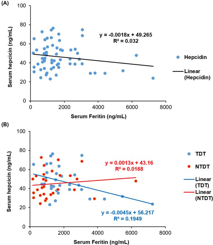
Figure 1. The correlation between serum hepcidin and serum ferritin
(A) Linear correlation between serum hepcidin and serum ferritin levels in all thalassemia patients (R2 = 0.032). (B) Linear correlation
between serum hepcidin and serum ferritin levels in TDT (R2 = 0.1949) and NTDT (R2 = 0.0168) patients.
study has a limitation that transferrin saturation was not measured. The values should have added to the explanation
of the high NTBI values in our patients.
Conclusions
No statistically significant correlations were observed between serum hepcidin with any variables including serum
ferritin, Hb, age, LPI, and number of blood transfusion units among the three groups of thalassemia patients. Likely,
the regulation of hepcidin in β-thalassemia patients was more affected by erythropoietic activity than iron overload.
Importantly, one limitation of the present study was that the erythropoietic activity was not measured.
Data Availability
The data used to support the findings of the present study are included within the article.
6 © 2021 The Author(s). This is an open access article published by Portland Press Limited on behalf of the Biochemical Society and distributed under the Creative Commons
Attribution License 4.0 (CC BY).Bioscience Reports (2021) 41 BSR20203352
https://doi.org/10.1042/BSR20203352
Competing Interests
The authors declare that there are no competing interests associated with the manuscript.
Funding
This work was supported by the Faculty of Medicine Endowment Fund, Chiang Mai University [grant number
FAC-MED-2556-01922].
Author Contribution
A.T. designed the research approach, obtained the research grant, analyzed data, and revised the manuscript. K.F. analyzed the
data, and then wrote and revised the manuscript. S.K. collected and analyzed the data, and wrote the manuscript. T.R., S.H.,
C.C.-A., E.R., L.N., and P.C. revised the manuscript and provided critical comments on the study. S.S. performed hepcidin tests
on the patients.
Downloaded from http://portlandpress.com/bioscirep/article-pdf/41/2/BSR20203352/904254/bsr-2020-3352.pdf by guest on 18 March 2021
Ethics Approval
The study protocol has been approved by the Research Ethical Committee for Human Study of Faculty of Medicine, Chiang Mai
University, Chiang Mai, Thailand (Study Code: FAC-MED-2556-01922). All patients gave their informed consent before their enroll-
ment in the study.
Abbreviations
BE, β-thalassemia/Hb E; BTI, β-thalassemia intermedia; BTM, β-thalassemia major; DHR, dihydrorhodamine; FI, fluorescence
intensity; Hb H + AE Bart’s, hemoglobin H + AE Bart’s disease; Hb, hemoglobin; HPLC, high performance liquid chromatog-
raphy; IQR, interquartile range; LPI, labile plasma iron; MOPS, 3-(N-morpholino)propanesulfonic acid; NTA, nitrilotriacetate;
NTBI, non-transferrin bound iron; NTDT, non-transfusion-dependent thalassemia; R, rhodamine; RBC, red blood cell; TDT,
transfusion-dependent thalassemia.
References
1 Wasi, P., Pootrakul, S., Pootrakul, P., Pravatmuang, P., Winichagoon, P. and Fucharoen, S. (1980) Thalassemia in Thailand. Ann. N.Y. Acad. Sci. 344,
352–363, https://doi.org/10.1111/j.1749-6632.1980.tb33674.x
2 Fucharoen, S. and Winichagoon, P. (1992) Thalassemia in SouthEast Asia: problems and strategy for prevention and control. Southeast Asian J. Trop.
Med. Public Health 23, 647–655
3 Forget, B.G. (2001) Molecular mechanisms of b thalassemia. In Disorders of Hemoglobin (Steinberg, M.H., Forget, B.G., Higgs, D.R. and Nagel, R.L.,
eds), pp. 252–276, Cambridge University Press, Cambridge, U.K.
4 Gardenghi, S., Marongiu, M.F., Ramos, P., Guy, E., Breda, L., Chadburn, A. et al. (2007) Ineffective erythropoiesis in beta-thalassemia is characterized
by increased iron absorption mediated by down-regulation of hepcidin and up-regulation of ferroportin. Blood 109, 5027–5035,
https://doi.org/10.1182/blood-2006-09-048868
5 Robson, K.J., Merryweather-Clarke, A.T., Cadet, E., Viprakasit, V., Zaahl, M.G., Pointon, J.J. et al. (2004) Recent advances in understanding
haemochromatosis: a transition state. J. Med. Genet. 41, 721–730, https://doi.org/10.1136/jmg.2004.020644
6 Schrier, S.L. (2002) Pathophysiology of thalassemia. Curr. Opin. Hematol. 9, 123–126, https://doi.org/10.1097/00062752-200203000-00007
7 Porter, J.B. (2001) Practical management of iron overload. Br. J. Haematol. 115, 239–252, https://doi.org/10.1046/j.1365-2141.2001.03195.x
8 Kohgo, Y., Ikuta, K., Ohtake, T., Torimoto, Y. and Kato, J. (2008) Body iron metabolism and pathophysiology of iron overload. Int. J. Hematol. 88, 7–15,
https://doi.org/10.1007/s12185-008-0120-5
9 Papanikolaou, G., Tzilianos, M., Christakis, J.I., Bogdanos, D., Tsimirika, K., MacFarlane, J. et al. (2005) Hepcidin in iron overload disorders. Blood 105,
4103–4105, https://doi.org/10.1182/blood-2004-12-4844
10 Rechavi, G. and Rivella, S. (2008) Regulation of iron absorption in hemoglobinopathies. Curr. Mol. Med. 8, 646–662,
https://doi.org/10.2174/156652408786241401
11 Ilkovaska, B., Kotevska, B., Trifunov, G. and Kanazirev, B. (2016) Serum hepcidin difference range, gender differences, menopausal dependence and
biochemical correlates in healthy subjects. J. IMAB 22, 1127–1131, https://doi.org/10.5272/jimab.2016222.1127
12 Roy, C.N., Mak, H.H., Akpan, I., Losyev, G., Zurakowski, D. and Andrews, N.C. (2007) Hepcidin antimicrobial peptide transgenic mice exhibit features of
the anemia of inflammation. Blood 109, 4038–4044, https://doi.org/10.1182/blood-2006-10-051755
13 Roetto, A., Papanikolaou, G., Politou, M., Alberti, F., Girelli, D., Christakis, J. et al. (2003) Mutant antimicrobial peptide hepcidin is associated with
severe juvenile hemochromatosis. Nat. Genet. 33, 21–22, https://doi.org/10.1038/ng1053
14 Gardenghi, S., Ramos, P., Follenzi, A., Rao, N., Rachmilewitz, E.A., Giardina, P.J. et al. (2010) Hepcidin and Hfe in iron overload in beta-thalassemia.
Ann. N.Y. Acad. Sci. 1202, 221–225, https://doi.org/10.1111/j.1749-6632.2010.05595.x
15 Gardenghi, S., Ramos, P., Marongiu, M.F., Melchiori, L., Breda, L., Guy, E. et al. (2010) Hepcidin as a therapeutic tool to limit iron overload and improve
anemia in beta-thalassemic mice. J. Clin. Invest. 120, 4466–4477, https://doi.org/10.1172/JCI41717
16 Ravasi, G., Pelucchi, S., Trombini, P., Mariani, R., Tomosugi, N., Modignani, G.L. et al. (2012) Hepcidin expression in iron overload diseases is variably
modulated by circulating factors. PLoS ONE 7, e36425, https://doi.org/10.1371/journal.pone.0036425
© 2021 The Author(s). This is an open access article published by Portland Press Limited on behalf of the Biochemical Society and distributed under the Creative Commons 7
Attribution License 4.0 (CC BY).Bioscience Reports (2021) 41 BSR20203352
https://doi.org/10.1042/BSR20203352
17 Pasricha, S.R., Frazer, D.M., Bowden, D.K. and Anderson, G.J. (2013) Transfusion suppresses erythropoiesis and increases hepcidin in adult patients
with beta-thalassemia major: a longitudinal study. Blood 122, 124–133, https://doi.org/10.1182/blood-2012-12-471441
18 Nemeth, E. (2010) Hepcidin in beta-thalassemia. Ann. N.Y. Acad. Sci. 1202, 31–35, https://doi.org/10.1111/j.1749-6632.2010.05585.x
19 Ganz, T., Olbina, G., Girelli, D., Nemeth, E. and Westerman, M. (2008) Immunoassay for human serum hepcidin. Blood 112, 4292–4297,
https://doi.org/10.1182/blood-2008-02-139915
20 Zipperer, E., Post, J.G., Herkert, M., Kundgen, A., Fox, F., Haas, R. et al. (2013) Serum hepcidin measured with an improved ELISA correlates with
parameters of iron metabolism in patients with myelodysplastic syndrome. Ann. Hematol. 92, 1617–1623,
https://doi.org/10.1007/s00277-013-1839-5
21 Singh, S., Hider, R.C. and Porter, J.B. (1990) A direct method for quantification of non-transferrin-bound iron. Anal. Biochem. 186, 320–323,
https://doi.org/10.1016/0003-2697(90)90088-Q
22 Cabantchik, Z.I., Glickstein, H., Milgram, P. and Breuer, W. (1996) A fluorescence assay for assessing chelation of intracellular iron in a membrane
model system and in mammalian cells. Anal. Biochem. 233, 221–227, https://doi.org/10.1006/abio.1996.0032
23 Pootrakul, P., Breuer, W., Sametband, M., Sirankapracha, P., Hershko, C. and Cabantchik, Z.I. (2004) Labile plasma iron (LPI) as an indicator of
Downloaded from http://portlandpress.com/bioscirep/article-pdf/41/2/BSR20203352/904254/bsr-2020-3352.pdf by guest on 18 March 2021
chelatable plasma redox activity in iron-overloaded beta-thalassemia/HbE patients treated with an oral chelator. Blood 104, 1504–1510,
https://doi.org/10.1182/blood-2004-02-0630
24 Haghpanah, S., Esmaeilzadeh, M., Honar, N., Hassani, F., Dehbozorgian, J., Rezaei, N. et al. (2015) Relationship between serum hepcidin and ferritin
levels in patients with thalassemia major and intermedia in Southern Iran. Iran Red Crescent Med. J. 17, e28343,
https://doi.org/10.5812/ircmj.17(5)2015.28343
25 Jagadishkumar, K., Yerraguntla, N. and Vaddambal, M.G. (2018) Serum hepcidin levels in children with beta thalassemia major. Indian Pediatr. 55,
911–912, https://doi.org/10.1007/s13312-018-1408-z
26 Chauhan, R., Sharma, S. and Chandra, J. (2014) What regulates hepcidin in poly-transfused beta-Thalassemia Major: erythroid drive or store drive?
Indian J. Pathol. Microbiol. 57, 39–42, https://doi.org/10.4103/0377-4929.130891
27 Jawad, M., Aftab, I., Saeed, M.T., Mumtaz, G., Iram, S. and Mohsin, S. (2016) Hepcidin levels in multi transfused β thalassemia major patients. J.
Rawalpindi Med. College 20, 206–208
28 Hershko, C., Graham, G., Bates, G.W. and Rachmilewitz, E.A. (1978) Non-specific serum iron in thalassaemia: an abnormal serum iron fraction of
potential toxicity. Br. J. Haematol. 40, 255–263, https://doi.org/10.1111/j.1365-2141.1978.tb03662.x
8 © 2021 The Author(s). This is an open access article published by Portland Press Limited on behalf of the Biochemical Society and distributed under the Creative Commons Attribution
License 4.0 (CC BY).You can also read