The combination of Demodex folliculorum and Aerobic Bacteria in the Etiopathogenesis of Chronic Blepharitis
←
→
Page content transcription
If your browser does not render page correctly, please read the page content below
JOURNAL OF
                                                                                                                                         Journal of

CONTEMPORARY MEDICINE
                                                                                                                                         Contemporary
                                                                                                                                         Medicine

DOI: 10.16899/jcm.791708
J Contemp Med 2021;11(2):142-146

Orjinal Araştırma / Original Article

        The combination of Demodex folliculorum and Aerobic
        Bacteria in the Etiopathogenesis of Chronic Blepharitis
                Kronik Blefarit Etyopatogenezinde Demodex folliculorum ve
                                Aerop Bakterilerin Birlikteliği
                                           Murat Cakmakliogullari1, Ahmet Ozbilgin2
                       1Karabük University Training and Research Hospital Ophthalmology Department, Karabük, Turkey
                          2Celal Bayar University School of Medicine Medical Parasitology Department, Manisa, Turkey

Abstract                                                                       Öz
Aim: This study was conducted to investigate the presence of                   Amaç: Bu çalışmada kronik blefarit tanısı almış hastalarda D.
thecombination of Demodex folliculorum and aerobic bacteria in                 folliculorum ile aerop bakteri birlikteliğini araştırmayı amaçladık.
patients with chronic blepharitis.
                                                                               Gereç ve Yöntem: Kronik blefarit tanısı alan 71 hastada, kirpik
Material and Method: Seventy-one patients diagnosed with                       epilasyonu ile alınan kirpiklerden hazırlanan preparatlar             ışık
chronic blepharitis were evaluated for the presence of D.
folliculorum by light microscope examination of samples prepared               mikroskobunda D. folliculorum varlığı açısından değerlendirildi.
from eyelashes collected by eyelash epilation. Culture samples                 Aynı zamanda bu hastaların kapak marjından kültür örnekleri alındı.
were also obtained from patients’ eyelid margins. Bacterial                    Kültürde üreyen baskın bakteri kolonilerinin tür tayinleri, BD Phoenix
strainsamong the predominant bacterial colonies grown in cultures              (BD Diagnostic Systems, Sparks, USA) tanımlama sistemi kullanılarak
were identified using the BD Phoenix identification system (BD                 yapıldı. Hastalar, D. folliculorum saptananlar Demodex pozitif
Diagnostic Systems, Sparks, USA). Patients were divided into two               saptanmayanlar ise Demodex negatif olmak üzere iki gruba ayrıldı.
groups, Demodex-positive and Demodex-negative,and compared
                                                                               Gruplar bakteri üremesi ve üreyen bakteri türlerine göre karşılaştırıldı.
according to bacterial production and bacterial strains produced.
Results: D. folliculorum was identified in 42 (59.1%) patients.                Bulgular: Hastaların 42’sinde (%59,1) D. folliculorum varlığı saptandı.
Comparison between Demodex-positive and -negative groups                       Demodex pozitif grupla Demodex negatif grup karşılaştırıldığında yaş
revealeda statistically significant increase in Demodex positivity             arttıkça Demodex pozitifliğinin de istatistiksel olarak arttığı saptandı.
with age. There was no significant relationship between gender                 Cinsiyet ile Demodex pozitifliği arasında anlamlı bir ilişki bulunmadı.
and Demodex positivity. The Demodex-positive group showed                      Demodex pozitif grupta alınan kültür örneklerinde bakteri üremesi
a statistically significantly higher bacterial growth in the culture           Demodex negatif gruba göre anlamlı yüksek bulundu. Her iki grupta
samples than the Demodex-negative group. Both groups
                                                                               en sık S. epidermidis üremesi olduğu saptandı. Demodex pozitif
exhibited a predominance of Staphylococcus epidermidis. S.
epidermidis (38.1% vs. 31.0%), Staphylococcus aureus (19.0% vs.                hastalarda Demodex negatif olanlara göre daha yüksek oranda
10.3%), and Corynebacterium spp. (16.7% vs. 6.9%) were detected at             görülen bakteri türlerinin S. epidermidis (%38,1; %31,0), S. aureus
higher rates in the Demodex-positive group than in the Demodex-                (%19,0; %10,3) ve Corinobacterium spp. (%16,7; %6,9) olduğu saptandı.
negative group. There was no statistically significant difference              Demodex pozitif ve Demodex negatif gruplarda üreyen bakteri türleri
between both groups regarding the presence of these bacterial                  karşılaştırıldığında türler arasında anlamlı bir fark bulunmadı.
species.
                                                                               Sonuç: Kronik blefaritli hastalarda sıklıkla D. folliculorum ile normal
Conclusions: Patients with chronic blepharitis could have a
                                                                               kapak florasında bulunan aerop bakterilerin birlikte mix enfeksiyon
mixedinfection site with the combination of D. folliculorum and
aerobic bacteria found in the normal eyelid flora.                             alanı oluşturabileceği düşüncesindeyiz.
Keywords: Chronic blepharitis, Demodex folliculorum, aerobic                   Anahtar Kelimeler: Kronik blefarit, D. folliculorum, aerop bakteriler,
bacteria, etiopathogenesis                                                     etyopatogenez

Corresponding (İletişim): Murat Cakmakliogullari, Karabük University Training and Research Hospital Ophthalmology Department, Karabük,
Turkey
E-mail (E-posta): drmurat3545@gmail.com
Received (Geliş Tarihi): 07.09.2020 Accepted (Kabul Tarihi): 17.12.2020
143 Journal of Contemporary Medicine
INTRODUCTION (Becton Dickinson, USA), eosin methylene blue agar (Becton
Demodex folliculorum and D. brevis are two strains of Demodex Dickinson, USA), and chocolate agar (Becton Dickinson, USA)
mites found in humans. They are most commonly found as and then incubated at 35°C for 24–48h. The strains found among
ectoparasites on the human skin and are also present in the the predominant bacterial colonies grown in the culture media
eyelid flora.[1] The majority of D. folliculorum species are located were identified using a fully automated BD Phoenix bacterial
in the infundibular section of hair follicles, whereas D. brevis are identification system (BD Diagnostic Systems, Sparks, USA).
located more deeply in the sebaceous gland and ductus.[2] Demodex Examination
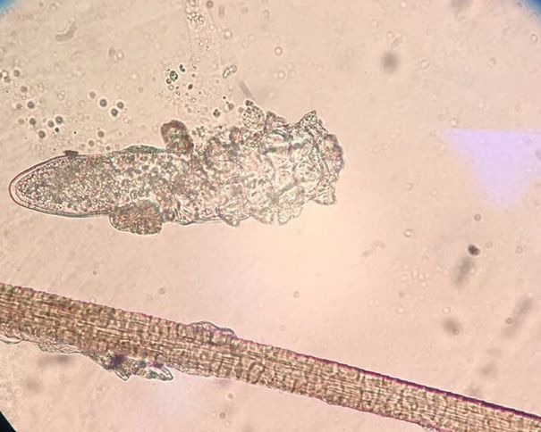
These mites can cause ocular disorders like blepharitis, Under biomicroscopy, six eyelashes were collected from the
conjunctivitis and keratitis.[3-5] Blepharitis is a frequently observed upper and lower eyelids of the patients by epilation. The
clinical condition, generally accompanied by a chronic course. eyelashes were sent to the microbiology laboratory in a sterile
Symptoms primarily include itching, burning, redness, feeling petri dish. The eyelash sampleswere mounted on slides and
of a foreign body in the eye, and flaking at the eyelash roots. prepared using saline according to the procedure described
It is generally diagnosed from the clinical appearance of the by English et al.[10] after which they were evaluated under a
eyelid and the accompanying symptoms.[6] Mites cause reactive light microscope at 10× and 40× magnification to detect the
hyperkeratinisation and epithelial hyperplasia by obstructing presence of D. folliculorum.
the sebaceous gland outlets and eyelash root follicles and play
Statistical Analysis
a role as a mechanical vector for the spread of bacteria.[7]
Statistical analyses were performed using the IBM SPSS version
The most common aerobic bacterial agents causing blepharitis
24.0 software (IBM Corporation, Armonk, NY, USA). Continuous
are Staphylococcus epidermidis, S. aureus, and Corynebacterium
variables were represented as mean±standard deviation,
spp., and the most common parasitic agent is D. folliculorum.[8]
and categorical data were represented as number (n) and
This widespread parasite is accepted as a saprophytic organism
percentage (%). In the analyses between groups of continuous
on the skin, and it is currently accepted as the pathogenic agent
variables, conformity of the data to normal distribution was
of chronic blepharitis; hence, when the parasite is determined,
assessed using the Kolmogorov–Smirnov test. Comparisons of
antiparasitic treatment is recommended.[9]
two groups of data with normal distribution were conducted
We conducted this study to investigate the presence of the using the t-test.The chi-square test was applied for comparisons
combination of D. folliculorum and aerobic bacteria in patients of categorical data. A value of p
Murat Cakmakliogullari, Demodex folliculorum and bacteria in blepharitis                                                                                    144

                                                                                In the culture samples obtained from the 71 patients with
    10x                                                                         chronic blepharitis, aerobic bacterial growth was detected
                                                                                in 59 (83.1%) patients and no bacterial growth was detected
                                                                                in 12 (16.9%) patients. According to the frequency of the
                                                                                bacterial growth, S. epidermidis (35.2%), S. aureus (15.5%), S.
                                                                                epidermidis other coagulase-negative Staphylococcus (CNS)
                                                                                (15.5%), Corynebacterium spp. (12.7%), and Streptococcus spp.
                                                                                (5.6%) were identified.
                                                                                Regarding the distribution of bacterial species, S.
                                                                                epidermidis (38.1% vs. 31.0%), S. aureus (19.0% vs.10.3%),
                                                                                and Corynebacterium spp.(16.7% vs. 6.9%) were observed
                                                                                at higher rates in the Demodex-positive group than in the
                                                                                Demodex-negative group. Streptococcus spp. (6.9% vs. 4.8%)
                                                                                and other CNS (20.7% vs.11.9%) were detected at higher
                                                                                rates in the Demodex-negative group than in the Demodex-
                                                                                positive group. However, there was no statistically significant
                                                                                difference between the patient groups withrespect to the
                                                                                bacterial species(p>0.05) (Table 3).

                                                                                Table 3. Comparisons between Demodex-positive and -negative groups
                                                                                with respect to bacterial species
    40x                                                                                                   Demodex- Demodex-         Total
                                                                                                           Negative Positive       (n=71)        p
                                                                                                            (n=29)   (n=42)
                                                                                                                      n    %     n     %     n     %
                                                                                S. epidermidis                        9   31.0   16   38.1   25   35.2   0.618*
                                                                                S. aureus                             3   10.3   8    19.0   11   15.5   0.506*
                                                                                Corynebacterium spp.                  2    6.9   7    16.7    9   12.7   0.239*
                                                                                Streptococcus spp.                    2    6.9   2    4.8     4   5.6    1.000*
                                                                                Other CNS                             6   20.7   5    11.9   11   15.5   0.338*
                                                                                * Chi-square test (Fisher’s exact test)

                                                                                DISCUSSION
                                                                                Blepharitis is a clinical condition frequently observed in the
                                                                                community and generally has a chronic course. Despite the
                                                                                presence of inflammation in the eyelash roots, the aetiology
                                                                                of blepharitis is still not completely understood.[6] It is believed
Figure 1. Microscopic eyelash examination of D. folliculorum at 10× and 40×
                                                                                that bacterial infections and inflammatory skin lesions such
                                                                                as atopic dermatitis together with D. folliculorum infestations
                                                                                play a role in the etiopathogenesis.[11]
Regarding bacterial growth, 90.5% of patients in the Demodex-
                                                                                The results of several studies support the relationship
positive group and 72.4% of those in the Demodex-negative
                                                                                between Demodex and blepharitis.[12-18] In a study conducted
group had bacterial growth in the cultures. The difference                      by Lee et al.[13] to determine the prevalence of Demodex,
between the two groups with respect to bacterial growth was                     positivity was determined at 70%, and a strong correlation
statistically significant (p=0.048) (Table 2).                                  was reported between the number of Demodex and the
                                                                                severity of the ocular disorder. In a case-controlled study,
Table 2. Comparisons between Demodex-positive and -negative patients            Biernat et al.[14] reported Demodex frequencies of 62.4% in
with respect to bacterial growth
                                                                                patients with chronic blepharitis and 24.3% in the control
                    Demodex-        Demodex-               Total
                  Negative (n=29) Positive (n=42)         (n=71)                group, with the difference being statistically significant.
                                                                         p
                                                                                In another meta-analysis, the probability of developing
                       n            %     n    %      n            %
                                                                                symptomatic blepharitis was reported to be 4.7-fold greater
Bacterial
growth (-)             8          27.6    4    9.5    12       16.9             in patients with Demodex in the eyelashes.[15] Consistent
                                                                       0.048*   with these findings in the literature, our study determined a
Bacterial             21          72.4    38   90.5   59       83.1             Demodex positivity of 59.1% in 42 of 71 patients with chronic
growth (+)
* Chi-square test (Fisher’s exact test)                                         blepharitis.
145 Journal of Contemporary Medicine
Although previous studies did not find a significant difference D. folliculorum causes direct damage to the follicular
in the frequency of D. folliculorum according to gender, it has epithelium within the eyelash follicles in the eyelid and eyelid
been found to increase with age.[13,14,19] Arici et al.[20] reported margin. The development of blepharitis due to the bacteria
that the presence of Demodex was not related to age and carried on the surface of Demodex mites triggers the host
gender. Demirmizrak et al.[21] found that there was a statistically immune response. The resulting mechanical blockage and
significant increase in male gender and the frequency of D. the delayed oversensitivity reaction cause inflammation in
folliculorum with increasing age. In the current study as well, the eyelid margin.[1] In a study conducted by Kim et al.[27] the
although there was no change in the frequency of D. folliculorum levels of IL-17, causing inflammation of the eyelid and ocular
according to gender, a positive correlation was found between surface, weredetected at a significantly higher rate in patients
the frequency of Demodex and age. with Demodex-infected blepharitis, indicating inflammatory
The eyelid margin is known to host normal bacterial flora events (27).
consisting of S. epidermidis, S. aureus, and, to a lesser degree, Due to the uncertainty in etiopathogenesis, the treatment
Corynebacterium spp. (22). In a study conducted by Groden et for blepharitis is confusing and ineffective, and the majority
al.[8] the commonly isolated bacteria in patients with chronic of cases become chronic. In blepharitis cases, bacterial
blepharitis were S. epidermidis (95.8%), Corynebacterium spp. infections and allergies are considered initially, due to which
(76.8%), Acinetobacter spp. (11.4%), and S. aureus (10.5%). antibacterial and steroid drops are generally used in empirical
Compared with the control group, the detection rates of S. treatment. If the cause of blepharitis is D. folliculorum
epidermidis and Corynebacterium spp. were significantly higher infestation, the patient would not benefit from this treatment
in patients with chronic blepharitis. Demler et al.[23] reported and the condition could become chronic. Pretreatment
a 52% D. folliculorum positivity rate in patients with chronic detection of the parasite may be beneficial, especially in
blepharitis and an increase in both Gram-negative and Gram- treatment-resistant chronic blepharitis patients.[28] As found
positive bacteria in the Demodex-positive patients. In a recent in the present study, D. folliculorum and bacterial agents are
study conducted by Zhu et al.[24] D. folliculorum frequency often detected together in patients with chronic blepharitis
(76.7%) was found to be significantly higher in patients with and it must be remembered that this creates a complicated
chronic blepharitis than in the control group (41.3%). Moreover, infection site, which must not be ignored in the treatment
the authors found no statistically significant increase in the process. Therefore, the use of tea tree oil is predominant in
density of the aerobic bacteria S. epidermidis and S. aureus in the treatment as it has been proven to have antiparasitic,
the presence of Demodex mites in the culture samples obtained antibacterial and anti-inflammatory effects in previous
from the eyelid margin and eyelashes. In the present study, S. studies.[29,30] In invivo and invitro studies conducted by Goa
epidermidis, other CNS, S. aureus, Corynebacterium spp., and et al.[31] it was observed that Demodex mites were effectively
Streptococcus spp. were detected in patients with chronic eliminated by treatment with tea tree oil.
blepharitis. Although bacterial growth was significantly higher A limitation of the present study was that the patients were
in Demodex-positive patients, no significant difference was not followed up after treatment. In future studies, changes in
observed between the bacterial strains. The most frequently the combination of bacterial strains and D. folliculorum should
isolated bacterium in patients with blepharitis patients has been be investigated after the application of various treatments,
reported to be S. epidermidis.[7,23] Consistent with this finding in which would helpin determining the treatment efficacy in
the literature, there was a predominance of S. epidermidis in more detail.
patients with chronic blepharitis in the present study.
Mites function as vectors, especially for Staphylococcus species
(25). In the current study, S. epidermidis (38.1% vs. 31.0%), S. CONCLUSION
aureus (19.0% vs.10.3%), and Corynebacterium spp.(16.7% vs. The results of this study suggest that in patients with
6.9%) were determined at higher rates in Demodex-positive chronic blepharitis, a mixed infection area is formed by the
patients than in Demodex-negative patients, but the difference combination of D. folliculorum and aerobic bacteria found in
was not statistically significant. This suggests that these the normal eyelid flora. It is beneficial to consider these results
bacteria, which are frequently found in the normal eyelid flora, during the initiation ofempirical treatment in patients with
settle more in the eyelash follicles through the mediation of chronic blepharitis.
Demodex mites and lead to the formation of a mixed infection.
Staphylococci are found in the normal eyelid flora and just like ETHICAL DECLARATIONS
mites, there is an increase in colonisation with age. The biofilm
Ethics Committee Approval: This study was approved by
layer formed by S. epidermidis in particular provides a suitable
the Clinical Research Ethics Committee of Karabük University
living and nutritional environment for Demodex mites. If the
Medical Faculty (no: 13, dated:31/08/2016)
cylindrical layer that forms with the accumulation of the biofilm
layer around the eyelash is accepted as a pathognomic finding Informed Consent: All patients signed the free and informed
for Demodex mites, there may be an association between S. consent form.
epidermidis and Demodex mites.[22,26] Referee Evaluation Process: Externally peer-reviewed.Murat Cakmakliogullari, Demodex folliculorum and bacteria in blepharitis 146
Conflict of Interest Statement: The authors have no conflicts 24. Zhu M, Cheng C, Yi H, Lin L, Wu K. Quantitative analysis of the bacteria in
of interest to declare. blepharitis with demodex infestation. Front. Microbiol. 2018; 9:1719
25. Shelley WB, Shelley ED, Burmeister V. Unilateral demodectic rosacea. J.
Financial Disclosure: This study was supported by Am. Acad. Dermatol.1989; 20:915–917.
Afyonkarahisar health sciences university medical faculties 26. Gao YY, Di Pascuale MA, Li W, Liu DT, Baradaran-Rafii A, Elizondo A, et
Fund (Project Number: 2020/396). al. High prevalence of Demodex in eyelashes with cylindrical dandruff.
Investig. Ophthalmol. Vis. Sci. 2005; 46:3089–3094.
Author Contributions: All of the authors declare that they 27. Kim JT, Lee SH, Chun YS, Kim JC. Tear cytokines and chemokines in
have all participated in the design, execution, and analysis of patients with Demodex blepharitis. Cytokine. 2011; 53:94–99.
the paper, and that they have approved the final version. 28. Tanrıverdi C, Demirci G, Balcı Ö, Odabaşı M, Özsütçü M. Investigation of
Demodex Parasitic Existence in Treatment-Resistant Chronic Blepharitis
Cases. Turkiye Parazitol Derg 2018; 42: 130-3.
REFERENCES 29. Messager S, Hammer KA, Carson CF, Riley TV. Assessment of the
antibacterial activity of tea tree oil using the European EN 1276 and EN
1. Liu J, Sheha H, Tseng SC. Pathogenic role of Demodex mites in blepharitis.
12054 standard suspension tests. J. Hosp. Infect. 2005; 59:113–125.
Curr.Opin. Allergy Clin. Immunol. 2010; 10:505–510.
30. Halcón L, MilkusK.Staphylococcus aureus and wounds: a review of tea tree
2. Nutting WB, Green AC. Hair follicle mites (Acari: Demodicidae) from
oil as a promising antimicrobial. Am. J. Infect. Control2004; 32:402–408.
Australian aborigines. Aust. J. Dermatol.1974; 15:10–14.
31. Gao YY, Di Pascuale MA, Li W, Baradaran-Rafii A, Elizondo A, Kuo CL et al. In
3. Kheirkhah A, Casas V, Li W, Raju VK, Tseng SC. Corneal manifestations of vitro and in vivo killing of ocular Demodex by tea tree oil. Br. J.Ophthalmol.
ocular demodex infestation. Am. J.Ophthalmol. 2007; 143:743–749. 2005; 89:1468–1473.
4. Luo X, Li J, Chen C, Tseng S, Liang L. Ocular demodicosis as a potential
cause of ocular surface inflammation. Cornea. 2017; 36:9–14.
5. Bhandari V, Reddy JK. Blepharitis: always remember demodex. Middle
East Afr. J.Ophthalmol. 2014; 21:317–320.
6. Kanski JJ, Bowling B. Clinical ophthalmology: A systematic approach.
Elsevier Health Sciences. 2011.
7. Baima B, Sticherling M. Demodicidosis revisited. Acta Derm.Venereol.
2002; 82:3–6.
8. Groden LR, Murphy B, Rodnite J, Genvert GI. Lid flora in blepharitis.
Cornea, 1991; 10:50–53.
9. Kamoun B, Fourati M, Feki J, Mlik M, Karray F, Trigui A et al. Blépharite à
Démodex: mytheouréalité. J. Fr.Ophtalmol. 1999; 22:525–527.
10. English FP, Nutting WB, Cohn D. Eyelid mite nests. Aust 3 Ophthaimol.
1982; 10:187–189.
11. Lindsley K, Matsumura S, Hatef E, Akpek EK. Interventions for chronic
blepharitis. Cochrane Database Syst. Rev.2012; 5, CD005556. doi:
10.1002/14651858.CD005556.pub2
12. Rusiecka-Ziólkowska J, Nokiel M, Fleischer M. Demodex - An old pathogen
or a new one? Adv. Clin. Exp. Med. 2014; 23:295–298.
13. Lee SH, Chun YS, Kim JH, Kim ES, Kim JC. The relationship between
Demodex and ocular discomfort. Investig. Ophthalmol. Vis. Sci.2010; 51:
2906–2911.
14. Biernat MM, Rusiecka-Ziółkowska J, Piątkowska E, Helemejko I, Biernat P,
Gościniak G. Occurrence of Demodex species in patients with blepharitis
and in healthy individuals: a 10-year observational study. Jpn. J.
Ophthalmol. 2018; 62, 628–633.
15. Zhao YE, Wu LP, Hu L Xu JR. Association of blepharitis with Demodex: a
meta-analysis. Ophthalmic Epidemiol. 2012; 19:95–102.
16. Eroglu S, Cakmakliogullari M, KalCakmakliogullari E. Is the presence of
Demodex folliculorumincreased with impaired glucose regulation in
polycystic ovary syndrome?J. Obstet. Gynaecol.2019; 1–5.
17. Gonzalez-Hinojosa D, Jaime-Villalonga A, Aguilar-Montes G, Lammoglia-
Ordiales L. Demodex and rosacea: Is there a relationship? Indian J.
Ophthalmol.2018; 66:36.
18. Patel NV, Mathur U, Gandhi A, & Singh M. Demodex
blepharokeratoconjunctivitis affecting young patients: A case
series. Indian J. Ophthalmol. 2020; 68:745.
19. Wesolowska M, Knysz B, Reich A, Blazejewska D, Czarnecki M, Gladysz
A et al. Prevalence of Demodex spp. in eyelash follicles in different
populations. Arch. Med. Sci. 2014; 10:319–324.
20. Arıcı KM, Sumer Z, Toker MI, Erdogan H, Topalkara A, Akbulut M. The
prevalence of Demodex folliculorumin blepharitis patients and the normal
population. Ophthalmic. Epidemiol. 2005; 12: 287–290.
21. Demirkazık M, Koltaş İS. Demodex Kaynaklı Blefarit Olguları. Turkiye
Parazitol Derg 2020; 44:21-24.
22. Rynerson JM, Perry HD. DEBS–a unification theory for dry eye and
blepharitis. Clin. Ophthalmol.(Auckland, NZ) 2016; 10:2455.
23. Demmler M, Möhring C, Klauss V. Blepharitis. Demodex
folliculorum, associated pathogen spectrum and specific
therapy. Ophthalmologe. 1997; 94:191–196You can also read