Characteristics of culprit lesions in young patients with metabolic syndrome and classic cardiovascular risk factors
←
→
Page content transcription
If your browser does not render page correctly, please read the page content below
EXPERIMENTAL AND THERAPEUTIC MEDICINE

        Characteristics of culprit lesions in young patients with
       metabolic syndrome and classic cardiovascular risk factors
                    FANGJIE HOU1,2, YUJIE ZHOU2, WEI LIU2, SHIWEI YANG2, ZHIJIAN WANG2,
                              XIAOTENG MA2, YU DU2, YA LI2,3 and JUN GUAN1

                     1
                     Department of Cardiology, Qingdao Municipal Hospital, Qingdao, Shandong 266000;
               2
                Department of Cardiology, Beijing Anzhen Hospital, Capital Medical University, Beijing 100029;
            3
              Department of Cardiology, Affiliated Hospital of Hebei University, Baoding, Hebei 071000, P.R. China

                                       Received June 25, 2019; Accepted January 6, 2020

                                                   DOI: 10.3892/etm.2020.8484

Abstract. The association between cardiovascular risk factors       smoking, type 2 diabetes (T2D), hypertension, hypercholester‑
(CVRFs) and characteristics of coronary plaque in young patients    olemia, family history and metabolic syndrome (MetS) (1,2).
has remained to be fully elucidated. Therefore, the present study   Imaging studies have demonstrated an association between
sought to determine the association between CVRFs and pheno‑        plaque phenotypes and CVRFs in middle‑aged and elderly
types of culprit coronary plaques revealed by optical coherence     patients with CHD (3‑5). However, the pathophysiology of
tomography (OCT) in young patients with stable coronary heart       atherosclerosis in young patients with CHD differs from that
disease (CHD) and acute coronary syndrome (ACS). OCT                in older patients (6). To date, the association between CVRFs
imaging pullback was performed at corresponding sites on            and the characteristics of culprit coronary plaque in young
123 lesions in 123 young patients (age, 36±7 years), including      patients has remained to be fully elucidated. Furthermore, the
those with stable CHD and ACS. Patients with analyzable             incidence of CHD has increased in young individuals. CHD
OCT images were classified as having thin‑cap fibroatheromas        may have serious consequences, including premature death
(TCFAs), plaque rupture, macrophage accumulation, calcified         and long‑term disability (7).
nodule, vasa vasorum, cholesterol crystal and erosion. TCFAs            Optical coherence tomography (OCT) has emerged as the
were more prevalent in patients with metabolic syndrome (MetS)      most accurate imaging modality for intracoronary evaluation,
than in those without MetS (P=0.020). Plaque rupture was more       with a resolution of 10‑20 µm (8). OCT has been widely used to
common in smokers than in non‑smokers (P=0.002). Multivariate       investigate atherosclerotic plaque microstructure, which may
analysis indicated that MetS was independently associated with      be a key factor in determining plaque stability (9). OCT find‑
TCFAs (P=0.041) and smoking was independently associated            ings are validated by histologic evaluation (10). In the present
with plaque rupture (P=0.006). Young patients with MetS were        study, the association between the phenotype of the culprit
demonstrated to have more extensive TCFAs and young smokers         atherosclerotic plaque as determined by OCT and CVRFs in
had a higher prevalence of culprit plaque rupture.                  young patients were assessed.

Introduction                                                        Patients and methods

Coronary heart disease (CHD) is correlated with well‑acknowl‑       Patients. The present study was a retrospective, single‑center
edged cardiovascular risk factors (CVRFs), including cigarette      study. Consecutive patients (age, 36±7 years; male 87%, female
                                                                    13%) who underwent OCT between April 2014 and March 2017
                                                                    in the Cardiology Department of Beijing Anzhen Hospital,
                                                                    including those with stable CHD and acute coronary syndrome
                                                                    (ACS), were selected. The exclusion criteria were a known history
Correspondence to: Dr Jun Guan, Department of Cardiology,
Qingdao Municipal Hospital,      1   Jiaozhou   Road,   Qingdao,
                                                                    of severe hepatic or renal dysfunction, an ongoing inflammatory
Shandong 266000, P.R. China                                         condition, familial hypercholesterolemia and arthritis. Patients
E‑mail: guanjun3582@163.com                                         with poor image quality, incomplete pullback, or missing data
                                                                    were also excluded. All of the patients provided informed consent
Dr Yujie Zhou, Department of Cardiology, Beijing Anzhen Hospital,
                                                                    and the study protocol was approved by the Ethics Committee of
Capital Medical University, 2 Anzhen Road, Beijing 100029,
P.R. China
                                                                    the Beijing Anzhen Hospital (Beijing, China).
E‑mail: azzyj12@163.com
                                                                    Definition of CVRFs. The definition of MetS was based on
Key words: vulnerable plaque, optical‑coherence tomography,         the criteria established in the Joint Scientific Statement (11).
premature coronary heart disease, coronary artery disease,          An adult with ≥3 of the following was deemed to have MetS:
cardiovascular risk factors                                         Waist circumference, ≥90 cm for males or ≥80 cm for females;
                                                                    triglycerides, ≥150 mg/dl; high‑density lipoprotein cholesterol,
2 HOU et al: PLAQUE CHARACTERISTICS AND CVRFS IN YOUTHS ≤40 mg/dl; systolic blood pressure (SBP), ≥130 mmHg and/or Table I. Baseline characteristics of the patients (n=123). diastolic blood pressure (DBP), ≥85 mmHg, or treated hyper‑ tension; and fasting blood glucose level, ≥100 mg/dl or treated Item Value T2D. Smoking was defined as current cigarette smoking. Hypertension was defined as SBP ≥140 mmHg and/or DBP Age (years) 36±7 (20‑45) ≥90 mmHg, or treated hypertension. T2D was defined Male sex 107 (87.0) as fasting blood glucose >126 mg/dl or treated T2D (a Family history of CHD 10 (8.1) diabetic diet or prescription of oral hypoglycemic agent). Smoking 67 (54.5) Hypercholesterolemia was defined as total cholesterol Hypertension 63 (51.2) >200 mg/dl or treated hypercholesterolemia. A family history Diabetes mellitus 22 (17.9) of coronary artery disease (CAD) was defined as premature Hypercholesterolemia 15 (12.2) CAD in a first‑degree relative (a male aged
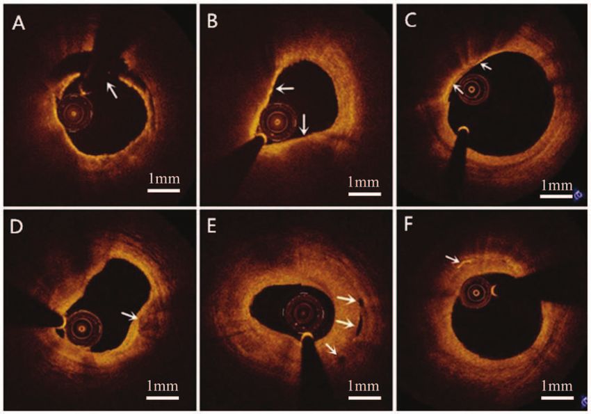
EXPERIMENTAL AND THERAPEUTIC MEDICINE                                                                       3

Figure 1. Representative optical coherence tomography images of different coronary plaque phenotypes (arrows). (A) Plaque rupture: The fibrous cap is broken
and the plaque content is partially washed away, leaving a cavity (11 to 3 o'clock). (B) Thin‑cap fibroatheroma: A large (>90̊) lipidic core and a thin (
4

Table II. Optical coherence tomography‑derived plaque characteristics according to cardiovascular risk factors.

                   Family history of CHD                                            Smoking                                        Hypertension                                  Diabetes mellitus                                Hypercholesterolemia                                 Metabolic syndrome
                ‑‑‑‑‑‑‑‑‑‑‑‑‑‑‑‑‑‑‑‑‑‑‑‑‑‑‑‑‑‑‑‑‑‑‑‑‑‑‑‑‑‑‑‑‑‑‑‑‑‑‑‑ ‑‑‑‑‑‑‑‑‑‑‑‑‑‑‑‑‑‑‑‑‑‑‑‑‑‑‑‑‑‑‑‑‑‑‑‑‑‑‑‑‑‑‑‑‑‑‑‑‑ ‑‑‑‑‑‑‑‑‑‑‑‑‑‑‑‑‑‑‑‑‑‑‑‑‑‑‑‑‑‑‑‑‑‑‑‑‑‑‑‑‑‑‑‑‑‑‑‑‑ ‑‑‑‑‑‑‑‑‑‑‑‑‑‑‑‑‑‑‑‑‑‑‑‑‑‑‑‑‑‑‑‑‑‑‑‑‑‑‑‑‑‑‑‑‑‑‑‑‑‑‑ ‑‑‑‑‑‑‑‑‑‑‑‑‑‑‑‑‑‑‑‑‑‑‑‑‑‑‑‑‑‑‑‑‑‑‑‑‑‑‑‑‑‑‑‑‑‑‑‑‑‑‑‑ ‑‑‑‑‑‑‑‑‑‑‑‑‑‑‑‑‑‑‑‑‑‑‑‑‑‑‑‑‑‑‑‑‑‑‑‑‑‑‑‑‑‑‑‑‑‑‑‑
                   Yes                No		 Yes                                            No		 Yes                                          No		 Yes                                           No		 Yes                                            No		 Yes                                           No
Item            (n=10) (n=113) P‑value (n=67) (n=56) P‑value (n=63) (n=60) P‑value (n=22) (n=101) P‑value (n=21) (n=102) P‑value (n=82) (n=41) P‑value

TCFA                5              82   0.155                          51              36   0.168                         49               38    0.112                       14               73   0.444                         15               72   1.000                          64              23             0.020
                 (50.0)          (72.6)		                            (76.1)          (64.3)		                           (77.8)           (63.3)		                          (63.6)           (72.3)		                           (71.4)           (70.6)		                            (78.0)          (56.1)
Macrophage          7              68   0.739                          45              30   0.141                         49               26
EXPERIMENTAL AND THERAPEUTIC MEDICINE 5
Table III. Univariate and multivariate analysis for TCFA and plaque rupture predictors.
A, Predictors of TCFA
Univariate analysis Multivariate analysis
‑‑‑‑‑‑‑‑‑‑‑‑‑‑‑‑‑‑‑‑‑‑‑‑‑‑‑‑‑‑‑‑‑‑‑‑‑‑‑‑‑‑‑‑‑‑‑‑‑‑‑‑‑‑‑‑‑‑‑‑‑‑‑‑‑‑‑‑‑‑‑‑‑‑‑‑‑‑‑‑‑‑‑‑ ‑‑‑‑‑‑‑‑‑‑‑‑‑‑‑‑‑‑‑‑‑‑‑‑‑‑‑‑‑‑‑‑‑‑‑‑‑‑‑‑‑‑‑‑‑‑‑‑‑‑‑‑‑‑‑‑‑‑‑‑‑‑‑‑‑‑‑‑‑‑‑‑‑‑‑‑‑‑‑‑‑‑‑‑
Factor RR 95% CI P‑value RR 95% CI P‑value
Gender 1.54 0.514‑4.610 0.440
Smoking 1.771 0.809‑3.877 0.153
Hypertension 2.026 0.917‑4.477 0.081 1.574 0.679‑3.650 0.291
Diabetes mellitus 0.671 0.254‑1.774 0.421
Hypercholesterolemia 1.042 0.369‑2.942 0.939
Metabolic syndrome 2.783 1.240‑6.246 0.013 2.421 1.038‑5.649 0.041
Aspirin 0.678 0.277‑1.660 0.395
Statins 0.542 0.216‑1.355 0.190
Beta blocker 0.827 0.343‑1.993 0.673
ACEI or ARB 1.210 0.499‑2.934 0.674
Insulin 0.607 0.097‑3.796 0.594
B, Predictors of plaque rupture
Univariate analysis Multivariate analysis
‑‑‑‑‑‑‑‑‑‑‑‑‑‑‑‑‑‑‑‑‑‑‑‑‑‑‑‑‑‑‑‑‑‑‑‑‑‑‑‑‑‑‑‑‑‑‑‑‑‑‑‑‑‑‑‑‑‑‑‑‑‑‑‑‑‑‑‑‑‑‑‑‑‑‑‑‑‑‑‑‑‑‑‑ ‑‑‑‑‑‑‑‑‑‑‑‑‑‑‑‑‑‑‑‑‑‑‑‑‑‑‑‑‑‑‑‑‑‑‑‑‑‑‑‑‑‑‑‑‑‑‑‑‑‑‑‑‑‑‑‑‑‑‑‑‑‑‑‑‑‑‑‑‑‑‑‑‑‑‑‑‑‑‑‑‑‑‑‑‑
Factor RR 95% CI P‑value RR 95% CI P‑value
Smoking 8.471 1.855‑38.690 0.006 8.301 1.813‑38.015 0.006
Hypertension 1.226 0.449‑3.352 0.691
Diabetes mellitus 0.531 0.113‑2.499 0.423
Hypercholesterolemia 0.566 0.120‑2.670 0.472
Metabolic syndrome 1.904 0.585‑6.205 0.285
Aspirin 0.380 0.082‑1.763 0.217
Statins 0.446 0.095‑2.081 0.304 2.064 0.422‑10.093 0.371
Beta blocker 0.328 0.071‑1.514 0.153
ACEI or ARB 0.285 0.062‑1.314 0.107
Insulin 4.250 0.658‑27.443 0.128
TCFA, thin‑cap fibroatheroma; ACEI, angiotensin‑converting enzyme inhibitor; ARB, angiotensin‑receptor blocker; RR, risk ratio.
with other CVRFs and MetS in patients diagnosed with isch‑ the aforementioned studies. The pathophysiology underlying
emic heart disease (age, 59±9 years) and indicated that MetS atherosclerosis and plaque characteristics differ between
patients had more frequent VH‑IVUS‑derived TCFAs within young and old patients with CHD (20). Furthermore, in the
the tree than non‑MetS patients. Similarly, the present study study by Yonetsu et al (19), selected 198 patients who had
suggested that patients with MetS had more frequent TCFAs nonculprit or nontarget coronary plaques with area stenosis
than patients without MetS. Zheng et al (3) also demonstrated >50% as measured by OCT; however, whether they are culprit
that T2D is independently associated with TCFAs. or non‑culprit may affect the characteristics of plaques (21).
Previous studies investigating features of coronary plaques The present study indicated that cigarette smoking is inde‑
in patients with MetS have provided conflicting results. pendently associated with plaque rupture. Cigarette smoking is
Specifically, a previous IVUS study demonstrated no signifi‑ associated with a high incidence of CVEs, including ACS (22).
cant association between the presence of TCFAs and MetS in It is also associated with a higher burden of necrotic cores in
patients with stable angina pectoris (age, 64.7±9.5 years) (18). coronary atherosclerotic plaques, which may be one of the
Another study using OCT indicated that coronary plaques mechanisms underlying the increased risk it poses for plaque
in patients with MetS (age, 60±11 years) and T2D (age, rupture and CVEs (23,24). By far, the most common risk factor
59±11 years) contain larger amounts of lipids, but neither MetS for early‑onset CHD is cigarette smoking (6), which increases
nor T2D was significantly associated with TCFAs (19). These the risk of plaque rupture. Cigarette smokers accounted for
conflicting results may have several reasons. First, the popula‑ 54.5% (n=67) of the patients of the present study. Cigarette
tion of the present study was significantly younger than that in smoking is thought to increase the burden of cardiovascular6 HOU et al: PLAQUE CHARACTERISTICS AND CVRFS IN YOUTHS
disease by inducing endothelial dysfunction, increasing the Competing interests
burden of coronary atherosclerosis and increasing the risk of
plaque rupture and CVEs (25). T2D was not significantly asso‑ The authors declare that they have no competing interests.
ciated with TCFAs or rupture in the present study. This may
be due to the larger number of patients with T2D than without References
(63.6% vs. 10.9%) using statins, which may reduce TCFAs and
plaque rupture (26). 1. Authors/Task Force Members: Piepoli MF, Hoes AW, Agewall S,
The universally acknowledged features of vulner‑ Albus C, Brotons C, Catapano AL, Cooney M, Corrà U,
Cosyns B, Deaton C, et al: 2016 European Guidelines on
able plaques currently include TCFAs, macrophage cardiovascular disease prevention in clinical practice: The
accumulation, calcified nodules, vasa vasorum and cholesterol Sixth Joint Task Force of the European Society of Cardiology
crystals (27‑31). The association between TCFAs and CVRFs and Other Societies on Cardiovascular Disease Prevention in
Clinical Practice (constituted by representatives of 10 societies
was described above. In the present study, macrophage accu‑ and by invited experts) Developed with the special contribution
mulation was more common in patients with hypertension and of the European Association for Cardiovascular Prevention &
cholesterol crystals were present more often in patients with a Rehabilitation (EACPR). Atherosclerosis 252: 207‑274, 2016.
2. Wilson PW, D'Agostino RB, Parise H, Sullivan L and Meigs JB:
family history of CHD and hypercholesterolemia. By contrast, Metabolic syndrome as a precursor of cardiovascular disease and
no significant correlation was observed between calcified type 2 diabetes mellitus. Circulation 112: 3066‑3072, 2005.
nodules or vasa vasorum and CVRFs in the patients of the 3. Zheng M, Choi SY, Tahk SJ, Lim HS, Yang HM, Choi BJ,
Yoon MH, Park JS, Hwang GS and Shin JH: The relationship
present study. The sample examined was not extracted from between volumetric plaque components and classical cardiovas‑
the general population but was rather composed of relatively cular risk factors and the metabolic syndrome a 3‑vessel coronary
young patients. Thus, the results may not be extrapolatable to artery virtual histology‑intravascular ultrasound analysis. JACC
Cardiovasc Interv 4: 503‑510, 2011.
the general population. In addition, the results may be affected 4. Rivera JJ, Nasir K, Cox PR, Choi E, Yoon Y, Cho I, Chun EJ,
by lifestyle factors, including diet and physical exercise. Choi S, Blumenthal RS and Chang HJ: Association of traditional
In conclusion, by using OCT evaluation, the present study cardiovascular risk factors with coronary plaque sub‑types assessed
by 64‑slice computed tomography angiography in a large cohort of
demonstrated that young patients with MetS had more extensive asymptomatic subjects. Atherosclerosis 206: 451‑457, 2009.
TCFAs and that young cigarette smokers were at increased risk 5. De Rosa R, Vasa‑Nicotera M, Leistner DM, Reis SM,
for culprit plaque rupture. Young patients with CHD should Thome CE, Boeckel J, Fichtlscherer S and Zeiher AM: Coronary
atherosclerotic plaque characteristics and cardiovascular risk
therefore actively control their body weight, blood lipids, blood factors‑insights from an optical coherence tomography study.
pressure and blood sugar levels, as well as quit smoking, so as Circ J 81: 1165‑1173, 2017.
to reduce the occurrence/risk of TCFAs and ruptures. 6. Aggarwal A, Srivastava S and Velmurugan M: Newer perspec‑
tives of coronary artery disease in young. World J Cardiol 8:
728‑734, 2016.
Acknowledgements 7. Kalantzi K, Korantzopoulos P, Tzimas P, Katsouras CS,
Goudevenos JA and Milionis HJ: The relative value of metabolic
syndrome and cardiovascular risk score estimates in premature
Not applicable. acute coronary syndromes. Am Heart J 155: 534‑540, 2008.
8. Rathore S, Terashima M, Matsuo H, Kinoshita Y, Kimura M,
Funding Tsuchikane E, Nasu K, Ehara M, Asakura Y, Katoh O and
Suzuki T: Association of coronary plaque composition and
arterial remodelling: A optical coherence tomography study.
No funding was received. Atherosclerosis 221: 405‑415, 2012.
9. Finn AV, Nakano M, Narula J, Kolodgie FD and Virmani R:
Concept of vulnerable/unstable plaque. Arterioscler Thromb
Availability of data and materials Vasc Biol 30: 1282‑1292, 2010.
10. Jang IK, Bouma BE, Kang DH, Park SJ, Park SW, Seung KB,
The datasets used and/or analyzed during the present study are Choi KB, Shishkov M, Schlendorf K, Pomerantsev E, et al:
Visualization of corona r y atherosclerotic plaques in
available from the corresponding author upon reasonable request. patients using optical coherence tomography: Comparison
with intravascular ultrasound. J Am Coll Cardiol 39: 604‑609,
Authors' contributions 2002.
11. Alberti KG, Eckel RH, Grundy SM, Zimmet PZ, Cleeman JI,
Donato KA, Fruchart JC, James WP, Loria CM, Smith SC
YZ and JG conceived the study. FH, WL, YD and YL collected Jr, et al: Harmonizing the Metabolic Syndrome: A Joint Interim
and analyzed the patients' general information. FH and WL wrote Statement of the International Diabetes Federation Task Force on
Epidemiology and Prevention; National Heart, Lung, and Blood
the manuscript. SY, XM and ZW analyzed the OCT images. All Institute; American Heart Association; World Heart Federation;
of the authors read and approved the final manuscript. International Atherosclerosis Society; and International
Association for the Study of Obesity. Circulation 120: 1640‑1645,
2009.
Ethics approval and consent to participate 12. Jia H, Dai J, Hou J, Xing L, Ma L, Liu H, Xu M, Yao Y, Hu S,
Yamamoto E, et al: Effective anti‑thrombotic therapy without
The Ethics Committee of the Beijing Anzhen Hospital (Beijing, stenting: Intravascular optical coherence tomography‑based
management in plaque erosion (the EROSION study). Eur Heart
China) approved the study protocol and all of the participants J 38: 792‑800, 2017.
provided written informed consent. 13. Tian J, Hou J, Xing L, Kim SJ, Yonetsu T, Kato K, Lee H, Zhang S,
Yu B and Jang IK: Significance of intraplaque neovascularisation
for vulnerability: Optical coherence tomography study. Heart 98:
Patient consent for publication 1504‑1509, 2012.
14. Nishimura S, Ehara S, Hasegawa T, Matsumoto K, Yoshikawa J
All of the participants provided written informed consent for and Shimada K: Cholesterol crystal as a new feature of coronary
vulnerable plaques: An optical coherence tomography study.
publication. J Cardiol 69: 253‑259, 2017.EXPERIMENTAL AND THERAPEUTIC MEDICINE 7
15. Tearney GJ, Regar E, Akasaka T, Adriaenssens T, Barlis P, 23. Abtahian F, Yonetsu T, Kato K, Jia H, Vergallo R, Tian J, Hu S,
Bezerra HG, Bouma B, Bruining N, Cho JM, Chowdhary S, et al: McNulty I, Lee H, Yu B and Jang IK: Comparison by optical
Consensus standards for acquisition, measurement, and reporting coherence tomography of the frequency of lipid coronary plaques
of intravascular optical coherence tomography studies: A report in current smokers, former smokers, and nonsmokers. Am J
from the International Working Group for Intravascular Optical Cardiol 114: 674‑680, 2014.
Coherence Tomography Standardization and Validation. J Am 24. Bolorunduro O, Cushman C, Kapoor D, Alexander K, Cuellar‑Silva J,
Coll Cardiol 59: 1058‑1072, 2012. Giri S, Robinson V and Ibebuogu UN: Comparison of coronary
16. Iribarren C, Go AS, Husson G, Sidney S, Fair JM, Quertermous T, atherosclerotic plaque burden and composition of culprit lesions
Hlatky MA and Fortmann SP: Metabolic syndrome and between cigarette smokers and non‑smokers by in vivo virtual
early‑onset coronary artery disease: Is the whole greater than its histology intravascular ultrasound. J Invasive Cardiol 27: 354‑358,
parts? J Am Coll Cardiol 48: 1800‑1807, 2006. 2015.
17. Iannaccone M, Quadri G, Taha S, D'Ascenzo F, Montefusco A, 25. Csordas A and Bernhard D: The biology behind the atherothrom‑
Omede P, Jang IK, Niccoli G, Souteyrand G, Yundai C, et al: botic effects of cigarette smoke. Nat Rev Cardiol 10: 219‑230, 2013.
Prevalence and predictors of culprit plaque rupture at OCT in 26. Gili S, Iannaccone M, Colombo F, Montefusco A, Amabile N,
patients with coronary artery disease: A meta‑analysis. Eur Calcagno S, Capodanno D, Scalone G, Rognoni A,
Heart J Cardiovasc Imaging 17: 1128‑1137, 2016. Omedè P, et al: Effects of statins on plaque rupture assessed by
18. Lee MG, Jeong MH, Kim DH, Lee KH, Park KH, Sim DS, optical coherence tomography in patients presenting with acute
Yoon NS, Yoon HJ, Kim KH, Park HW, et al: Can metabolic coronary syndromes: Insights from the optical coherence tomog‑
syndrome predict the vulnerable plaque in patients with stable raphy (OCT)‑FORMIDABLE registry. Eur Heart J Cardiovasc
angina pectoris? Virtual histology‑intravascular ultrasound Imaging 19: 524‑531, 2018.
analysis. J Cardiol 59: 266‑274, 2012. 27. Kume T and Uemura S: Current clinical applications of coronary
19. Yonetsu T, Kato K, Uemura S, Kim BK, Jang Y, Kang SJ, Park SJ, optical coherence tomography. Cardiovasc Interv Ther 33: 1‑10, 2018.
Lee S, Kim SJ, Jia H, et al: Features of coronary plaque in 28. Lee T, Mintz GS, Matsumura M, Zhang W, Cao Y, Usui E,
patients with metabolic syndrome and diabetes mellitus assessed Kanaji Y, Murai T, Yonetsu T, Kakuta T and Maehara A:
by 3‑vessel optical coherence tomography. Circ Cardiovasc Prevalence, predictors, and clinical presentation of a calcified
Imaging 6: 665‑673, 2013. nodule as assessed by optical coherence tomography. JACC
20. Barbero U, Scacciatella P, Iannaccone M, D'Ascenzo F, Niccoli G, Cardiovasc Imaging 10: 883‑891, 2017.
Colombo F, Ugo F, Colangelo S, Mancone M, Calcagno S, et al: 29. Gonzalez L and Trigatti BL: Macrophage apoptosis and necrotic core
Culprit plaque characteristics in younger versus older patients development in atherosclerosis: A rapidly advancing field with clinical
with acute coronary syndromes: An optical coherence tomog‑ relevance to imaging and therapy. Can J Cardiol 33: 303‑312, 2017.
raphy study from the FORMIDABLE registry. Catheter 30. Janoudi A, Shamoun FE, Kalavakunta JK and Abela GS: Cholesterol
Cardiovasc Interv 92: E1‑E8, 2018. crystal induced arterial inflammation and destabilization of
21. Trusinskis K, Juhnevica D, Strenge K and Erglis A: iMap intra‑ atherosclerotic plaque. Eur Heart J 37: 1959‑1967, 2016.
vascular ultrasound evaluation of culprit and non‑culprit lesions 31. Taruya A, Tanaka A, Nishiguchi T, Matsuo Y, Ozaki Y,
in patients with ST‑elevation myocardial infarction. Cardiovasc Kashiwagi M, Shiono Y, Orii M, Yamano T, Ino Y, et al: Vasa
Revasc Med 14: 71‑75, 2013. vasorum restructuring in human atherosclerotic plaque vulner‑
22. Erhardt L: Cigarette smoking: An undertreated risk factor for ability: A clinical optical coherence tomography study. J Am
cardiovascular disease. Atherosclerosis 205: 23‑32, 2009. Coll Cardiol 65: 2469‑2477, 2015.You can also read