Effect of combined Intravitreal Injections of Bevacizumab and Triamcinolone Acetonide vs intravitreal Bevacizumab in Diffuse Diabetic Macular Edema
←
→
Page content transcription
If your browser does not render page correctly, please read the page content below
IOSR Journal of Dental and Medical Sciences (IOSR-JDMS)
e-ISSN: 2279-0853, p-ISSN: 2279-0861.Volume 13, Issue 6 Ver. IV (Jun. 2014), PP 01-06
www.iosrjournals.org

  Effect of combined Intravitreal Injections of Bevacizumab and
 Triamcinolone Acetonide vs intravitreal Bevacizumab in Diffuse
                    Diabetic Macular Edema
                        1
                            Ambade Rakhee, 2Ambade Ajay, 3Sagdeo M
           1
            MD, Associate professor, Department of physiology, NKP Salve Institute of Medical Sciences and
                                Research center, Digdoha hills, Higna, Nagpur-440019
                2
                  Mch(opthal),DOMS,Consultant, Ambade Eye Hospital, Indora Square Nagpur-440017,
             3
               MD, Prof. and Head, Department of physiology, NKP Salve Institute of Medical Sciences and
                                Research center, Digdoha hills, Higna, Nagpur-440019

Abstract
Purpose: To evaluate and compare the efficacy of two treatment strategies intravitreal anti-
VEGF(bevacizumab-IVB) and steroid (triamcinolone acetonide-IVTA) combination in comparison to
intravitreal anti-VEGF (IVB) in treatment of diabetic macular edema.
Material and Methods: 98 eyes of 81 subjects with diabetic macular edema were included in this prospective,
randomized, interventional study, divided into two groups of 49 each. Group A was treated with
bevacizumab(0.05ml; 1.25mg) and Group B (IVB+IVTA)with bevacizumab and triamcinolone at 0 ,1 and 2
months respectively. Each eye was evaluated at baseline, at 3 months and at 6 months for central macular
thickness (CMT), BCVA (logMAR) and intraocular pressure. Result: Parameters identified for comparative
study included CMT, BCVA (logMAR) and intraocular pressure. The difference in mean CMT between the two
groups at the end of 3 months was significant (Grp. A 360.59±92.44 vs. Grp. B 345.10±76.62) and much more
so at the end of 6 months (Grp A 332.24±91.78 vs. Grp B 292.82±88.84), the other criteria like BCVA (logMAR)
also showed marked improvement in Grp B at the end of 6months.The rise in IOP was found to be significant at
the end of study period in Grp B.
Conclusion: In the treatment of diffuse diabetic macular edema, using a combination of IVB+IVTA is definitely
more effective than using IVB alone, though caution has to be exercised in patient selection and close IOP
monitoring is required in patients treated with IVTA.
Key words: Diffuse diabetic macular edema, bevacizumab, intra-vitreal triamcinolone acetonide, central
macular thickness, intraocular pressure.

                                             I.     Introduction
          Diabetic retinopathy (DR) is an important cause of acquired visual loss and impairment in working age
group worldwide.[1-4]The Salisbury Eye Evaluation Study showed that diabetic retinopathy was the third most
important cause for visual impairment.[1]Diabetic macular edema (DME) is a manifestation of diabetic
retinopathy that produces loss of central vision .[2]In the Early Treatment Diabetic Retinopathy Study (ETDRS),
focal photocoagulation of eyes with clinically significant macular edema (CSME) reduced the risk of moderate
visual loss by approximately 50%.[5]In spite of treatment, 12% of treated eyes developed moderate visual loss.
Furthermore, central retinal thickening remained in approximately 40% and 25% of treated eyes after 12 months
and 36 months, respectively.[5-7]Recently, a series of studies suggested post-laser release of inflammatory
factors, accumulation of leucocytes in the non-photocoagulated posterior pole, and up-regulation of angiogenic
growth factors, such as vascular endothelial growth factor (VEGF), play a role in the pathogenesis of the
edema.[9–13] VEGF is up-regulated in diabetic retinopathy,[12,13] so administration of some kind of anti VEGF
agent seems a logical option. Several studies are currently evaluating the role of anti- VEGF agents for the
treatment of ocular disease associated with choroidal and/or retinal neovascularization and exudative processes,
especially age-related macular degeneration [14,16] and diabetic retinopathy.[17-22] Corticosteroids also may work
through multiple mechanisms of action. They are known to reduce vascular permeability, reduce blood–retinal
barrier breakdown, down-regulate VEGF production, and inhibit some matrix metalloproteinase.[9, 10, 23, 24]Some
studies have evaluated this drug effect in DME. [22, 23]There are many factors that are involved in pathogenesis of
DME, so many alternatives may be suggested for these patients (pharmacologic or surgical). The increase in
retinal capillary permeability and subsequent retinal edema may be the result of a breakdown of the blood–
retinal barrier mediated in part by VEGF.
          Intravitreal bevacizumab has been effective in cases with center involved DME in the improvement of
visual acuity, reduction of macular edema, fibro vascular proliferation in retinal NV and resolution of vitreous
                                             www.iosrjournals.org                                          1 | Page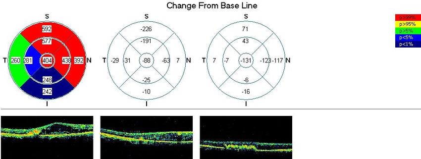
Effect of combined Intravitreal Injections of Bevacizumab and…
hemorrhage, but in cases with center involved DME refractory to focal grid laser studies have shown that IVTA
has superior efficacy than IVB.[21-23]Available literature on the subject indicates that adding intravitreal steroid
to intravitreal anti-VEGF agent may intensify and/or consolidate effect of both agents.
Thus, the purpose of this study is to evaluate the efficacy and safety of the combined effect of
triamcinolone acetonide and bevacizumab in comparison to Avastin in the management of DME. The aims of
our study were to compare, using an interventional case series design, the efficacy of intravitreal injection of
bevacizumab and triamcinolone combination for reducing foveal thickness, and to evaluate the visual prognosis
and anatomic alterations of macular edema using spectral domain OCT
Materials and method
It is a prospective, randomized, comparative interventional case series of 98 eyes of 81 subjects with
diabetic macular edema. This study was conducted in accordance with ethical standards and the Helsinki
declaration. The patients were fully informed on the risks and the benefits of treatments, and accordingly written
informed consents were obtained.
The study duration was of six month and included patients of type-II diabetes above forty years of age
hailing from central India.
A detailed systemic evaluation including medical history, blood pressure, serum HbA1c (glycosylated
hemoglobin) levels, renal profiles and complete ocular examination was performed for each enrolled patient.
Ocular examination included best corrected visual acuity (BCVA), intraocular pressure (IOP), presence of lens
opacities using the Lens Opacities Classification System III (LOCS III), fundus examination, and macular
thickness measurement by the optical coherence tomography (optovue–spectral domain OCT).
Inclusion criteria –Patients with very severe NPDR to high-risk PDR with clinically significant macular
edema (CSME –ETDRS definition) were considered for enrollment into the study. The central macular
thickness (CMT)> 300µ on cross hair protocol (SD – OCT) reported in the central 1 mm macular thickness map
was taken as the mean retinal thickness of the macula. OCT criteria included more than 50% of area with
cystoid changes on Emm5 protocol. Pre-operative assessment of all the patients included the best-corrected
visual acuity, applanation tonometry and fundus examination.
The exclusion criteria were macular edema related to recent intraocular surgery or other procedures,
vitreous traction (based on OCT), history of any treatment for diabetic retinopathy at any time or anticipating
the need for pan retinal laser photocoagulation (PRP)in the 6 months following randomization, uncontrolled
glaucoma, steroid responders, recent history of arterial thromboembolic event, and poorly controlled
hypertension, use of systemic steroids and/or systemic anti-VEGF.
All intravitreal injections were performed using a standard protocol under topical anesthesia and sterile
operating conditions. Bevacizumab (Avastin; Genentech, Inc., South San Francisco, CA, USA)(0.05 ml; 1.25
mg) was injected superotemporally using 30 G needle through pars-plana. Simultaneously Triamcinolone
acetonide (Trilon, Ajanta Pharma India) (0.05 ml; 2 mg) was injected in a separate syringe using 30 G needle
inferotemporally through pars-plana for the IVTA+IVB group. Central retinal artery was assessed after
injection. Post operative anti-glaucoma medication was started for all patients. Patients were followed at 24
hours post-operatively and weekly thereafter for the assessment of anterior chamber reaction and/or intraocular
pressure (IOP). Two more such injections were repeated after an interval of one month. Therefore a total of
three intravitreal injections of Avastin were given in group A and IVB+IVTA in group B. Best-corrected visual
acuity, funduscopy, fluorescein angiography, and posterior segment OCT were conducted at baseline, 3rd month
and 6th month post-operatively. Complications like cataract formation, vitreous hemorrhage, and
endophthalmitis were recorded.
Statistical analysis
The primary efficacy outcomes in the study were CMT, BCVA (logMAR) and IOP at 3 and 6 months
as compared to baseline. Repeated measure analysis of variance (ANOVA) was performed to evaluate the
statistical significance of change of each parameter with time in each treatment group. Assumption of sphericity
was evaluated using Mauchly’s test for each parameter. When the assumption was violated, Greenhouse-Geisser
(< 0.75) or Huynh-Feldt (> 0.75) corrections were used to decide statistical significance of difference across
time. Upon significant, post hoc analysis was carried out following Bonferroni correction. Visual acuity was
correlated with CMT at 3 and 6 months using Pearson’s correlation coefficient.
Results
A total of 98 eyes of 81 patients with diabetic macular edema were included in this study.Equal number
i.e., 49 eyes were randomly assigned to Avastin (Group A)and IVTA+IVB(Group B) groups. Summary statistics
for baseline characteristics in terms of age, macular thickness, visual acuity and IOP between two groups were
compared as shown in Table 1.The mean age of the patients in Avastin group was 54.73 ± 11.91 years, whereas
www.iosrjournals.org 2 | PageEffect of combined Intravitreal Injections of Bevacizumab and…
in IVTA+IVB group it was 58.18 ± 11.22 years; and the mean difference was statistically insignificant with P-
value of 0.151. Another baseline characteristic, duration of diabetes, in Avastin group had a mean of 11.34 ±
6.69 years, while in IVTA+IVB group it was 10.97 ± 4.65 years and the difference was statistically insignificant
(P-value: 0.765). Further, mean CMT in Avastin group (478.10 ± 142.78 µm) differed insignificantly from that
of IVTA+IVB (474.71 ± 96.29 µm) with a P-value of 0.889. Mean IOP before treatment in Avastin group
(15.10 ± 1.74 mm Hg) and in IVTA+IVB (15.26 ± 1.38 mmHg) also differed insignificantly (P-value: 0.654).
BCVA expressed in terms of logMAR (minimum angle resolution) showed statistically insignificant difference
between Avastin (0.82 ± 0.14) and IVTA+IVB (0.86 ± 0.09) with P-value of 0.096.
Table 1: Baseline characteristics of patients in two treatment groups
Treatment type
Characteristics Avastin IVTA + IVB P-value
No. of Eyes 49 49
Age in years [Mean ±SD] 54.73 ± 11.91 58.18 ± 11.22 0.151
Duration of Diabetes [Mean ± SD] 11.34 ± 6.69 10.97 ± 4.65 0.765
CMT (µm) [Mean ± SD] 478.10 ± 142.78 474.71 ± 96.29 0.889
BCVA [Mean(log MAR) ± SD] 0.82 ± 0.14 0.86 ± 0.09 0.096
IOP (mm Hg) [Mean ± SD] 15.10 ± 1.74 15.26 ± 1.38 0.642
After ascertaining the baseline features of two groups, the treatment effect within each group was
evaluated considering CMT,BCVA(logMAR) and IOP as dependents and using repeated measure one-way
analysis of variance. The post-treatment effects for each of these parameters were compared with the baseline in
respective groups.
Central macular thickness
In Avastin group, CMT differed significantly across time points (F=40.527; P< 0.0001) with a
Greenhouse-Geisser correction in repeated measure ANOVA (Table 2). Post hoc tests using Bonferroni
correction revealed that mean CMT decreased significantly at month 3 and 6 when compared with the baseline
with P< 0.0001 (Table 3). However, the difference between month 3 and 6 differed insignificantly (P = 0.062).
On similar lines, CMT changes were evaluated in IVTA+IVB group. Repeated measure ANOVA with
Greenhouse-Geisser correction revealed statistically significant reduction in mean CMT (F=109.03; P< 0.0001)
across time points. Post hoc analysis also showed significant difference between all pair wise comparisons
(Table 3). Figure 1 shows the line plots for mean CMT across time points for two groups. The difference in the
mean CMT between two groups at the end of 6 months was statistically significant according to t-test for
independent samples (P= 0.034).
Visual acuity
In Avastin group, repeated measure ANOVA showed significant improvement in visual acuity using
Huynh-Feldt correction (F=40.988; P< 0.0001) across time points (Table 2). Subsequent post hoc analysis
suggested significant difference in all pair wise comparisons (Table 3). Similar was the observation in
IVTA+IVB group. The Greenhouse-Geisser correction in repeated measure analysis revealed significant
increase in visual acuity (F=112.55; P< 0.0001) and further post hoc analysis showed highly significant
difference between all pair wise comparisons (Table 3). Line plots showing the change in the mean logMAR
score for two groups are shown in Figure 1. At the end of six months, the difference of mean logMAR score
between two groups was statistically insignificant as per t-test for independent samples (P=0.341).
Intraocular Pressure
The post-treatment change in IOP was analyzed in both the groups using repeated measure ANOVA.
Analysis revealed that in Avastin group, the increase in IOP with time was insignificant as indicated by P-value
of 0.061 (F=3.057) after using Huynh-Feldt correction. A marginal increase in post-treatment mean IOP with
reference to baseline is evident through Figure 1. However, in the IVTA+IVB group, IOP followed assumption
of sphericity (P=0.151) and showed significant increase after treatment as compared to baseline (F=40.142;
PEffect of combined Intravitreal Injections of Bevacizumab and…
Table 2: Statistical significance of difference in different parameters using repeated measure one-way ANOVA
Mauchly's
Treatment Time scale Sphericity test F value DF P value
Parameter group Baseline Month 3 Month6 (p-value)
Central macular Avastin1 478.10 ± 360.59 ± 332.24 ± < 0.0001
thickness (CMT) 142.78 92.44 91.78
2
IVTA+IVB 474.71 ± 345.10 ± 292.82 ± < 0.0001 40.527 1.35 < 0.0001
96.29 76.62 88.84 4†
Visual acuity Avastin1 0.82 ± 0.64 ± 0.52 ± < 0.0001 109.03 1.44 < 0.0001
(VA)[log(MAR)] 0.14 0.19 0.27 1†
2
IVTA+IVB 0.86 ± 0.72 ± 0.57 ± < 0.0001 40.988 1.58 < 0.0001
0.09 0.18 0.26 8‡
Intra-ocular pressure Avastin1 15.10 ± 15.45 ± 15.22 ± 0.003 112.55 1.41 < 0.0001
(IOP) 1.74 1.90 1.80 7†
IVTA+IVB2 15.26 ± 16.83 ± 16.79 ± 0.151 3.057 1.68 0.061
1.38 1.84 1.79 9‡
†Greenhouse-Geisser correction (< 0.75); ‡Huynh-Feldt correction (> 0.75); *No correction since sphericity
assumption holds; 1n=49; 2n=49
Table 3: Post-hoc test for pair wise statistical significance between time points
Treatment Paired comparison*
Parameter group Baseline vs Month 3 Baseline vs Month 6 Month 3 vs Month 6
Central macular thickness (CMT) Avastin < 0.0001 < 0.0001 0.062
IVTA+IVB < 0.0001 < 0.0001 0.002
Visual acuity (VA) Avastin < 0.0001 < 0.0001 0.001
IVTA+IVB < 0.0001 < 0.0001 < 0.0001
Intra-ocular pressure (IOP) Avastin NA NA NA
IVTA+IVB < 0.0001 < 0.0001 0.999
*Bonferroni correction
Figure 1: Line plots showing mean parameter values for two groups across time points.
www.iosrjournals.org 4 | PageEffect of combined Intravitreal Injections of Bevacizumab and…
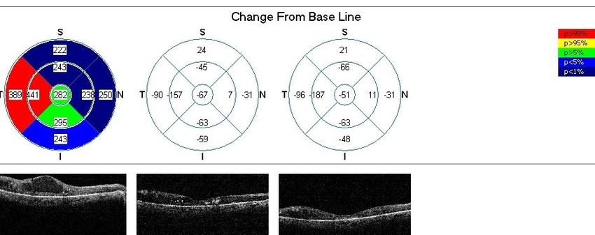
Picture 1. Macular thickness change seen in a patient treated with Intravitreal Bevacizumab
Picture 2 Macular thickness change seen after combine Intravitreal Bevacizumab and Triamcinolone
acetonide
Discussion
DME is one of the major causes of visual impairment in patients with diabetic retinopathy. It has been
characterized by inflammation, including intravitreous induction of pro-inflammatory cytokine, intraretinal expression of
pro-inflammatory caspases and mediators, and therefore, many clinical investigators have found that Intravitreal injection of
a corticosteroid like triamcionoloneacetonide may reduce macular edema. Although the reduction effect of triamcinolone on
macular edema improves visual function, recurrence of macular edema was often observed within 24 weeks after treatment
and IOP was occasionally increased in this therapy. Therefore, safer and longer-acting therapy for DME was sought for.
Paccola et al.[3o]showed that a single intravitreal injection of triamcinolone acetonide has advantage over Avastin in the
short-term management of refractory DME ,specially with regards to changes in CMT. In the present study the beneficial
effect of triamcinolone was maintained by repeating IVTA+IVB injection at one month interval for further two months.
Since it was reported that the vitreous level of VEGF increased and correlated with the severity of macular edema in DME
patients, anti-VEGF therapy is expected to show a dramatic reduction of DME.Haritiglou et al.[23] reported a significant
reduction in macular thickness at 2(15%),6(17%) and 12weeks(25%) following monthly IVTA in patients with DME. This
feature of IVTA is emulated in this study as well. In a study by Kriechbaum et al.(Diabetic Retinopathy Research Group) [29]
3 injections of 2.5mg bevacizumab and one 8mg triamcinolone were compared. After 6 month,visual rehabilitation was
comparable but reduced in 12th month in triamcinolone group due to factors such as cataract formation.Our study shows a
synergistic effect in group IVTA+IVB & which sustains till the end of the study. Though this effect was not statistically
significant between groups but the percentage reduction of CMT at 3 rdmonth in Avastin group was, 25.73% and in
IVTA+IVB group, was 34.81%. This indicates that the combination has synergic effect on reduction of CMT. The aim of
treatment is to gain visual acuity and the IVTA+IVB group show statistically significant improvement over IVB group.
One of the most important side effects of triamcinolone is raised IOP for this reason steroid responders were
excluded and a lower dose of steroid was used in the study .Therefore, as expected, the dynamic change of IOP in the
bevacizumab-injected eye was “safer” than that in the triamcinolone-injected eye. Still, careful observation is needed to
perform Intravitreal injection because of the reported cases of endopthalmitis and systemic side effect. But since the doses of
triamcinolone was half of the regular previous studies the expected IOP bounce was not seen in IVTA+IVB group.
The CMT reduction & visual acuity gain in IVTA+IVB group is better & statistically significant but close
observation is required for control of IOP including selection of patient specially to exclude patient with uncontrolled
glaucoma .
www.iosrjournals.org 5 | PageEffect of combined Intravitreal Injections of Bevacizumab and…
Thus it is suggested that DME can be well controlled & effectively managed by IVTA+IVB injection.
In this study cataract formation in triamcinolone group has not been considered. This is because of a lesser duration of study
and a lower dose of steroid used.
Conclusion - This study demonstrates the synergistic effect of reduction in central macular thickness resulting in better
visual acuity in patients of diffuse DME treated with combine IVTA+IVB. The beneficial effect can be sustained for a
longer period of time when steroid and anti-VEGF are combined. The result of this study cannot be generalized, adequate
patient selection and careful monitoring of patient for any uncontrolled rise in intraocular pressure is required during the
post-operative period.
References:
[1]. Munoz B,westsk, rubinGS,etal .cause of blindnessand visual impairmentin population of older americans ;The
Salisburyeyeevalution study .Arch Ophthalmol 2000:118;819-25
[2]. Klein r, klein BE, Moss SE, Davis MD ,Demets DL. The Wisconsin epidemiologic study of diabetic retinopathy .4diabetic
macular edema .ophthalmology 1984,91;1464-74
[3]. Moss se ,klein R, Klein BE. Ten year incidence of visual loss in a diabetic population. Ophthalmology 1994; 101: 1061-70
[4]. Moss SE, Klein R, Klein BE. The 14 year incidence of visual loss in a diabetic population. Ophthalmology 1998; 105:998-
1003.
[5]. Early Treatment Diabetic retinopathy Study Research Group. Photocoagulationfor diabetic macular edema, Report No.1
Arch Ophthalmol. 1985;103:1796-1806
[6]. Early Treatment Diabetic retinopathy Study Research Group. Treatment , techniques, & clinical guidelines for diabetic
macular edema, report no.2. Ophthalmology. 1987;94:761-774
[7]. Early Treatment Diabetic retinopathy Study Research Group. Focal Photocoagulation treatment of diabetic macular edema,
Report No. 9.Arch Ophthalmol. 1991;98:766-85
[8]. Morgan CM, Schatz H. Atrophic creep of the retinal pigment after focal macular photocoagulation. Ophthalmology.
1989;96:96-103
[9]. Schatz H, Madeira D, McDonald RN. Progressive enlargement of laser scars following grid laser photocoagulation for diffuse
diabetic macular edema. Arch Ophthalmol.1991;109:1549-1551
[10]. Luttrull JK, Musch DC, Mainster MA. Subthreshold diode micropulse photocoagulation for the treatment of clinically
significant diabetic macular edema. Br J Ophthalmol. 2005;89:74-80
[11]. Sinclair S, Alaniz ,Presti P. Laser treatment of diabetic macular edema: comparision of ETDRS-level treatment with threshold
level treatment by using high contrast discriminant center visual field testing.SeminOphthalmol. 1999;14:214-222
[12]. Shimura M, Yasuda K, Nakazawa T, Tamai M. Effective treatment of temporal grid pattern photocoagulation in patients with
diffuse diabetic macular edema. Ophthalmic Surg Lasers Imaging. 2004;35:270-280
[13]. Olk R J, Akduman L. Minimal intensity diode laser (810 nanometer) photocoagulation (MIP) for diffuse diabetic macular
edema (DDME). SeminOphthalmol. 2001;16:25-30
[14]. Shimura M, Yasuda K, Shiono K. Pretreatment of posterior subtenon injection of triamcinolone acetonide has beneficial
effects for grid pattern photocoagulation against diffuse diabetic macular edema. Br J Ophthalmol. 2007;91:449-454
[15]. Funatsu H, Yamashita H, Ikeda T, Mimura T, Eguchi S, Hori S. Vitreous levels of interleukin-6 & vascular endothelial groth
factor are related to diabetic macular edema. Ophthalmology. 2003;110:1690-1696
[16]. Funatsu H, Yamashita H, Sakata K, et al. Vitreous level of vascular endothelial growth factor & intracellular adhesion
molecule 1 are related to diabetic macular edema. Ophthalmology. 2005;112:806-816
[17]. Jonas JB, Kressig I, Sofker A, Degenring RF. Intrvitreal injection of triamcinolone for diffuse diabetic macular edema. Arc
Ophthalmol. 2003;121:57-61
[18]. Sutter Fk, Simpson JM, Gilles MC, Intravitreal triamcinolone for diabetic macular edema that persist after laser treatment: 3
month efficacy & safety results of prospective, randomized, double-masked, placebo-comtrolled clinical trial.
Ophthalmology. 2004;111:2044-2049
[19]. Massin P, Andren F, Haouchine B, et al. Intravitreal triamcinolone acetonide for diabetic macular edema-preliminary results
of a prospective controlled trial. Ophthalmology. 2004;111:218-225
[20]. Martidis A, Duker JS, Greenberg PB, et al. Intravitreal triamcinolone for refractory diabetic macular edema. Ophthalmology.
2002;109:920-927
[21]. Verma LK, Vivek MB, Kumar A, Tewari HK, Venkatesh P. A prospective controlled trial to evaluate the adjunctive role of
posterior sub tenon triamcinolone in the treatment of diffuse diabetic macular edema. J Ocular PharmacolTher. 2004;20:277-
284
[22]. Arevalo JF, Fromow-Guerra J, Quiroz-mercado H, et al. Primary intravitrealbevacizumab (avastin) for diabetic macular
edema. Ophthalmology. 2007;114:743-750
[23]. Haritoglou C, Kook D, Neubauer A, et al. Intravitrealbevacizumab (avastin) therapy for persistent diffuse diabetic macular
edema. Retina. 2006;26:999-1005
[24]. Kang SW, Sa HS, Cho HY, Kim JI. Macular grid phocoagulation after intravitrealacetonide for diffuse diabetic macular
edema. Arch Ophthalmol. 2006;124:653-658
[25]. Massin P, Vicaut E, Haouchine B, Erginay A, Paques M, Gaudric A. Reproducibility of retinal mapping using optical
coherence tomography. Arch Ophthalmolol. 2001;119:1135-1142
[26]. Rivellese M, George A, Sulkes D, Reichel E, Puliafito C. Optical coherence tomography after laser photocoagulation for
clinically significant macular edema. Ophthalmic Surg Lasers. 2000;31:192-197
[27]. Moss SE, Klein BEK. The incidence of visual loss in a diabetic population. Ophthalmology. 1988;95:1340-1348
[28]. McMeel JW, Trempe CL, Franks EB. Diabetic Retinonopathy. Trans Sect Ophthalmol Am AcdOphthalmolOtolaryngol.
1977;83:476-487
[29]. Katharina Kriechbaum, MDet, al.; the Diabetic Retinopathy Research Group ViennaIntravitrealBevacizumabvs
Triamcinolone for Treatment of Diabetic Macular Edema(PUBMED)
[30]. Paccola L, Costa RA, Folgosa MS, Barbosa JC, Scott IU, Jorge R.Intravitreal triamcinolone versus bevacizumab for treatment
of refractory diabetic macular oedema (IBEME study).(PUBMED).
www.iosrjournals.org 6 | PageYou can also read