Subthreshold Micropulse Laser vs. Conventional Laser for Central Serous Chorioretinopathy: A Randomized Controlled Clinical Trial - Frontiers
←
→
Page content transcription
If your browser does not render page correctly, please read the page content below
ORIGINAL RESEARCH
                                                                                                                                                 published: 16 July 2021
                                                                                                                                        doi: 10.3389/fmed.2021.682264

                                              Subthreshold Micropulse Laser vs.
                                              Conventional Laser for Central
                                              Serous Chorioretinopathy: A
                                              Randomized Controlled Clinical Trial
                                              Lijun Zhou 1† , Kunbei Lai 1† , Ling Jin 1 , Chuangxin Huang 1 , Fabao Xu 1 , Yajun Gong 1 ,
                                              Longhui Li 1 , Zhe Zhu 2 , Lin Lu 1 and Chenjin Jin 1*
                                              1
                                                  State Key Laboratory of Ophthalmology, Zhongshan Ophthalmic Center, Sun Yat-sen University, Guangzhou, China,
                                              2
                                                  Department of Medicine, Herbert Irving Comprehensive Cancer Center, Columbia University, New York, NY, United States

                                              Purpose: To investigate the effectiveness and safety of 577-nm subthreshold micropulse
                                              laser (SML) on acute central serous chorioretinopathy (CSC).
                                              Methods: One hundred and ten patients with acute CSC were randomized to receive
                                              SML or 577-nm conventional laser (CL) treatment. Optical coherence tomography and
                            Edited by:        best-corrected visual acuity (BCVA) were performed before and after treatment.
                      Gemmy Cheung,
                Singapore National Eye
                                              Results: At 3 months, the complete resolution of subretinal fluid (SRF) in 577-nm
                     Center, Singapore        SML group (72.7%) was lower than that in CL group (89.1%) (Unadjusted RR, 0.82;
                        Reviewed by:          P = 0.029), but it was 85.5 vs. 92.7% at 6 months (unadjusted RR, 0.92; P = 0.221).
                       Miguel Rechichi,
                                              The mean LogMAR BCVA significantly improved, and the mean central foveal thickness
               Centro Polispecialistico
                    Mediterraneo, Italy       (CFT) significantly decreased in the SML group and CL group (all P < 0.001) at 6 months.
                   Alessandro Meduri,         But there was no statistical difference between the two groups (all P > 0.05). In the SML
            University of Messina, Italy
                                              group, obvious retinal pigment epithelium (RPE) damage was shown only in 3.64% at 1
                    *Correspondence:
                             Chenjin Jin
                                              month but 92.7% in the CL group (P < 0.001).
                jinchj@mail.sysu.edu.cn
                                              Conclusions: Although 577-nm SML has a lower complete absorption of SRF
     † These   authors have contributed       compared with 577-nm CL for acute CSC at 3 months, it is similarly effective as 577-nm
                    equally to this work
                                              CL on improving retinal anatomy and function at 6 months. Importantly, 577-nm SML
                                              causes less damage to the retina.
                    Specialty section:
          This article was submitted to       Keywords: subthreshold micropulse laser, subretinal fluid, central foveal thickness, central serous
                        Ophthalmology,        chorioretinopathy, conventional laser photocoagulation
                a section of the journal
                   Frontiers in Medicine

           Received: 18 March 2021            INTRODUCTION
            Accepted: 08 June 2021
            Published: 16 July 2021           Central serous chorioretinopathy (CSC) is a common macular condition affected mainly in
                             Citation:        middle-aged patients. It is characterized by a serous neuroepithelium detachment with or without
  Zhou L, Lai K, Jin L, Huang C, Xu F,        retinal pigment epithelium (RPE) detachment (1). The acute CSC is considered self-limited and
   Gong Y, Li L, Zhu Z, Lu L and Jin C        usually resolves spontaneously within 3 to 6 months (2, 3). Therefore, observation is often
(2021) Subthreshold Micropulse Laser
                                              recommended as the current care for acute CSC (1). However, spontaneous resolution does not
    vs. Conventional Laser for Central
          Serous Chorioretinopathy: A
                                              always occur, and 30–50% of the patients with CSC experienced recurrence. Even 5% of patients
  Randomized Controlled Clinical Trial.       progressed to chronic CSC, resulting in permanent damage in visual acuity (4–6). What’s more, the
                Front. Med. 8:682264.         outer nuclear layer and photoreceptor could be injured as long as subretinal fluid (SRF) is present
     doi: 10.3389/fmed.2021.682264            (7). Based on the above conditions, some proper treatments for acute CSC are reasonable.

Frontiers in Medicine | www.frontiersin.org                                           1                                             July 2021 | Volume 8 | Article 682264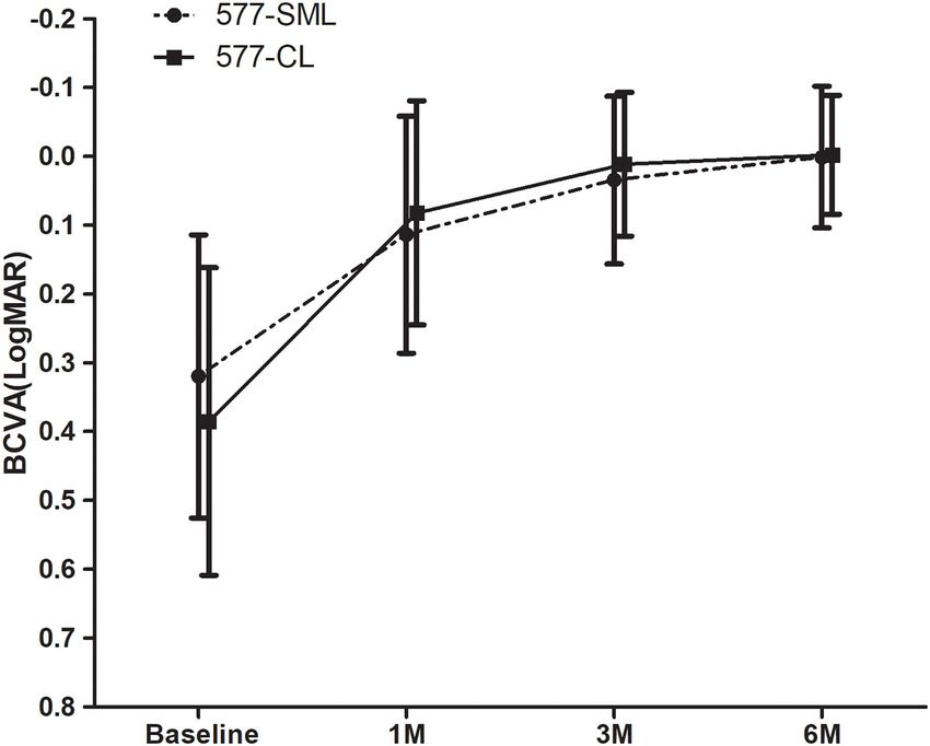
Zhou et al.                                                                                                Subthreshold Micropulse Laser for CSC

    Photodynamic therapy (PDT) and conventional laser (CL) are            Study Protocol
the mainly proved methods. Although PDT is effective for CSC, it          All patients received complete eye examinations at baseline
causes ischemia and atrophy of the choroid (8). Besides, it is off-       and followed up at 1, 3, and 6 months after treatment.
label and expensive for most patients, particularly in developing         Best-corrected visual acuity was measured using the decimal
countries. CL can seal the leakage and accelerate the resolution          chart and was converted to the logarithm of the minimum
of SRF, but it is not favorable for the leakage close to the fovea        angle of resolution (LogMAR) for statistical analysis. Fundus
because it usually leads to retinal scars and scotoma (9, 10),            angiography (Spectralis HRA + OCT; Heidelberg Engineering,
which significantly impaired visual function. Therefore, less or          Germany) was performed to determine the leakage spot and
non-damage treatment is need.                                             to exclude other maculopathies at baseline. SD-OCT and
    A 577-nm subthreshold micropulse laser (SML) is a                     fundus autofluorescein (FAF, Spectralis HRA + OCT; Heidelberg
reliable and cost-effective treatment. Furthermore, the 577-nm            Engineering, Germany) were performed at baseline and each
wavelength is yellow light and is outside the absorption spectrum         visit. The central foveal thickness (CFT) was defined as the
of retinal xanthophylls, which potentially facilitates treatment          distance from the neurosensory retina’s inner surface to the
close to the fovea (11). Recently, it has been reported that SML          inner surface of the choroid at the fovea measured by OCT.
treatment is useful for the CSC without apparent retinal damage           RPE change was assessed using the FFA at the 1-month visit
(12, 13) and better than observation for acute CSC (14). However,         after laser treatment. RPE was categorized into: no RPE damage
the patients in previous studies are almost chronic CSC (12, 15),         (no changes at the treatment area), mild RPE damage (focally
and there is no prospective report that compared the efficacy             rough RPE but no obvious laser spot), and obvious RPE damage
of the SML with CL for acute CSC. Therefore, we conducted a               (presence of clear laser spots).
clinical trial to compare the effectiveness of 577-nm SML with a
577-nm CL to treat active acute CSC.                                      Interventions
                                                                          CL group was treated with a 577-nm laser (Supra 577Y Laser
                                                                          System; Quantel Medical, Clermont-Ferrand, France) using a
MATERIALS AND METHODS                                                     continuous-wave model with a 100-µm spot diameter, a 0.1-s
                                                                          duration, and 80–120 mW power. A slight gray spot was the
Study Design                                                              endpoint of CL. The micropulse mode of the 577-nm laser was
This was a single-center, randomized, controlled trial of 577-nm
                                                                          used for the SML group. The micropulse treatment parameters
SML vs. 577-nm CL to treat acute CSC, which was registered
                                                                          were standardized for all patients, with 100 µm spot size, 200 ms
on ClinicalTrials.gov (identifier: NCT02784665). The study was
                                                                          duration, and a 5% duty cycle. The titration was individualized
carried out at Zhongshan Ophthalmic Center (ZOC), Sun Yat-sen
                                                                          and operated in the normal retina outside the vascular arcades.
University in China, from June 2016 to March 2018. Patients were
                                                                          The titration power was started at 600 mW with a monospot
randomized at a ratio of 1:1 into the 577-nm SML group and 577-
                                                                          micropulse model and increased gradually until a just visible
nm CL group by block randomization, with a block size of 10. The
                                                                          minimal graying reaction was seen as the threshold burn. Then
randomization sequence was generated using a computerized
                                                                          the laser power was reduced to 50% as the treatment power
randomization stable. All subjects were masked to the treatment
                                                                          of SMPL. Titration power ranged from 800 to 1,200 mW.
allocation groups and gave informed consent before treatment.
                                                                          Hence, the treatment power was between 400 and 600 mW.
The study was adhered to the tenets of the Declaration of Helsinki
                                                                          The micropulse laser in a dense pattern overlaid the leakage
and approved by the Ethics Committee of ZOC.
                                                                          points, and the number of micropulse spots was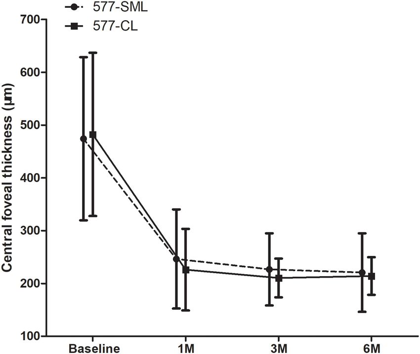
Zhou et al.                                                                                             Subthreshold Micropulse Laser for CSC

 FIGURE 1 | Study design flow chart.

577-nm CL group, with 80% power at a 2-sided α of 0.05 to detect        missing values at 3 months and the final endpoint. Results were
a difference between two groups, estimating 20% loss to follow-         presented as the mean (SD) or median (IQR) for the continuous
up. The sample size was calculated with PASS 11 (NCSS Statistical       variables and frequency for categorical variables. Baseline data
Software, Kaysville, UT).                                               comparisons between the two groups were performed by a 2-
    According to the intention-to-treat analysis principles that        tailed t-test for continuous variables with normal distribution,
all randomized participants were included, we used the last             the Wilcoxon rank-sum test for continuous variables with non-
observation carried forward (LOCF) method to impute the                 normal distribution, the chi-square test, or Fisher exact test

Frontiers in Medicine | www.frontiersin.org                         3                                     July 2021 | Volume 8 | Article 682264
Zhou et al. Subthreshold Micropulse Laser for CSC
TABLE 1 | Baseline demographics and clinical characteristics by treatment group (N = 110).
Characteristic 577 nm SML group (n = 55.50%) 577 nm CL group (n = 55.50%) P-value for comparing SML and CL
Age, mean (SD), years 41.2 (6.00) 41.1 (5.30) 0.894†
Sex, No. (%)
Male 47 (85.5%) 46 (83.6%) 0.792*
Female 8 (14.5%) 9 (16.4%)
Eye, No. (%)
Right 29 (52.7%) 33 (60.0%) 0.442*
Left 26 (47.3%) 22 (40.0%)
Duration of symptoms median (IQR), months 2.00 (1.00–4.00) 3.00 (1.00–5.00) 0.519‡
BCVA (logMAR), mean(SD) 0.32 (0.21) 0.39 (0.22) 0.113†
CFT, mean(SD), µm 474 (154) 482 (157) 0.780†
SD, Standardized deviation; IQR, Interquartile range; BCVA, Best-corrected visual acuity; LogMAR, Logarithm of the minimum angle of resolution; CFT, Central foveal thickness.
* Pearson χ2 test, † Independent-sample T-test, ‡ Wilcoxon rank-sum test.
TABLE 2 | Intention-to-treat analysis of potential determinants of SRF at 3-months follow-up.
Variable Simple Regression† (n = 110)‡ Multiple Regression (n = 110)‡
Relative risk (95% CI) P-Value Relative risk (95% CI) P-Value
SML group (CL group as reference) 0.82 (0.68–0.98) 0.033 0.78 (0.66–0.93) 0.005
Age, year 0.99 (0.97–1.01) 0.199 0.99 (0.97–1.01) 0.253
Male sex 0.99 (0.77–1.28) 0.969 – –
Right eye 1.19(0.98–1.45) 0.077 1.05 (0.86–1.27) 0.657
Duration of symptom, months 0.98 (0.93–1.03) 0.363 – –
Baseline BCVA (LogMAR) 0.43 (0.25–0.75) 0.003 0.44 (0.25–0.16) 0.004
Baseline CFT, µm 0.99 (0.99–1.01) 0.061 0.99 (0.99–1.04) 0.485
† Variableswith P < 0.20 in the simple regression analysis were included in the multiple regression model. Relative risk and 95% CI were estimated using the generalized linear model
with the option of Poisson regression. ‡ Included five missing data.
for categorical variables. The complete absorption of SRF at eight patients (7.27%) were lost to follow-up: six patients were
a 3-month follow-up, the primary outcome, with a relative too busy to follow up, one patient moved to another hospital, and
risk (RR) and 95% confidence intervals (CIs), was calculated one patient transferred to another place (Figure 1). All missing
and compared. data were imputed with the LOCF approach for the intention-to-
For the potential prognostic factors at baseline, variables treat analysis.
with p < 0.20 level in a simple regression model were added The baseline demographic and clinical characteristics were
to the multiple regression model. All statistical analysis was balanced between the two groups and are summarized in Table 1.
performed using SAS statistical software, version 9.4 (SAS More specifically, there was no significant difference between the
Institute Inc., Cary, NC). A p-value withZhou et al. Subthreshold Micropulse Laser for CSC
FIGURE 2 | The change of mean LogMAR-BCVA. The BCVA of all patients showed a statistically significant improvement at each visit compared with the baseline in
each group (p < 0.05). Whereas, there was no significant difference between the 577-nm SML group and the 577-nm CL group (p > 0.05). LogMAR, logarithm of the
minimum angle of resolution; BCVA, best-corrected visual acuity. SML, subthreshold micropulse laser. CL, conventional laser.
duration, and baseline CFT, were not significantly associated with the visual acuity (LogMAR) was markedly improved from 0.39
the complete resolution of SRF (Table 2). ± 0.22 at baseline to 0.08 ± 0.16 at 1 month, 0.01 ± 0.11 at 3
The patients who were still present SRF involved in the macula months, and 0.00 ± 0.09 at 6 months (all p < 0.001). The change
at a the 3-month follow-up received the same intervention as the of BCVA (LogMAR) in the SML group was lower than that in
baseline: 14 patients in the SML group and five patients in the the CL group with an unadjusted difference (mean, 0.09; 95% CI,
CL group. And the SRF resolution of all patients was assessed 0.02–0.17; p = 0.017). However, there was no statistical difference
again as the other outcome at the final endpoint (6-month follow- concerning the change of BCVA (LogMAR) at the final endpoint
up). The complete resolution of SRF reached 85.5% (47/55) in between the two treatment groups (unadjusted mean difference,
the SML group and 92.7% (51/55) in the CL group, but there was 0.07; 95% CI, −0.01 to 0.15; p = 0.093) (Figure 2).
no significant difference between the two groups (unadjusted RR,
0.92; 95% CI, 0.81–1.05; P = 0.221).
Changes of Central Foveal Thickness
In the 577-nm SML group, the mean CFT decreased significantly
Changes in Visual Acuity from 474 ± 154 µm at baseline to 246 ± 93.8 µm at 1 month, 227
After treatment, the mean visual acuity (LogMAR) had a ± 68.4 µm at 3 months, and 221 ± 74.4 µm at 6 months (all p
statistically significant improvement from baseline to the < 0.001). The mean CFT was 482 ± 155 µm at baseline in the
endpoint in the two groups. The mean BCVA in the 577-nm SML 577-nm CL group and decreased remarkably to 226 ± 77.1 µm
group was 0.11 ± 0.17 at 1 month, 0.03 ± 0.12 at 3 months, at 1 month, 210 ± 36.6 µm at 3 months, and 214 ± 35.6 µm
and 0.00 ± 0.10 at 6 months, respectively, all of which was at 6 months (all p < 0.001). However, there was no statistical
significantly improved compared with the visual acuity of the difference for the change of CFT at the 3-month visit (unadjusted
baseline (0.32 ± 0.21) (all p < 0.001). In the 577-nm CL group, mean difference, −24.4; 95% CI, −83.8 to 35.1; p = 0.418) and
Frontiers in Medicine | www.frontiersin.org 5 July 2021 | Volume 8 | Article 682264Zhou et al. Subthreshold Micropulse Laser for CSC
FIGURE 3 | The change of mean CFT during baseline and visits. CFT showed a statistically significant reduction at each visit compared with the baseline in each
group (p < 0.05). Whereas, there was no significant difference between 577-nm SML group and 577-nm CL group (p > 0.05). CFT, central foveal thickness. SML,
subthreshold micropulse laser. CL, conventional laser.
at the 6-month visit (unadjusted mean difference, −14.7; 95% lower complete absorption rate in the short-term (at 3 months),
CI, −74.4 to 44.8; p = 0.625) between the two treatment groups compared with that in the CL group. But after retreatment, 577-
(Figure 3). nm SML can reach a similar effect on improving the functional
and anatomical outcomes of eyes with acute CSC. Importantly,
Safety 577-nm SML scarcely damaged RPE compared with CL.
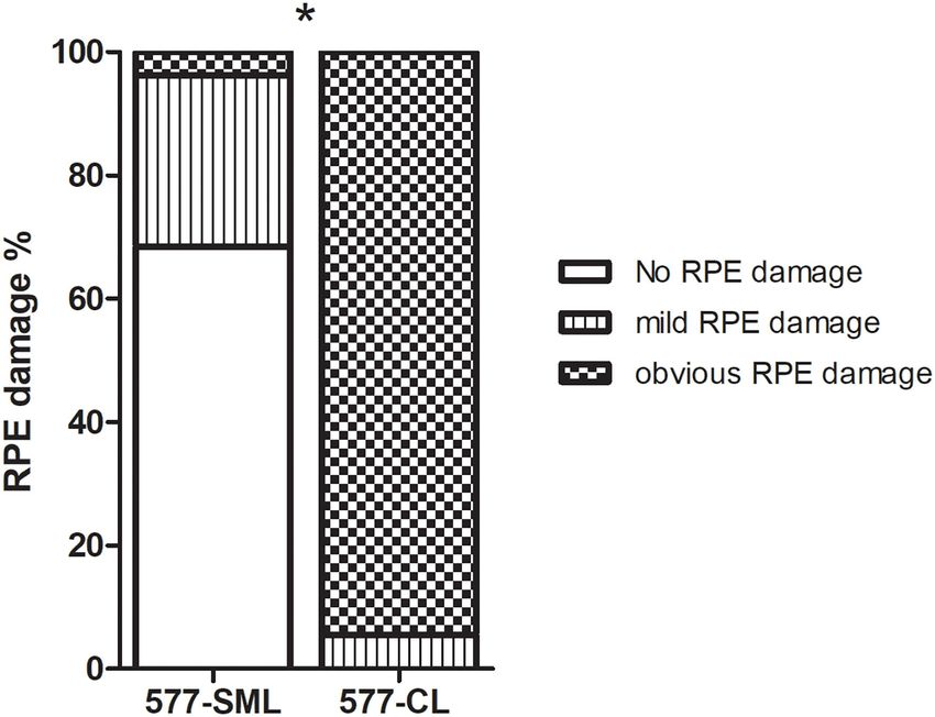
Based on the FAF images, RPE damage was evaluated at a the The conventional laser has been applied to retinal disease
1-month follow-up. In the 577-nm SML group, 69.1% (38/55) for many years, and it works by thermal energy to the RPE
patients showed no RPE damage, 27.3% (15/55) had mild RPE (16). Nevertheless, except for the proven effect, it has inherent
damage, and only 3.64% (2/55) showed obvious RPE damage. adverse effects that often destroy the adjacent tissue, such as
The corresponding data in the 577-nm CL group were 0.00% the inner retina and photoreceptors, due to thermal energy
(0/55), 7.27% (4/55), and 92.7% (51/55), respectively (Figure 4). conduction. Therefore, it is not suitable for subfoveal and
The patients who received conventional wave laser treatment had juxtafoveal leaks (1, 17). In the present study, we showed
more significant RPE damage than the patients who received similar findings of the CL treatment, as reported previously.
subthreshold micropulse laser treatment (p < 0.001). Besides, In the 577-nm CL group, SRF was absorbed entirely in 92.7%
during the 6-month follow-up, choroidal neovascularization was (51/55) of patients, and CFT decreased prominently from 482
not seen on OCT imaging in all patients. ± 15 µm at baseline to 214 ± 35.6 µm 6 months (p < 0.001).
Accordingly, visual acuity improved significantly from 0.39 ±
DISCUSSION 0.22 LogMAR at baseline to 0.00 ± 0.09 LogMAR (p < 0.001)
after treatment. But evident laser scar (RPE damage) was seen
To the best of our knowledge, this study is the first prospective in 51 (92.7%) patients, significantly higher than that of 577-
randomized controlled trial on the comparison of 577-nm SML nm SML.
with 577-nm CL for acute CSC. Our study showed that the SRF Minimizing the adverse reactions of CL treatment, new
of patients with acute CSC in the 577-nm SML group had a modalities have emerged over the years (18). SML, described in
Frontiers in Medicine | www.frontiersin.org 6 July 2021 | Volume 8 | Article 682264Zhou et al. Subthreshold Micropulse Laser for CSC FIGURE 4 | The comparison of eyes with RPE change on FAF imaging at 1 month after treatments in the 577-nm SML group and 577-nm CL group. Error bars represent standard errors of the mean. SML, subthreshold micropulse laser. CL, conventional laser. RPE, retinal pigment epithelium. FAF, fundus autofluorescence. *P < 0.05. detail by Dorin in 2003, is a newer method for macular disease, in our study were acute CSC, the RPE reaction to SML would especially in diabetic macular edema and chronic CSC in recent be better. Second, their follow-up was relatively shorter from years (19–22). SML is composed of a train of repetitive ultrashort 2 to 6 weeks but 12–24 weeks in our study. Additionally, our laser pulses that has “on-time” and “off-time” and produces a results showed that repeated treatment of SML was available for sublethal cellular thermal effect. The SML mechanism in treating patients with persistent SRF because of less RPE damage. The CSC may be due to the activation of RPE biological response findings have shown that SML treatment has fewer side effects, rather than merely thermal coagulation of RPE (23). As shown even repeated use. in our study, only 3.64% of eyes had obvious RPE damage after There is a challenge for SML treatment, especially in SML treatment, significantly less than CL treatment. power selection. Power titration is an essential step in SML Recent retrospective studies have demonstrated that 577-nm treatment, and it is usually outside the vascular arcades. The SMPL could achieve the equivalent effect as conventional laser method of titration was reported in several clinical studies but without RPE damage (13). It has been reported that in (12, 15, 25). It was performed in a single spot pattern, and chronic CSC, the complete resolution rate of the SRF varied from the power was increased gradually until a just visible spot 33 to 75% in previous studies using SML (15, 21, 24). In our was seen. This power was the threshold burn. Then the research, the result was better with a complete resolution rate laser power was reduced to 50% for the actual treatment of 72.7% at 3 months after the first treatment and 85.5% at 6 power in our study. However, the real treatment power can’t months after retreatment. There might be two reasons for the fit any retina because retinal epithelial cells and choroidal faster absorption ratio. First, patients in previous studies were melanocytes vary from location. Thus, it was explained that RPE chronic CSC and might have diffuse damaged RPE that would damage was seen in two patients of our study. So, it should slow down the absorption of subretinal fluid. While the patients be used more judiciously when the leakage was just at the Frontiers in Medicine | www.frontiersin.org 7 July 2021 | Volume 8 | Article 682264
Zhou et al.                                                                                                                      Subthreshold Micropulse Laser for CSC

macular fovea. If you do, it is better to use
Zhou et al.                                                                                                                         Subthreshold Micropulse Laser for CSC

15. Yadav NK, Jayadev C, Mohan A, Vijayan P, Battu R, Dabir S, et al.                    24. Scholz P, Ersoy L, Boon CJ, Fauser S. Subthreshold micropulse laser (577 nm)
    Subthreshold micropulse yellow laser (577 nm) in chronic central serous                  treatment in chronic central serous chorioretinopathy. Ophthalmologica.
    chorioretinopathy: safety profile and treatment outcome. Eye. (2015) 29:258–             (2015) 234:189–94. doi: 10.1159/000439600
    64; quiz 265. doi: 10.1038/eye.2014.315                                              25. van Dijk EHC, Fauser S, Breukink MB, Blanco-Garavito R, Groenewoud
16. Ficker L, Vafidis G, While A, Leaver P. Long-term follow-up of a prospective             JMM, Keunen JEE, et al. Half-dose photodynamic therapy versus high-density
    trial of argon laser photocoagulation in the treatment of central serous                 subthreshold micropulse laser treatment in patients with chronic central
    retinopathy. Br J Ophthalmol. (1988) 72:829–34. doi: 10.1136/bjo.72.11.829               serous chorioretinopathy: The PLACE Trial. Ophthalmology. (2018)
17. Iacono P, Battaglia Parodi M, Falcomata B, Bandello F. Central serous                    125:1547–55. doi: 10.1016/j.ophtha.2018.04.021
    chorioretinopathy treatments: a mini review. Ophthalmic Res. (2015) 55:76–           26. Zhou L, Chong V, Lai K, Huang C, Xu F, Gong Y, et al. A pilot prospective
    83. doi: 10.1159/000441502                                                               study of 577-nm yellow subthreshold micropulse laser treatment with
18. Dorin G. Subthreshold and micropulse diode laser photocoagulation. Semin                 two different power settings for acute central serous chorioretinopathy.
    Ophthalmol. (2003) 18:147–53. doi: 10.1076/soph.18.3.147.29812                           Lasers Med Sci. (2019) 34:1345–51. doi: 10.1007/s10103-019-
19. Ohkoshi K, Yamaguchi T. Subthreshold micropulse diode laser                              02721-8
    photocoagulation for diabetic macular edema in Japanese patients. Am J               27. Liew G, Quin G, Gillies M, Fraser-Bell S. Central serous chorioretinopathy:
    Ophthalmol. (2010) 149:133–9. doi: 10.1016/j.ajo.2009.08.010                             a review of epidemiology and pathophysiology. Clin Exp Ophthalmol. (2013)
20. Lavinsky D, Sramek C, Wang J, Huie P, Dalal R, Mandel Y, et al. Subvisible               41:201–14. doi: 10.1111/j.1442-9071.2012.02848.x
    retinal laser therapy: titration algorithm and tissue response. Retina. (2014)
    34:87–97. doi: 10.1097/IAE.0b013e3182993edc                                          Conflict of Interest: The authors declare that the research was conducted in the
21. Lavinsky D, Palanker D. Nondamaging photothermal therapy for the retina:             absence of any commercial or financial relationships that could be construed as a
    initial clinical experience with chronic central serous retinopathy. Retina.         potential conflict of interest.
    (2015) 35:213–22. doi: 10.1097/IAE.0000000000000340
22. Scholz P, Altay L, Fauser S. A review of subthreshold micropulse                     Copyright © 2021 Zhou, Lai, Jin, Huang, Xu, Gong, Li, Zhu, Lu and Jin. This is an
    laser for treatment of macular disorders. Adv Ther. (2017) 34:1528–                  open-access article distributed under the terms of the Creative Commons Attribution
    55. doi: 10.1007/s12325-017-0559-y                                                   License (CC BY). The use, distribution or reproduction in other forums is permitted,
23. Sramek C, Mackanos M, Spitler R, Leung LS, Nomoto H, Contag                          provided the original author(s) and the copyright owner(s) are credited and that the
    CH, et al. Non-damaging retinal phototherapy: dynamic range of heat                  original publication in this journal is cited, in accordance with accepted academic
    shock protein expression. Invest Ophthalmol Vis Sci. (2011) 52:1780–                 practice. No use, distribution or reproduction is permitted which does not comply
    7. doi: 10.1167/iovs.10-5917                                                         with these terms.

Frontiers in Medicine | www.frontiersin.org                                          9                                                July 2021 | Volume 8 | Article 682264
You can also read