Quality of Endodontic Treatment and Prevalence of Apical Radiolucencies in a Bulgarian Subpopulation: a CBCT Analysis
←
→
Page content transcription
If your browser does not render page correctly, please read the page content below
Folia Medica 63(1):81-7
                                                                                                                          DOI: 10.3897/folmed.63.e52204

                                                                 Original Article

Quality of Endodontic Treatment
and Prevalence of Apical Radiolucencies
in a Bulgarian Subpopulation: a CBCT Analysis
Teodora Karteva1, Neshka A. Manchorova-Veleva1, Ekaterina Karteva1, Donka Keskinova2,
Petya Kanazirska3, Georgi Jordanov3, Stoyan Vladimirov1
1 Department of Operative Dentistry and Endodontics, Faculty of Dental Medicine, Medical University of Plovdiv, Plovdiv, Bulgaria
2 Department of Applied and Institutional Sociology, Paisii Hilendarski University of Plovdiv, Plovdiv, Bulgaria
3 Department of Imaging Diagnostics, Dental Allergology and Physiotherapy, Medical University of Plovdiv, Faculty of Dental Medicine, Plovdiv, Bulgaria

Corresponding author: Teodora Karteva, Department of Operative Dentistry and Endodontics, Faculty of Dental Medicine, Medical University of
Plovdiv, 3 Hristo Botev Blvd., Plovdiv, Bulgaria; E-mail: tedy.karteva@gmail.com; Tel.: +359 879 851 091

Received: 18 Mar 2020 ♦ Accepted: 2 July 2020 ♦ Published: 28 Feb 2021

Citation: Karteva T, Manchorova-Veleva NA, Karteva E, Keskinova D, Kanazirska P, Jordanov G, Vladimirov S. Quality of endodontic
treatment and prevalence of apical radiolucencies in a Bulgarian subpopulation: a CBCT analysis. Folia Med (Plovdiv) 2021;63(1):81-7.
doi: 10.3897/folmed.63.e52204.

Abstract
Introduction: The advent of Cone Beam Computed Tomography (CBCT) in endodontics has enhanced the diagnosis of periapical
radiolucencies and the assessment of endodontically treated teeth.
Aim: The purpose of this study was to assess the prevalence of periapical radiolucencies in a Bulgarian subpopulation and the quality
of previous endodontic treatment using CBCT scans.
Materials and methods: This study included 2795 roots from 160 Large FOV CBCT which were evaluated by two independent
examiners using two scoring systems: CBCT-PAI and PESS.
Results: The inter-examiner agreement spanned from strong to almost perfect (0.892 and 0.983). The prevalence of periapical lesions
according to the two scoring systems was 23.1% and 12.9 %, respectively. The prevalence of endodontically treated teeth was high
(34.1%). Sixty-five percent of them presented with signs of periapical radiolucencies, while only 1.4% of all non-treated roots had a
periapical lesion. A significant association between periapical disease, poor quality of the root canal filling and inadequate coronal seal
was found (p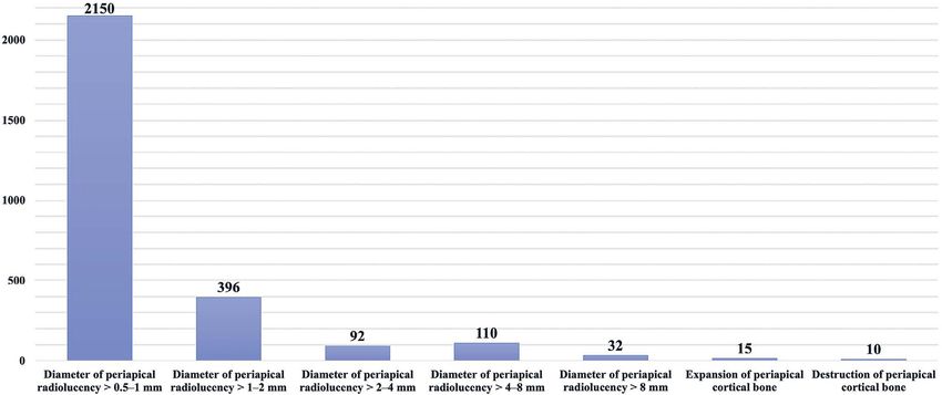
T. Karteva et al
Recent epidemiologic research documents that the pre- Imaging Software. Monitor settings concerning brightness
valence of periapical radiolucencies varies among patients and contrast were adjusted to the preferences of the exa-
aged 20 to 30 by 33%, aged 30 to 40 by 40%, aged 40 to miners. A slice thickness of 0.2 mm to 0.5 mm was used
50 by 48%, 50 to 60 by 57%, and in patients older than 60 for the multiplane views in accordance with the examiner’s
years of age by 62%.3 The prevalence of chronic apical pe- preferences.
riodontitis in Bulgaria is high - between 2.0% and 18%.4 A total of 2795 roots (1843 teeth) were examined. The
Furthermore, the prevalence of periapical radiolucencies in root was adopted as the unit of observation and each root
teeth with root canal treatment is also very high - 71.3%.4 was scored with two previously published indices. The
Therefore, chronic periapical pathosis is regarded as a so- CBCT-PAI by Estrela et al.10 scores the size of the lesion,
cially and economically significant disease and a public its location, and relationship with the roots of the tooth
health problem. (Table 1). The PESS index by Venskutonis et al.18 is a
The contemporary method that is most widely used in complex scoring system evaluating lesion size, root canal
day-to-day clinical practice for evaluation of the periapi- treatment quality and possible complications. It consists of
cal area is the periapical radiography (conventional or di- two scoring systems – COPI, the complex periapical index
gital).5-7 However, it provides a two-dimensional view of (Table 2), designed for the identification and classification
three-dimensional (3D) structures.8-10 Several studies have of periapical bone lesions, and ETTI, the endodontical-
reported on the limitations of periapical radiography in de- ly treated tooth index (Table 3), designed for endodontic
tecting periapical lesions due to bone characteristics, lesion treatment quality evaluation. For the assessment of the en-
location, morphologic variations, surrounding bone den- dodontic treatment, the scores were distributed in the fol-
sity, x-ray angulations and radiographic contrast.5,8,11 His- lowing categories: the scores of L2, L3 and L4 were labeled
tologic analysis is considered the most accurate diagnostic as an inadequate length of the root canal filling, the scores
method but its application is limited to endodontic surgery of H2 – as an inadequate quality of the root canal filling and
only due to its invasive nature.12 the scores of CS2 – as an inadequate coronal restoration.
Recently, CBCT has been successfully implemented in
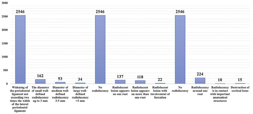
the field of endodontics as it provides a 3D reconstruction Table 1. Cone beam computed tomography periapical index
of the anatomical structures. Several studies have confir- scores (CBCTPAI)
med the increased accuracy of CBCT in detecting periapi-
cal lesions in comparison with conventional radiographic Quantitative Bone Alterations in Mineral
techniques.13-17 Therefore, CBCT can be beneficial to en- Score
Structures
dodontic diagnosis. The purpose of this study was to des-
0 Intact periapical bone structures
cribe the periapical health status of a Bulgarian subpopula-
tion via CBCT measurements and estimate the prevalence Diameter of periapical radiolucency > 0.5–1
1
of disease and treatment. mm
2 Diameter of periapical radiolucency > 1–2 mm
3 Diameter of periapical radiolucency > 2–4 mm
MATERIALS AND METHODS 4 Diameter of periapical radiolucency > 4–8 mm
5 Diameter of periapical radiolucency > 8 mm
Cases Selection Score (n) + E* Expansion of periapical cortical bone
Score (n) + D* Destruction of periapical cortical bone
The retrospective study was approved by the institutional The variables E (expansion of cortical bone) and D (destruction
review board of the Medical University of Plovdiv, with of cortical bone) were added to each score when either of these
accordance to the ethical standards of the Declaration of conditions was detected in the CBCT analysis.
Helsinki and with a waiver of informed consent due to the
design of the study. Large FOV CBCT images of 120 pa-
tients were selected from the database of a dental radiology
Statistical analysis
laboratory in Plovdiv, Bulgaria. The patients included in the
study were aged 18-64 (mean age 48.5 years) with a mini- Data were typed into a spreadsheet, and SPSS software
mum of 10 teeth. (version 17; SPSS Inc., Chicago, IL) was used to perform
the analysis. The Cohen kappa was calculated to assess
Imaging methods and analysis the inter-examiner agreement for each parameter for the
indexes. The Mann-Whitney test was used as the univari-
The CBCT images were obtained with the Planmeca Pro- ate approach to detect statistically significant differences
Max 3D Max dental X-ray unit. The scans’ parameters were between the categories. Correspondence analysis was used
voxel size of 0.200 & 0.200 & 0.200 mm, 15 bits. The CBCT for the assessment of the association of the quality of en-
volumetric data were evaluated by two independent and dodontic treatment and status of the apical periodontium.
calibrated examiners with the Planmeca Romexis Dental The level of significance adopted was 1%.
82 Folia Medica I 2021 I Vol. 63 I No. 1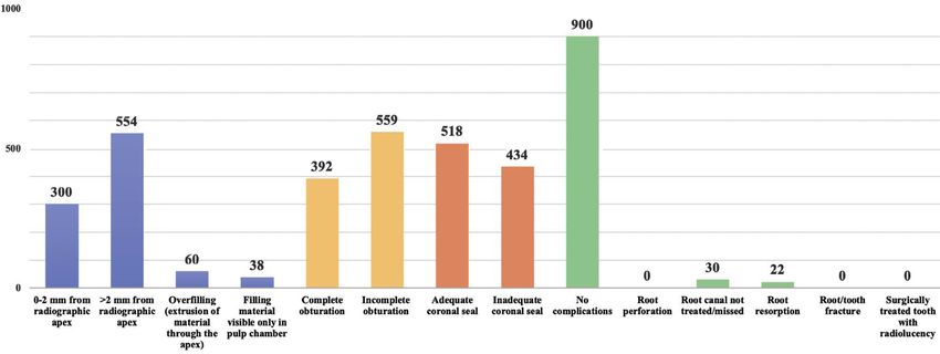
Quality of Endodontic Treatment and Prevalence of Apical Radiolucencies

Table 2. The Complex Periapical Index (COPI)

Score       S (Size of the radiolucent lesion)
S0          Widening of the periodontal ligament not exceeding two times the width of the lateral periodontal ligament
S1          The diameter of small well-defined radiolucency up to 3 mm
S2          Diameter of medium well-defined radiolucency 3-5 mm
S3          Diameter of large well-defined radiolucency >5 mm
            R (Relationship between root and radiolucent lesion)
R0          No radiolucency, when widening of the periodontal ligament is not exceeding two times the width of the lateral peri-
            odontal ligament
R1          Radiolucent lesion appears on one root
R2          Radiolucent lesion appears on more than one root
R3          Radiolucent lesion with involvement of furcation
            D (location of bone destruction)
D0          No radiolucency, when widening of the periodontal ligament is not exceeding two times the width of the lateral peri-
            odontal ligament
D1          Radiolucency around one root
D2          Radiolucency is in contact with important anatomical structures
D3          Destruction of cortical bone

Table 3. The endodontically treated tooth index (ETTI)

Score       L (length of the root canal filling
L0          0-2 mm from radiographic apex
L1          >2 mm from radiographic apex
L2          Overfilling (extrusion of material through the apex)
L3          Filling material visible only in pulp chamber
            H (homogeneity of the root canal filling)
H1          Complete obturation (homogenous appearance of the root canal filling)
H2          Incomplete obturation (voids or porous appearance of the root canal filling)
            CS (coronal seal)
CS1         Adequate (coronal restoration appears intact radiographically)
CS2         Inadequate (detectable radiographic signs of overhangs, open margins, recurrent caries, or lost coronal restoration
            CF (complications/failures)
CF0         No complications
CF1         Root perforation
CF2         Root canal not treated/missed
CF3         Root resorption
CF4         Root/tooth fracture
CF5         Surgically treated tooth with radiolucency

RESULTS
                                                                    apical radiolucency, while only 1.4% of all non-endodonti-
An overall inter-examiner agreement value of 0.94 was               cally treated roots were assessed to have a periapical lesion.
found for the measurements, thus indicating almost perfect          The Mann-Whitney test showed a significant association
agreement. The distribution of the CBCTPAI and PESS re-             between periapical status and the length of the root canal
sults is shown on Figs 1, 2, 3. Periapical lesions were asses-      filling with scores of L2, L3 or L4 (p
T. Karteva et al Figure 1. Distribution of CBCTPAI results. Figure 2. Distribution of COPI results. Figure 3. Distribution of ETTI results. 84 Folia Medica I 2021 I Vol. 63 I No. 1
Quality of Endodontic Treatment and Prevalence of Apical Radiolucencies

DISCUSSION                                                      which is compliant with the accepted standards. The cases
                                                                examined in this study do not represent a statistically sig-
Radiographically detectable periapical radiolucencies mark      nificant sample of the Bulgarian adult population. Nevert-
the presence of a chronic destructive inflammatory process.     heless, the results provide an insight into the poor quality
These chronic lesions are either of primary or secondary        of endodontic treatment and the prevalence of periapical
endodontic origin and patients generally do not present         radiolucencies.
with any symptoms. Even persistent endodontic lesions
can provide functionality and comfort to the patients and
they may never seek further treatment until complications       CONCLUSIONS
arise.19 Therefore, the cases chronic apical periodontitis
tend to accumulate in given population. Effective screening     The present study revealed high prevalence of periapi-
methods are needed for their successful diagnosis and ti-       cal radiolucencies in endodontically treated teeth among
mely treatment.                                                 the Bulgarian subpopulation. Our findings underline the
   The high proportion of endodontically treated teeth pre-     need of strict post-treatment endodontic evaluation proto-
sented with periapical radiolucency which correlates with       cols and continuous post-operative monitoring and care.
previous reports.19-21 These results can be due to a variety    Within the limitations of the present study, it was conclu-
of factors. While endodontically compromised teeth are re-      ded that the quality of endodontic treatment in Bulgaria is
tained successfully through root canal treatment as it is the   not up to the accepted clinical standards.
effective way to reduce the symptoms and treat periapical
disease, unsuccessful treatments do not always present as
functional or symptomatic failures and accumulate over          Acknowledgments
time in a population.22 Furthermore, radiographic scree-
ning and cross-sectional studies are an effective way of as-    The support of Grant No 07/2018 from Medical University –
sessing the prevalence, severity and origin of the problem.     Plovdiv is acknowledged.
However, these snapshot studies cannot distinguish bet-
ween healing and persistent lesions.19 One of the limitati-
ons of the present retrospective study is that our evaluation   REFERENCES
was only based solely on the radiographic analysis. No data
about the dental history of the patients were available.        1.  Stashenko P, Teles R, D’souza R. Periapical inflammatory responses
   The overall prevalence of periapical disease in the Bul-         and their modulation. Crit Rev Oral Biol Med 1998; 9(4):498–521.
garian subpopulation was higher than previous reports           2. Ahmed GM, El-Baz AA, Hashem AAR, et al. Expression levels of ma-
based on conventional radiographic techniques.4 This can            trix metalloproteinase-9 and gram-negative bacteria in symptomatic
be explained with higher sensitivity of CBCT in the detec-          and asymptomatic periapical lesions. J Endod 2013; 39(4):444–8.
tion of periapical radiolucencies.23 The prevalence also va-    3. Ørstavik D. Endodontic treatment of apical periodontitis. In: Ørstavik
ried according to the two scoring systems. This is due to           D, editor. Essential Endodontology: Prevention and Treatment of
the different disease threshold criteria in their design. The       Apical Periodontitis. 3rd ed. 2019; 313–44.
conventional strict criteria for endodontic success on con-     4. Gusyiska AZ. [Orthogradic treatment of chronic apical periodontitis –
ventional two-dimensional radiographs include a complete            biological approaches] [PhD dissertation] Medical University, Sofia
absence of periradicular radiolucency along with the re-es-         2012. [In Bulgarian]
tablishment of a normal periodontal ligament space and a        5. Estrela C, Bueno MR, Leles CR, et al. Accuracy of cone beam com-
defined lamina dura. established in clinical practice.24 With       puted tomography and panoramic and periapical radiography for de-
the advent of the new high-resolution three-dimensional             tection of apical periodontitis. J Endod 2008; 34(3):273–9.
images new, universal criteria need to be established for the   6. Özen T, Kamburoğlu K, Cebeci ARI, et al. Interpretation of chemi-
successful evaluation of treatment success and prevention           cally created periapical lesions using 2 different dental cone-beam
of unnecessary treatment.                                           computerized tomography units, an intraoral digital sensor, and con-
   An evaluation of the quality of pervious endodontic tre-         ventional film. Oral Surg Oral Med Oral Pathol Oral Radiol Endod
atment was performed with the ETTI scoring system. The              2009; 107(3):426–32.
high resolution CBCT images provided for the comprehen-         7. Patel S, Dawood A, Whaites E, et al. New dimensions in endodontic
sive assessment of any factors that could have impacted tre-        imaging: part 1. Conventional and alternative radiographic systems.
atment success. Our results indicate that incomplete obtu-          Int Endod J 2009; 42(6):447–62.
ration of the root canals and a compromised coronal seal        8. Huumonen S, Orstavik D. Radiological aspects of apical periodonti-
increase the risk of secondary infection of the root canal          tis. Endodontic Topics 2002; 1(1):3–25.
system and are associated the development of periapical di-     9. Patel S, Dawood A, Mannocci F, et al. Detection of periapical bone
sease. The results are in accordance with previous reports          defects in human jaws using cone beam computed tomography and
and underline the need for optimized treatment strate-              intraoral radiography. Int Endod J 2009; 42(6):507–15.
gies.25-30 The best treatment results were associated with a    10. Estrela C, Bueno MR, Azevedo BC, et al. A new periapical index based
root canal obturation ending 0–2 mm short of the apex,              on cone beam computed tomography. J Endod 2008; 34(11):1325–31.

Folia Medica I 2021 I Vol. 63 I No. 1                                                                                                85
T. Karteva et al
11. Halse A, Molven O, Fristad I. Diagnosing periapical lesions - dis- 2011; 112(1):136–42.
agreement and borderline cases. Int Endod J 2002; 35(8):703–9. 21. Ahmed I, Ali R, Mudawi A. Prevalence of apical periodontitis and
12. Laux M, Abbott PV, Pajarola G, Nair PNR. Apical inflammatory root frequency of root-filled teeth in an adult Sudanese population. Clin
resorption: a correlative radiographic and histological assessment. Int Exp Dent Res 2017; 3(4):142–7.
Endod J 2000; 33(6):483–93. 22. Pak JG, White SN. Pain prevalence and severity before, during,
13. Cotton T, Geisler T, Holden D, et al. Endodontic applications of cone- and after root canal treatment: a systematic review. J Endod 2011;
beam volumetric tomography. J Endod 2007; 33(9):1121–32. 37(4):429–38.
14. Nair MK, Nair UP. Digital and advanced imaging in endodontics: a 23. Strindberg LZ. The dependence of the results of pulp therapy on
review. J Endod 2007;33(1):1–6. certain factors; an analytic study based on radiographic and clinical
15. Velvart P, Hecker H, Tillinger G. Detection of the apical lesion and follow-up examinations. Acta odontologica Scandinavica Supple-
the mandibular canal in conventional radiography and computed to- mentum 1956; 10(1):20–7.
mography. Oral Surg Oral Med Oral Pathol Oral Radiol Endod 2001; 24. Ray HA, Trope M. Periapical status of endodontically treated teeth
92(6):682–8. in relation to the technical quality of the root filling and the coronal
16. Lofthag-Hansen S, Huumonen S, Gröndahl K, et al. Limited cone- restoration. Int Endod J 1995; 28(1):12–8.
beam CT and intraoral radiography for the diagnosis of periapical 25. Sjögren U, Hägglund B, Sundqvist G, et al. Factors affecting the long-
pathology. Oral Surg Oral Med Oral Pathol Oral Radiol Endod 2007; term results of endodontic treatment. J Endod 1990; 16(10):498–504.
103(1):114–9. 26. Ng Y-L, Mann V, Rahbaran S, et al. Outcome of primary root ca-
17. Nakata K, Naitoh M, Izumi M, et al. Effectiveness of dental computed nal treatment: systematic review of the literature – Part 1. Effects
tomography in diagnostic imaging of periradicular lesion of each root of study characteristics on probability of success. Int Endod J 2007;
of a multirooted tooth: a case report. J Endod 2006; 32(6):583–7. 40(12):921–39.
18. Venskutonis T, Plotino G, Tocci L, et al. Periapical and endodontic 27. Gomes AC, Nejaim Y, Silva AI, et al. Influence of endodontic treat-
status scale based on periapical bone lesions and endodontic treat- ment and coronal restoration on status of periapical tissues: a cone-
ment quality evaluation using cone-beam computed tomography. J beam computed tomographic study. J Endod 2015; 41(10):1614–8.
Endod 2015; 41(2):190–6. 28. Song M, Park M, Lee C-Y, et al. Periapical status related to the qual-
19. Pak JG, Fayazi S, White SN. Prevalence of periapical radiolucency and ity of coronal restorations and root fillings in a Korean population. J
root canal treatment: a systematic review of cross-sectional studies. J Endod 2014; 40(2):182–6.
Endod 2012; 38(9):1170–6. 29. Gillen BM, Looney SW, Gu L-S, et al. Impact of the quality of coronal
20. Özbaş H, Aşcı S, Aydın Y. Examination of the prevalence of periapi- restoration versus the quality of root canal fillings on success of root
cal lesions and technical qulity of endodontic treatment in a Turkish canal treatment: a systematic review and meta-analysis. J Endod 2011;
subpopulation. Oral Surg Oral Med Oral Pathol Oral Radiol Endod 37(7):895–902.
86 Folia Medica I 2021 I Vol. 63 I No. 1Quality of Endodontic Treatment and Prevalence of Apical Radiolucencies Качество эндодонтического лечения и распространённость апикальных рентгенопрозрачностей в подгруппе населения Болгарии: анализ КЛКТ Теодора Картева1, Нешка А. Манчорова-Велева1, Екатерина Картева1, Донка Кескинова2, Петя Каназирска3, Георги Йорданов3, Стоян Владимиров1 1 Кафедра оперативного зуболечения и эндодонтии, Факультет дентальной медицины, Медицинский университет – Пловдив, Пловдив, Болгария 2 Кафедра прикладой и отраслевой социологии, Пловдивский университет „Паисий Хилендарски“, Пловдив, Болгария 3 Кафедра рентгенологии, дентальной аллергологиия и физиотерапии, Медицинский университет – Пловдив, Пловдив, Болгария Адрес для корреспонденции: Теодора Картева, Кафедра оперативного зуболечения и эндодонтии, Факультет дентальной медицины, Медицинский университет – Пловдив, бул. „Христо Ботев“ № 3, Пловдив, Болгария; E-mail: tedy.karteva@gmail.com; Тел.: +359 879 851 091 Дата получения: 18 марта 2020 ♦ Дата приемки: 2 июля 2020 ♦ Дата публикации: 28 февраля 2021 Образец цитирования: Karteva T, Manchorova-Veleva NA, Karteva E, Keskinova D, Kanazirska P, Jordanov G, Vladimirov S. Quality of endodontic treatment and prevalence of apical radiolucencies in a Bulgarian subpopulation: a CBCT analysis. Folia Med (Plovdiv) 2021;63(1):81-7. doi: 10.3897/folmed.63.e52204. Резюме Введение: Внедрение конусно-лучевой компьютерной томографии (КЛКТ) в эндодонтии способствует диагностике периа- пикальной рентгенопрозрачности и оценке зубов, подвергшихся эндодонтическому лечению. Цель: Целью этого исследования было вычислить частоту периапикальных рентгенопрозрачностей в подгруппе населения Болгарии и качество предшествующего эндодонтического лечения с использованием КЛКТ-сканирования. Материалы и методы: Это исследование включало 2795 корней из 160 изображений КЛКТ с большим полем обзора, кото- рые были оценены двумя независимыми экспертами с двумя различными системами оценки: КЛКТ-PAI и PESS. Результаты: Соответствие оценок специалистов колебалось от высокого до почти полного (0.892 и 0.983). Частота периа- пикальных поражений по двум системам оценки составила 23.1% и 12.9% соответственно. Частота случаев эндодонтического лечения зубов была высокой (34.1%). У 65 из них установлено наличие периапикальных рентгенопрозрачностей, в то время как только 1.4% всех необработанных корней имели периапикальные поражения. Были обнаружены значимые связи между периапикальным заболеванием, плохим качеством пломбирования корневых каналов и неправильной коронковой пломби- ровкой (p
You can also read