EXTENDED APICAL PERIODONTAL CYST IN MAXILLA: CASE REPORT
←
→
Page content transcription
If your browser does not render page correctly, please read the page content below
UNINGÁ Journal, v. 58, eUJ3691, 2021
                                                                         doi.org/10.46311/2318-0579.58.eUJ3691

    EXTENDED APICAL PERIODONTAL CYST IN MAXILLA: CASE REPORT
       CISTO PERIODONTAL APICAL EXTENSO EM MAXILA: RELATO DE CASO

Rebeca Ferreira Moreira1* , Onaldo Aguiar2 , Thainá Araújo Pacheco Brito3
1
  Centro Universitário UniFTC, Salvador, BA, Brasil.
2
  Universidade de São Paulo, São Paulo, SP, Brasil.
3
  Universidade Federal da Bahia, Salvador, BA, Brasil.
*rebecaferreiramoreira@hotmail.com

ABSTRACT

The apical periodontal cyst represents the most common inflammatory pathology among odontogenic
cysts, corresponding to about 60% of the maxilla and mandible cysts. It is associated with the apex
of a devitalized tooth and is generally asymptomatic, affecting more frequently the anterior region of
the maxilla. A 44-year-old leukoderma patient presented with a large asymptomatic lesion located in
the anterior region of the maxilla. On clinical examination, it was possible to observe an increase in
volume associated with the erasure of the gingivolabial fold, in addition to a negative pulp vitality
test in dental units # 13, # 12, # 11, # 21 and # 22. Radiographically, it appears as a well-defined
unilocular radiolucent image surrounding the apexes of the respective dental units. An aspiration
puncture was performed, showing bloody secretion, reinforcing the suspected suspicion of apical
periodontal cyst. The proposed treatment was two aspirations per week for 15 days followed by
enucleation, the 45-day postoperative panoramic radiograph showed bone neoformation. Thus, it is
possible to verify that the surgical approach consists of a resolutive therapeutic option, mainly in
extensive lesions.

Keywords: Apical periodontal cyst. Oral Surgery. Oral Pathology.

RESUMO

O cisto periodontal apical representa a patologia de natureza inflamatória mais comum entre os cistos
odontogênicos, correspondendo a cerca de 60% dos cistos da maxila e mandíbula. Está associado ao
ápice de um dente desvitalizado e geralmente é assintomático, acometendo com mais frequência a
região anterior da maxila. Paciente 44 anos, leucoderma, apresentou uma lesão de grande proporção
assintomática localizada em região anterior da maxila. Ao exame clínico foi possível observar
aumento de volume associado ao apagamento do sulco gengivolabial, além de teste de vitalidade
pulpar negativo nas unidades dentárias #13, #12, #11, #21 e #22. Radiograficamente, apresenta-se
como uma imagem radiolúcida unilocular bem definida circundando os ápices das respectivas
unidades dentárias. Foi realizada uma punção aspirativa, evidenciando secreção piossanguinolenta,
reforçando a suspeita diagnóstica de cisto periodontal apical. Foram realizadas duas punções
aspirativas por semana, durante duas semanas, seguidas de injeção intralesional de Rifocina. Após a
quarta sessão, foi realizada abordagem cirúrgica para enucleação da lesão. A radiografia panorâmica
pós-operatória de 45 dias evidenciou neoformação óssea. Desta forma, é possível constatar que a
abordagem cirúrgica consiste em uma opção terapêutica resolutiva, principalmente em lesões
extensas.

Palavras-chave: Cisto Bucal. Cisto Periodontal Apical. Patologia Bucal.

       Revista UNINGÁ, v. 58, eUJ3691, 2021.       Received: August, 28th, 2020; Accepted: April, 26th, 2021.
EXTENDED APICAL PERIODONTAL CYST IN MAXILLA: CASE REPORT
INTRODUCTION
The apical periodontal cyst, one of the types of root cyst, is defined as a pathological cavity
internally lined by epithelium and externally by fibrous connective tissue, containing within it a semi-
fluid or fluid material and is associated with the apex of a devitalized tooth. It is classified as an
inflammatory odontogenic cyst due to its coating derived from the proliferation of Malassez epithelial
remains, induced by an inflammatory stimulus, resulting from the infection of the root canals due to
a pulp necrosis (LIN; RICUCCI; KAHLER, 2017; MARTINS et al., 2018).
Root cysts correspond to 60% of odontogenic cysts, with the anterior region of the maxilla
being the most affected. There is a predominance of males between the third and sixth decade of life,
with leukoderma patients being more affected than melanoderms in a 2: 1 ratio (JUNQUEIRA et al.,
2011; DANTAS et al., 2014; COMIM et al., 2017).
As for the pathogenesis of the root cyst, three phases are described: initial, cystic formation
phase and growth phase. The formation of the cystic cavity originates from a pre-existing periapical
granuloma due to the degeneration and central death of fibroblast cells, collagen fibrils, endothelial
and capillary cells (SANTOS et al., 2011).
Periapical cysts are usually asymptomatic, however, when the lesion reaches large
proportions, signs of tenderness may be observed. The lack of response to pulp tests is a characteristic
of all apical periodontal cysts. Radiographically, a radiolucent unilocular image is observed, rounded
or oval, circumscribed by a well-defined radiopaque line that surrounds the apex of the tooth.
Difficulty in differentiating radicular cyst from apical granuloma has been observed on radiography
(LIN; RICUCCI; KAHLER, 2017; RESENDE et al., 2017)
It is necessary for the diagnosis of the root cyst the absence of pulp vitality of the involved
unit. Other complementary methods can also be used for the differential diagnosis, such as
electrophoretic analysis, computed tomography and aspiration of the cystic content. All surgical
specimens must be sent for histopathological examination to define the definitive diagnosis
(JUNQUEIRA et al., 2011; LIN; RICUCCI; KAHLER, 2017). Treatment varies from the endodontics
of the dental unit involved to the surgical approach (PINTO et al., 2016; SILVA et al., 2018).
The aim of the present study is to report the clinical case of a large radicular cyst, treated with
decompression followed by enucleation.
CASE REPORT
Female patient, leucoderma, 44 years old, attended a private dental office in the city of
Salvador-BA complaining of an increase in volume in the anterior region of the maxilla, lip and nasal
base for two months, progressing with dyspnea. On intraoral physical examination, the gingivolabial
sulcus was erased due to the presence of softened swelling (Figure 1a).
On intraoral examination, there was an increase in volume in the anterior region of the maxilla,
with normal mucosa color and smooth surface. The presence of intraoral fistula was observed, with
drainage of purulent secretion. The sensitivity test was negative for units # 13, # 12, # 11, # 21 and #
22.
Panoramic radiography showed a radiolucent, unilocular lesion, with regular contours,
between units # 13 and # 22 (Figure 1b). It is possible to observe that the root canal of the unit # 12
is subobutured, an inadequate intra-root pin in unit # 11, as well as external root resorption in units
11, 21 and 22.
3D computed tomography showed an extensive lesion in the anterior region of the maxilla,
without compromising the nasal cavity or maxillary sinus. The contours are regular, with partially
defined limits, measuring about 3.4 x 3.1 x 0.5 cm (Figures 2a and 2b).
Page 2 of 6Moreira; Aguiar; Brito
Figure 1 - Swelling in the anterior region of the maxilla, with erasure of the gingivolabial
sulcus (A). Panoramic radiography: lesion involving the dental apexes of unit # 13 and # 22
(B)
Source: the authors.
Figure 2 - 3D tomography of the maxilla, showing the limits of the lesion (A). Computed
tomography showing the size of the lesion and involvement of the buccal and palatal bone
plates (B)
Source: the authors.
An aspiration puncture was performed with a 20ml syringe to check the nature of the lesion's
content, resulting in 15ml of purulent and bloody content, with the diagnostic hypothesis of an apical
periodontal cyst being extensive. The therapeutic approach was four aspiration punctures (two per
week) using a 20 ml syringe with a 40 x 12 needle, promoting a decrease in osmotic pressure, followed
by intralesional injection of Rifocin, in order to avoid infectious complications at the site. In the last
section, the presence of liquid content was noted, which was collected and sent to the laboratory for
culture with antibiogram, showing a negative result. The patient was then submitted to enucleation
of the lesion (Figures 3a, 3b and 3c), and the piece was sent for histopathological analysis.
Figure 3 - Transoperative cystic enucleation
Notes: Semi-lunar flap for access to the lesion (A). Surgical store after enucleation (B).
Synthesis of the flap, providing closure by first intention (C)
Source: the authors.
Page 3 of 6EXTENDED APICAL PERIODONTAL CYST IN MAXILLA: CASE REPORT
Histopathological examination showed fragments of a cystic capsule covered by stratified
squamous epithelium, showing several irregular Rusthon corpuscles. The capsule consisted of fibrous
connective tissue with variable density, well vascularized and exhibiting a chronic inflammatory
infiltrate. In view of these findings, the histopathological diagnosis of apical periodontal cyst was
issued.
A new thermal test with Endo Ice spray was performed on the dental elements involved,
showing the absence of pulp vitality in elements 11, 12, 13, 21 and 22. The patient was instructed on
the need to perform endodontic treatment of the units involved.
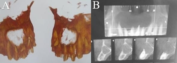
Panoramic radiography was performed 45 days after the operation, where it was possible to
observe radiopacity compatible with bone neoformation in the region (Figure 4). The patient is being
monitored and maintained with the team.
Figure 4 - Panoramic radiography (45 days control)
Source: the authors.
Note: Image suggestive of new bone formation.
DISCUSSION
The apical periodontal cyst behaves as an inflammatory response due to an infection of the
root canals. Radiographically, the image is described as a circumscribed, oval radiolucent, involving
the apex of the devitalized tooth (KESHARWANI et al., 2020). Epidemiological data show that
women over the third decade of life are more affected, with a prevalence of the maxillary region of
52% to 68% (DANTAS et al., 2014; LIN; RICUCCI; KAHLER, 2017). This information coincides
with the diagnosis, location of the lesion and the patient's age in the reported case.
The treatment of the root cyst is widely discussed, with alternatives being endodontic
treatment with or without apicectomy, extraction of the involved tooth, decompression,
marsupialization or enucleation with primary closure, for example. The treatment of choice depends
on a number of factors such as size and location of the lesion, proximity of the cyst to vital teeth and
adjacent noble structures (lower alveolar canal, mental foramen, infaorbital foramen, nasal cavity and
maxillary sinus), as well as behavior cyst, its aggressiveness and expansion. It is worth mentioning
that computed tomography is an important ally that helps to identify these aspects (JUNQUEIRA et
al., 2011; ABOULHOSN et al., 2019).
In the pathogenesis of the root cyst three phases are considered. The mechanisms involved are
not yet widely known, however, in the initial phase, bacterial infectious agents irritate the periapex
giving rise to periapical granuloma. On the other hand, during the deposition of root dentin, Hertwig's
Page 4 of 6Moreira; Aguiar; Brito
epithelial sheath is disorganized, leaving the epithelial remains of Malassez. The presence of
inflammatory cytokines seems to stimulate the division of these epithelial cells in order to separate
the inflammatory stimulus from the surrounding bone (LIN; RICUCCI; KAHLER, 2017;
CARVALHO et al., 2020).
In the stage of cystic formation, the central region of the granuloma ends up not receiving the
necessary nutrients for the metabolism of the lesion, which triggers the degeneration and death of the
cells causing a cavity inside the proliferative tissue. The growth of the lesion results in an increase in
osmotic pressure, causing the products of necrosis and epithelial desquamation to lead to the
accumulation of albumin proteins within the cystic cavity, promoting the attraction of liquids from
tissue spaces into the cavity (OLIVEIRA et al., 2011; SANTOS et al., 2011; CARVALHO et al.,
2020).
In the growth phase, the interstitial enlargement that compresses the cystic fibrous wall, blood
vessels and cells, resulting in metabolic and mechanical stress. This results in the release of products
derived from arachidonic acid, such as prostaglandins, being important inducers of peripheral bone
resorption. With this bone resorption, the cyst stabilizes, hydrostatic pressure decreases, resuming a
new cycle of protein accumulation, fluid attraction and new peripheral bone resorption (CARVALHO
et al., 2020; GUARALDI; HERINGER, 2020).
True cysts are lesions that have complete lining epithelium, and the cystic cavity is detached
from the teeth. They are self-sustaining lesions and it is unlikely that their repair will occur only with
endodontic treatment, requiring surgical complementation, especially in cases where endodontic
treatment did not promote tissue repair. When the extraction of the unit involved is chosen as a form
of treatment, curettage of the apical tissues should always be performed (JUNQUEIRA et al., 2011;
LIN; RICUCCI; KAHLER, 2017; MENDONÇA et al., 2017; COSTA et al., 2020).
In this case, the conservative approach was recommended through aspiration puncture because
a drain for maintaining the surgical window could cause discomfort for the patient, in addition to
becoming a site for the accumulation of bacterial plaque, and anti-aesthetic in the case of the patient
in question. Decompression stands out in the surgical field for the treatment of large injuries; it can
reduce internal pressure by removing fluid from the lesion, triggering the gradual reduction of the
lesion. Usually performed when there is a risk to the adjacent noble structures (RODRIGUES et al.,
2017; ABOULHOSN et al., 2019; GUARALDI; HERINGER, 2020).
The treatment instituted in the present case proved to be satisfactory, since there was bone
repair verified on the panoramic control radiography.
FINAL CONSIDERATIONS
Periapical lesions are commonly found in the dental surgeon's clinical practice. Therefore, the
professional must be able to detect and treat these lesions, and a detailed anamnesis, knowledge of
the clinical, radiographic, histopathological and etiopathogenic characteristics of the disease is
essential. We can consider that the treatment instituted was satisfactory, minimizing the risk of
damage to anatomical structures, in addition to facilitating the bone repair mechanism.
REFERENCES
ABOULHOSN, M. et al. Decompression and enucleation of a mandibular radicular cyst, followed
by bone regeneration and implant-supported dental restoration. Case Reports in Dentistry, v. 2019,
p. 1-8, 2019.
CARVALHO, G. A. O. et al. Etiopathogenesis and diagnosis of inflammatory odontogenic cysts:
literature review. Research, Society and Development, v. 9, n. 7, p. 1-21 , 2020.
Page 5 of 6EXTENDED APICAL PERIODONTAL CYST IN MAXILLA: CASE REPORT
COMIM, L. et al. Cisto periapical de grandes proporções na região anterior da maxila: Relato de
caso. Salusvita, v. 36, n. 2, p. 501-508, 2017.
COSTA, G. P. et al. Enucleação de cistoperiapical associado a tratamento endodôntico: relato de
caso. Archives of Health Investigation, v. 8, n. 9, p. 1-5, 2020.
DANTAS, R. M. X. et al. Enucleação de cisto radicular maxilar associado à apicectomia: relato de
caso. Revista de Cirurgia e Traumatologia Buco-maxilo-facial, v. 14, n. 3, p. 21-26, 2014.
GUARALDI, K. S.; HERINGER, E. M. Tratamento do cisto periapical pela técnica de
marsupialização. Cadernos de Odontologia do UNIFESO, v. 1, n. 2, p. 1-20, 2020.
JUNQUEIRA, R. B. et al. Tomografia computadorizada de feixe cônico como instrumento
complementar de diagnóstico e planejamento cirúrgico de cisto radicular: relato de um caso clínico.
Revista de Odontologia da UNESP, v. 40, n. 6, p. 338-343, 2011.
KESHARWANI, P. et al. Massive radicular cyst involving multiple teeth in pediatric mandible- A
case report.Journal of family medicine and primary care. Journal of Family Medicine and Primary
Care, v. 9, n. 2, p. 1253-1256, 2020.
LIN, L. M.; RICUCCI, D.; KAHLER, B. Radicular cysts review. JSM Dental Surgery, v. 2, n. 2, p.
1011-1017, 2017.
MARTINS, A. K. et al. CISTO PERIAPICAL: REVISÃO. Anais de Odontologia, v. 3, n. 1, p. 11-
12, 2018.
MENDONÇA, D. W. R. et al. Tratamento cirúrgico de cisto radicular em maxila: relato de caso.
Archives of Health Investigation, v. 6, n. 8, p. 1-5, 2017.
OLIVEIRA, D. H. I. P. et al. Cisto residual com grande dimensão: relato de caso e revisão da
literatura. Revista de Cirurgia e Traumatologia Buco-maxilo-facial, v. 11, n. 2, p. 21-26, 2011.
PINTO, G. N. S. et al. Marsupialização como tratamento definitivo de cistos odontogênicos: relato
de dois casos. Revista da Faculdade de Odontologia-UPF, v. 20, n. 3, p. 361-366, 2016.
RESENDE, M. A. P. et al. Tratamento cirúrgico e conservador de cisto periapical de grande
proporção: relato de caso. HU Revista, v. 43, n. 2, p. 191-196, 2017.
RODRIGUES, J. T. et al. Influence of surgical decompression on the expression of inflammatory and
tissue repair biomarkers in periapical cysts. Oral surgery, Oral Medicine, Oral Pathology and
Oral Radiology, v. 124, n. 6, p. 561-567, 2017.
SANTOS, L. C. S. et al. Histopathological study of radicular cysts diagnosed in a Brazilian
population. Brazilian Dental Journal, v. 22, n. 6, p. 449-454, 2011.
SILVA, R. N. F. et al. Tratamento de cisto radicular de grande extensão: relato de caso clínico.
Revista Odontológica do Brasil Central, v. 27, n. 80, p. 52-56, 2018.
Page 6 of 6You can also read