2D Browsing Software and 3D PDF of Canine Ear Based on Real Color Sectioned Images - SciELO
←
→
Page content transcription
If your browser does not render page correctly, please read the page content below

Int. J. Morphol.,
38(1):147-152, 2020.

                 2D Browsing Software and 3D PDF of Canine
                   Ear Based on Real Color Sectioned Images

                             Software de Navegación 2D y PDF en 3D del Oído Canino
                                    Basado en Imágenes Seccionadas en Color Real

                                                                 Jin Seo Park

PARK, J. S. 2D browsing software and 3D PDF of canine ear based on real color sectioned images. Int. J. Morphol., 38(1):147-152,
2020.

        SUMMARY: Dog ear is very important because of disease vulnerability. Therefore, gross anatomy and sectional anatomy on CT
and MRI of the dog ear should be mastered by veterinarian. The purpose of this research was to present the digital atlases which high
quality sectioned images and 3D models of detailed structures of dog ear could be displayed freely. In the sectioned images of a female
beagle, ear structures were reconstructed by surface modeling to make 3D models. The sectioned images and 3D models were put into
the browsing software and PDF file, respectively. Using the browsing software and the PDF file, gross and radiological anatomy of dog
ear could be learned easily and accurately. The auditory tube of a dog was placed anterior to the tympanic cavity unlike human. The
tensor tympani muscle of a dog was connected to the dorsal wall of the tympanic cavity with the malleus. No remarkable difference in the
auditory ossicles, semicircular ducts, facial nerve, and endolymphatic duct was observed between dogs and humans. The software and
the PDF file will be provided to other researchers freely to help contribute to veterinary research and education.

        KEY WORDS: Cross sectional anatomy; Dogs; Ear; Three-dimensional imaging; Visible human project.

INTRODUCTION

         A dog ear is known evolutionarily, to be highly                  the ear and surroundings (e.g., brain and skin) (Badea et al.,
developed to compensate for their relatively poor eyesight                2008; Russo et al., 2002). In Germany, dog sectioned images
(Altman and Kalmykova, 1986). Therefore, ear diseases and                 were created, but the images do not illustrate the genuine
frequent operations are important for the quality of a dog´s              body color because formalin and colored resin were
life. This is the reason veterinarians should know the dog                previously injected. Moreover, their intervals (1 mm) are
ear anatomy in detail (Garosi et al., 2003; Little et al., 1991).         too thick to identify the minute structures of the ear (Bottcher
However, the existing atlases and textbooks including                     and Maierl, 1999).
photographs of dissected dogs are not enough to grasp ear
components (Miller et al., 2013; Budras, 2007). On magnetic                     To understand perfectly anatomy of dog ear, three
resonance images (MRIs) for diagnosing a disease, it is                   dimensional (3D) models are indispensable. However,
difficult to interpret not only ear ossicles but also cochlea             because there were no high quality 2D images in which
and semicircular ducts of the dog ear (Harran et al., 2012).              anatomical details of the dog ear could be observed, the 3D
On computed tomography (CTs) of dog ear, the cavity and                   models of dog ear, could not be found anywhere.
duct in temporal bone can be barely distinguished (Schlegel
et al., 2010; Ostertag and Weigel, 1982; Kaufman et al.,                         Meanwhile, we produced the sectioned images of the
1981). Also, the micro computed tomography (micro CTs)                    dog (intervals 0.2 mm; pixel size 0.1 mm), which have been
show only bones and some blood vessels in gray scale and                  elaborated for the Visible Korean (Park et al., 2014). The
do not demonstrate the ear components realistically (Liao                 aim of this study is to present the browsing software
et al., 2016; Badea et al., 2008). In addition, a small specimen          including high quality real color sectioned images and the
of micro CTs does not allow simultaneous observations of                  3D PDF file including 3D models of dog ear for veterinary

Department of Anatomy, Dongguk University School of Medicine, Dongdae-ro 123, Gyeongju-si, Republic of Korea.
This study was funded by the Ministry of Trade, Industry and Energy (MOTIE) and Korea Institute for Advancement of Technology (KIAT) through the
International Cooperative R&D program (Grant number: N0002249).

                                                                                                                                          147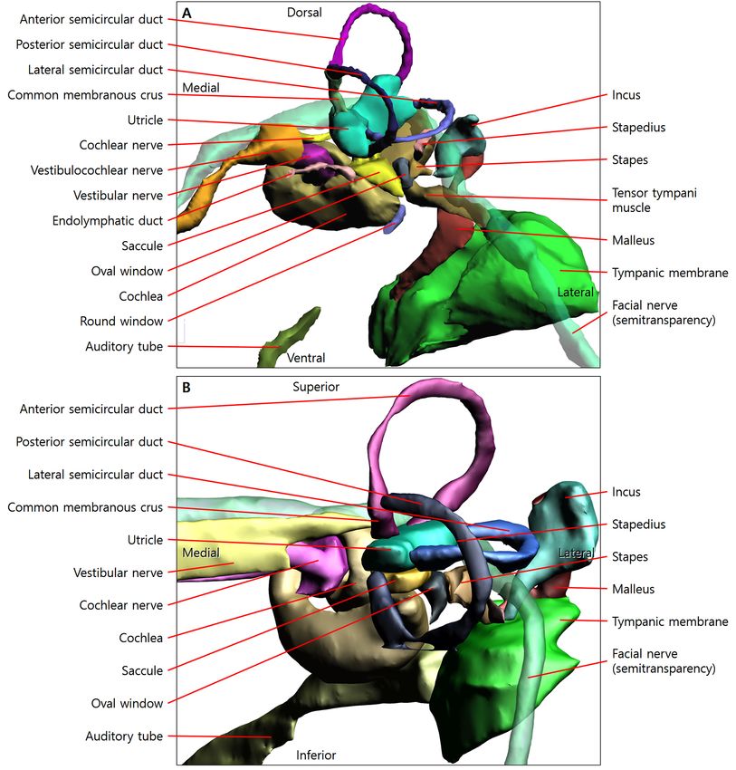
PARK, J. S. 2D browsing software and 3D PDF of canine ear based on real color sectioned images. Int. J. Morphol., 38(1):147-152, 2020.

anatomy. The original planes were stacked to produce two                                        In Mimics, the 3D models of STL files were exported
other orthogonal planes, where the ear components were then                              as PDF file. After running the PDF file, list of segmented
observed closely in the three kinds of planes. Furthermore,                              structures and 3D models were shown in left and right
based on the sectioned images of ear, 3D models of ear                                   windows respectively (Fig. 2b) (Shin et al., 2012; Park &
structures were made.                                                                    Jung, 2016).

MATERIAL AND METHOD
                                                                                         RESULTS

       In the previous study, frontal sectioned images
(intervals 0.2 mm; resolution 2,076 X 4,088; pixel size 0.1                                     According to the observation of the sectioned images
mm; color depth, 48 bit color; file format, TIFF) have been                              and 3D models using the browsing software and PDF file
made of a female beagle (height 460 mm; width 160 mm;                                    (Fig. 2), the ear anatomy of dog could be described in the
length 711 mm; 1 years old) (Park et al., 2014).                                         following ways.

        Among the frontal sectioned images of whole body,                                       In the external acoustic meatus of a dog, the wall
only 151 ones including the right ear were remained. The                                 consisted mostly of cartilage (Figs. 1d, 3a). This was different
excessive margins were cropped to achieve an image                                       from the external acoustic meatus of humans that contained
resolution of 356 X 267 (intervals 0.2 mm; pixel size 0.1                                both cartilage and temporal bone (Moore et al., 2017).
mm) (Fig. 1a,c). After increasing the pixel size from 0.1
mm to 0.2 mm, the images were stacked to make dorsal and                                         Prominently, the tympanic bulla of a dog, similar to
sagittal planes (intervals 0.2 mm; pixel size 0.2 mm; file                               wall of tympanic cavity of human, had relatively large space
format, TIFF) (Fig. 1d,f) using house development software.                              (Figs. 1a-c, 3c-e) and it was located in the mastoid process
                                                                                         (Fig. 3b), while mastoid air cell was located in the mastoid
        We decided to make 3D models of 26 ear structures                                process of human (Moore et al., 2017).
(Table I). First work was segmentation. In the cropped
sectioned images (intervals 1.0 mm; pixel size 0.1 mm), 24                                      The auditory tube of a dog was placed anterior to the
structures were outlined semiautomatically using Magnetic                                tympanic bulla (Figs. 1e,f, 3c,d) unlike human. It was
Lasso Tool or Lasso Tool in Photoshop CC 2015 (Adobe                                     because the snout and pharynx of a dog were situated in
Systems, Inc., San Jose, CA, USA). The outlines of skin                                  front of the middle ear (Fig. 3a,b).
and bone used one of previous study (Park & Jung, 2016).
The Inner spaces of outlined structures were automatically                                       The tensor tympani muscle of dog connected the
filled with a specific color using Automate - Batch Tool in                              medio-dorsal wall of the tympanic bulla (Figs. 1a,b, 3d) like
Photoshop to make segmented images of 26 structures.                                     human. However, the muscle did not connect the auditory
                                                                                         tube (Fig. 3c,d), while the muscle of human connected the
       In browsing software, already made in Visible Korean                              auditory tube with the malleus. It was probably because the
(Park & Jung, 2016), the cropped sectioned images and the                                dog’s tympanic bulla was big and the auditory tube was
segmented images of 1.0 mm intervals and 0.1 mm pixel                                    located too rostral (Fig. 3c,d). The stapedius muscle
size were put into each folder. After running the browsing                               connected the caudal wall of the tympanic bulla with the
software, cropped sectioned images with names of                                         stapes in both dog (Fig. 3e) and human.
segmented structures were shown in main window. Area of
cropped sectioned images in frontal head and segmented                                           From medial surface of the tympanic membrane to
images were shown in sub-window (Fig. 2a).                                               ovals window connected malleus, incus, and stapes (Figs.
                                                                                         1b,c, 4a) like human (Fig. 4b). The facial nerve of dog passed
Second work was 3D reconstruction. The segmented                                         between incus and stapes like that of human (Fig. 4).
images were automatically reconstructed by surface modeling
using Mimics version 17.01 (Materialise, Leuven, Belgium)                                        Direction of the semicircular ducts of dog were ante-
(Park et al., 2013) to make 3D models (Figs. 3-5). We already                            rior, posterior, and lateral sides (Figs. 3f, 4a, 5b) like human
made the 3D models of dog skin and bone in the previous                                  (Figs. 4b, 5d), whereas brainstem laid in a prone position
study (Park & Jung, 2016). In the 3D models of dog skin and                              (Fig. 5a) unlike human (Fig. 5d). The common membranous
bone, 24 structures of this study were assembled and saved as                            crus was made of the anterior and posterior semicircular ducts
stereolithography (STL) files using Mimics (Shin et al., 2012).                          (Figs. 3f, 4a) like human (Fig. 4b). The cochlea made two
The 3D models of 26 structures were totally made.                                        and half turns around its axis (Fig. 3d).
148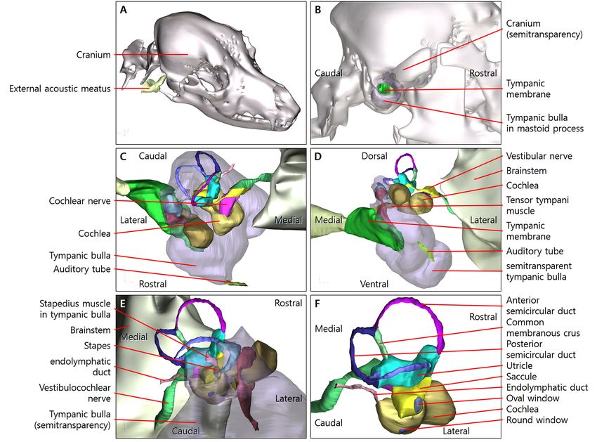
PARK, J. S. 2D browsing software and 3D PDF of canine ear based on real color sectioned images. Int. J. Morphol., 38(1):147-152, 2020.

Table I. Twenty six structures of the right ear segmented in the sectioned images and reconstructed to make 3D models.
 Region                  Structures
 External ear            Skin, External acoustic meatus, Tympanic membrane
 Middle ear              Tympanic bulla, Oval window, Round window, Stapes, Incus, Malleus, Tensor tympani muscle, Stapedius
                         muscle, Auditory tube
 Internal ear            Utricle, Saccule, Anterior semicircular duct, Posterior semicircular duct, Lateral semicircular duct, Cochlear
                         duct, Common membranous crus
 Other                   Bone, Brainstem, Facial nerve, Vestibulocochlear nerve, Vestibular nerve, Cochlear nerve, Endolymphatic duct

   Fig. 1. Sectioned images of dog ear from rostral to caudal. (A-C) In the sectioned images of frontal view, middle ear structures including
   large tympanic bulla can be shown. (D-F) In the sectioned images of dorsal view, external ear and parts of middle ear can be shown.

   Fig. 2. Browsing software and 3D PDF files. (A) In main window of the browsing software, the sectioned images of frontal view
   can be displayed continuously using scroll-bar or play-buttons of left side. (B) In the PDF file, 3D models of dog ear with skin and
   bone (cranium) can be shown three-dimensionally.

                                                                                                                                                              149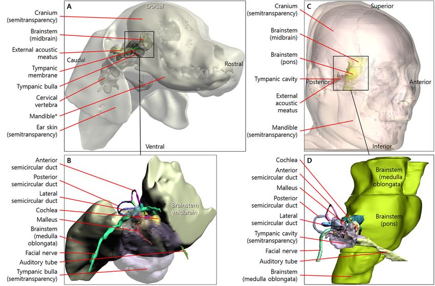
PARK, J. S. 2D browsing software and 3D PDF of canine ear based on real color sectioned images. Int. J. Morphol., 38(1):147-152, 2020.

      Fig. 3. Three dimensional models in PDF file. (A) The external acoustic meatus of a dog do not consist of bone
      (cranium). (B) The mastoid process of dog is full of the tympanic bulla (lateral view). (C) The auditory tube is
      placed at rostral of the tympanic bulla which have relatively large space (ventral view). (D) The tensor tympani
      muscle connect the medio-dorsal wall of the tympanic bulla and do not connect the auditory tube (frontal view). (E)
      The stapedius muscle is located in the caudal wall of the tympanic bulla with the stapes (caudal-lateral view). (F)
      The anterior and posterior semicircular ducts share a common membranous crus. The endolymphatic duct arise
      from the medial wall of the cochlea (caudal-lateral view).

        The endolymphatic duct arises from the medial wall                               3D models, and consequently we are easily intelligible the
of the cochlea; this duct ended in an endolymphatic sac on                               direction and position of ear structures (Fig. 3). In case of
the caudal surface of the petrous portion of the temporal                                micro CTs, its thickness is only 0.035 mm or more (Lee et
bone (Fig. 3e-f), where it was in contact with the dura mater                            al., 2013; Shin et al., 2013), while the thickness of the
(Miller et al., 2013).                                                                   sectioned images in this study is 0.2 mm. When looking at
                                                                                         the thickness and resolution, the sectioned images of this
                                                                                         study could never be better than micro CTs. However, the
DISCUSSION                                                                               sectioned images almost show the structures of body in real
                                                                                         color, while the micro CTs show bone and some blood vessels
                                                                                         of gray color in small specimens.
       The strong point of the Visible Korean Project is the
sectioned images which both large specimens and small                                            By sectioned images of this study, ear of the dog and
specimens with real color and high resolution could be shown                             human can be described evolutionarily. Even if it evolves
(Park et al., 2009; Park et al., 2014). For example, the images                          from the Precambrian era to the present, equilibrium sense
enable tracing of the vestibulocochlear nerve that passes from                           of all living things on earth must be identical because the
saccule and utricle in the internal ear (small specimens) to                             direction of earth´s gravity is always identical for everyone.
the brainstem (large specimen) through internal acoustic                                 Therefore, the direction of semicircular ducts of dog and
meatus (Fig. 1). Furthermore, Visible Korean Project have                                human was identical (Fig. 4). In evolution, the biggest
150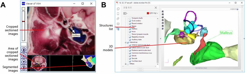
PARK, J. S. 2D browsing software and 3D PDF of canine ear based on real color sectioned images. Int. J. Morphol., 38(1):147-152, 2020.

                                                                                                             difference of the lower and higher animals
                                                                                                             is the walking and position of their head. A
                                                                                                             dog walks on four legs and the head
                                                                                                             including brainstem is placed in front of its
                                                                                                             trunk; consequently, a spinal cord, brainstem,
                                                                                                             cerebellum, and cerebrum of dog lie in a
                                                                                                             prone position (Fig. 5a). As human evolve,
                                                                                                             they walk two legs (orthograde posture) and
                                                                                                             the head is placed at superior side of the
                                                                                                             trunk; consequently, human’s nervous system
                                                                                                             including brainstem stands upright (Fig. 5c).
                                                                                                             The comparison is valuable because the
                                                                                                             morphology of the human ear has been well
                                                                                                             studied (Figs. 4b, 5d) (Park et al., 2013).

                                                                                                           Fig. 4. Comparison of dog of this study and
                                                                                                           human of previous study (Park et al., 2013). (A)
                                                                                                           In the dog and (B) human, direction of
                                                                                                           semicircular ducts, shape and position of auditory
                                                                                                           ossicles, and facial nerve between incus and
                                                                                                           stapes are almost identical.

Fig. 5. Comparison of dog of this study and human of previous study (Park et al., 2013). Direction of semicircular ducts of (B)
the dog and (D) human is identical. (B) Brainstem of dog lie in a prone position, while (D) brainstem of human stands upright.

                                                                                                                                                       151
PARK, J. S. 2D browsing software and 3D PDF of canine ear based on real color sectioned images. Int. J. Morphol., 38(1):147-152, 2020.
This study presented the sectioned images and 3D Badea, C. T.; Johnston, S.; Johnson, B.; Lin, M.; Hedlund, L. W. & Johnson, G.
A. A dual micro-CT system for small animal imaging. Proc. SPIE Int. Soc.
models of dog where detailed structures of the ear were Opt. Eng., 6913:691342, 2008.
shown (Figs. 1, 3). Also, this study presents the browsing Böttcher, P. & Maierl, J. Macroscopic cryosectioning: a simple new method for
software and PDF file which the sectioned images and 3D producing digital, three-dimensional databases in veterinary anatomy. Anat.
models could be shown easily (Fig. 2). Furthermore, Histol. Embryol., 28(2):97-102, 1999.
Budras, K. D. Anatomy of the Dog. 5th rev. ed. Hannover, Schlütersche
characteristic morphology of the dog ear was compared with Verlagsgesellschaft, 2007.
a human ear. In the next study, we will compare dog with Garosi, L. S.; Dennis, R. & Schwarz, T. Review of diagnostic imaging of ear
human minutely. This is intended to stimulate the diseases in the dog and cat. Vet. Radiol. Ultrasound, 44(2):137-46, 2003.
Harran, N. X.; Bradley, K. J.; Hetzel, N.; Bowlt, K. L.; Day, M. J. & Barr, F.
development of educational material (e.g. atlas book) and MRI findings of a middle ear cholesteatoma in a dog. J. Am. Anim. Hosp.
virtual simulation (e.g. virtual surgery) of dog and human Assoc., 48(5):339-43, 2012.
ears. The browsing software and 3D PDF file of dog ear are Kaufman, H. H.; Cohen, G.; Glass, T. F.; Huchton, J. D.; Pruessner, J. L.; Ostrow,
P. T.; Andia-Waltenbaugh, A. M. & Dujovny, M. CT atlas of the dog brain.
distributed free of charge at neuroanatomy.kr. J. Comput. Assist. Tomogr., 5(4):529-37, 1981.
Lee, J. Y.; Shin, K. J.; Kim, J. N.; Yoo, J. Y.; Song, W. C. & Koh, K. S. A
morphometric study of the semicircular canals using micro-CT images in
three-dimensional reconstruction. Anat. Rec. (Hoboken), 296(5):834-9, 2013.
ACKNOWLEDGMENTS Liao, S. H.; Zhu, X. H.; Xie, J.; Sohodeb, V. K. & Ding, X. Influence of trabecular
bone on peri-implant stress and strain based on micro-CT finite element
modeling of beagle dog. BioMed, 2016, 3926941, 2016.
Funding: This study was funded by the Ministry of Little, C. J.; Lane, J. G.; Gibbs, C. & Pearson, G. R. Inflammatory middle ear
disease of the dog: the clinical and pathological features of cholesteatoma, a
Trade, Industry and Energy (MOTIE) and Korea Institute complication of otitis media. Vet. Rec., 128(14):319-22, 1991.
for Advancement of Technology (KIAT) through the Miller, M. E.; Evans, H. E. & Delahunta, A. Miller's Anatomy of the Dog. St.
International Cooperative R&D program (Grant number: Louis, Elsevier Saunders, 2013.
Moore, K. L.; Dalley, A. F. & Agur, A. M. R. Clinically Oriented Anatomy.
N0002249). Philadelphia, Lippincott Williams & Wilkins, 2017.
Ostertag, C. B. & Weigel, K. Three-dimensional CT scanning of the dog brain.
J. Comput. Assist. Tomogr., 6(5):1036-7, 1982.
PARK, J. S. Software de navegación 2D y PDF en 3D del oído canino Park, H. S.; Chung, M. S.; Shin, D. S.; Jung, Y. W. & Park, J. S. Accessible and
basado en imágenes seccionadas en color real. Int. J. Morphol., 38(1): informative sectioned images, color-coded images, and surface models of
147-152, 2020. the ear. Anat. Rec. (Hoboken), 296:1180-6, 2013.
Park, H. S.; Shin, D. S.; Cho, D. H.; Jung, Y. W. & Park, J. S. Improved sectioned
images and surface models of the whole dog body. Ann. Anat., 196:352-9,
RESUMEN: La oreja del perro es importante debido a la vul- 2014.
nerabilidad de enfermedad. Por lo tanto, el veterinario debe conocer ple- Park, J. S. & Jung, Y. W. Software for browsing sectioned images of a dog body
namente la anatomía macroscópica y la anatomía seccional en la TC y la and generating a 3D model. Anat. Rec. (Hoboken), 299: 81-7, 2016.
RM del oído del perro. El objetivo de esta investigación fue presentar los Park, J. S.; Chung, M. S.; Shin, D. S.; Har, D. H.; Cho, Z. H.; Kim, Y. B.; Han,
atlas digitales que podían mostrar imágenes seccionadas de alta calidad J. Y. & Chi, J. G. Sectioned images of the cadaver head including the brain
y modelos 3D de estructuras detalladas de orejas de perro. En las imáge- and correspondences with ultrahigh Field 7.0 T MRIs. Proc. IEEE,
nes seccionadas de una hembra Beagle, las estructuras de las orejas se 97(12):1988-996, 2010.
reconstruyeron mediante modelado de superficie con el objetivo de crear Russo, M.; Covelli, E. M.; Meomartino, L.; Lamb, C. R. & Brunetti, A. Computed
tomographic anatomy of the canine inner and middle ear. Vet. Radiol.
modelos 3D. Las imágenes seccionadas y los modelos 3D se colocaron
Ultrasound, 43(1):22-6, 2002.
en un software de navegación y un archivo PDF. El uso de software de Schlegel, K.; Parry, A. T.; Lamb, C. R.; Kneiss, S.; Probst, A.; Tichy, A. &
navegación y el archivo PDF permiten un aprendizaje fácil y preciso de Mayrhofer, E. X-ray and CT morphology of atlas variants in the dog. Berl.
la anatomía macroscópica y radiológica de la oreja de perro. El músculo Munch. Tierarztl. Wochenschr., 123(9-10):425-30, 2010.
tensor del tímpano de un perro estaba conectado a la pared dorsal de la Shin, D. S.; Chung, M. S.; Park, J. S.; Park, H. S.; Lee, S.; Moon, Y. L. & Jang,
cavidad timpánica con el martillo. No se observaron diferencias nota- H. G. Portable document format file showing the surface models of cadaver
bles en los huesecillos auditivos, los conductos semicirculares, el nervio whole body. J. Korean Med. Sci., 27(8):849-56, 2012.
facial y el conducto endolinfático entre perros y humanos. El software y Shin, K. J.; Lee, J. Y.; Kim, J. N.; Yoo, J. Y.; Shin, C.; Song, W. C. & Koh, K. S.
el archivo PDF estarán disponibles libremente para los investigadores Quantitative analysis of the cochlea using three-dimensional reconstruction
based on microcomputed tomographic images. Anat. Rec. (Hoboken),
para ayudar en la investigación y educación veterinaria.
296(7):1083-8, 2013.
PALABRAS CLAVE: Anatomía de sección transversal; Pe-
rros; Oreja; Imágenes tridimensionales; Proyecto humano visible. Corresponding author:
Jin Seo Park
Department of Anatomy
Dongguk University School of Medicine
REFERENCES
Dongdae-ro 123, Gyeongju-si
REPUBLIC OF KOREA
Altman, J. A. & Kalmykova, I. V. Role of the dog's auditory cortex in Received: 11-07-2019
discrimination of sound signals simulating sound source movement. Hear. Accepted: 13-08-2019
Res., 24(3):243-53, 1986.
Email: park93@dongguk.ac.kr
152You can also read