BIOLOGICAL SCIENCES - Index Copernicus
←
→
Page content transcription
If your browser does not render page correctly, please read the page content below
4                                   BIOLOGICAL SCIENCES / «Colloquium-journal» #9(96), 2021

                                    BIOLOGICAL SCIENCES
                                                Kotsyumbas G. I., Khalaniia M. R., Zolototska O.B.
                Stepan Gzhytskyi National University of Veterinary Medicine and Biotechnologies Lviv
                                                             DOI: 10.24412/2520-6990-2021-996-4-8
    MORPHOFUNCTIONAL FEATURES OF CHANGES IN THE PANCREAS IN WET AND DRY
                   FORMS OF FELINE INFECTIOUS PERITONITIS

     Abstract
     The article presents the results of macroscopic, histological and histochemical studies of the pancreas of cats
in wet and mixed forms of FIP. A pathological examination of 18 carcasses of cats aged from 3 months to 5.5
years, in which lifelong feline infectious peritonitis was diagnosed (based on history, clinical signs, morphological
and biochemical analysis of blood, ultrasound, Rivalt test and rapid test). Samples of the pancreas were taken,
which were fixed in 10% aqueous solution of neutral formalin, solutions of Carnois, Buena and 96 ° ethyl alcohol.
Histoxylin and eosin-stained histoscripts were prepared according to McManus, Brachet, Van Gieson and exam-
ined under a microscope.
     It was found that the most severe structural and functional changes in the pancreas of dead cats were devel-
oped in the stroma of the organ, in the arterio-venular system, receptors, excretory ducts, both in wet and dry
forms of FIP. The development of pancreatosis in the pancreas of cats in the wet form found, which was charac-
terized by disorganization of connective tissue, the development of fibrinoid necrosis of the walls of arterioles,
disseminated venous thrombosis, stasis in the capillaries, obstruction and obstruction of the excretory ducts. The
detected morphological changes indicate the development of irreversible vascular-stromal dystrophies in the pan-
creas.
     Productive pancreatitis has developed in the pancreas of cats in dry form, which in cats at a shorter course
of the disease was characterized by round-cell infiltration of the stroma, dystrophicnecrobiotic processes of paren-
chymal cells, obstruction of the interparticle ducts. Fibrosis progressed over a longer course in cats, which was
expressed in the growth of interstitial connective tissue fibers, atrophy of the parenchymal lobes, productive ne-
crotic vasculitis, and occlusion of the interparticle excretory ducts.

     Keywords: cats, infectious peritonitis, FIP, pathomorphology, pancreas, exocrinocytes, vascular stromal dys-
trophies, pancreatosis, pancreatitis.

      Introduction. Feline infectious peritonitis (FIP) is    cats with infectious peritonitis. Thus, it will help to un-
caused by an RNA genomic virus from the family                derstand the morphogenesis of changes in wet and dry
Coronaviridae, genus Coronavirus. The disease, cur-           forms and to supplement the diagnosis of the disease.
rently, still remains one of the incurable diseases of cats        The aim of the study was to investigate the patho-
[1,2]. Infectious peritonitis affects domestic and wild       logical, histological and histochemical changes in the
cats, most often young animals under 3 years of age.          pancreas of cats in wet and dry forms of FIP.
The disease is characterized by a long incubation pe-
riod, the absence of severe symptoms in the early stages                  Materials and methods of research.
of the disease, and the progression of pathological pro-           Pathological examination of 18 corpses of cats
cesses in the organs and tissues of patients with FIP in      aged from 3 months to 7 years, in which lifelong FIP
cats often leads to death.                                    was diagnosed (based on medical history, clinical
      Symptoms in ill cats, depending on the clinical and     signs, morphological and biochemical analysis of
anatomical form varies widely. Cats with weak cellular        blood, ultrasound diagnosis, Rivalt test and rapid diag-
immunity develop a wet (effusive) form, and in a mod-         nosis). Selected samples of the pancreas were fixed in
erate reaction - a dry (non-effusive) form of FIP. [3,4,      10% aqueous solution of neutral formalin, Carnua so-
5]. The morphogenesis of changes in pathogenic pro-           lution, Buena. Then performed dehydration, filling the
cesses by FIP in various organs and systems of cats is        tissue with paraffin. Histocuts on a sled microtome MS-
not yet fully understood.                                     2 were made from paraffin blocks. Histographs were
      There are relatively few publications in the avail-     stained with hematoxylin and eosin, and a
able literature on the coverage of morphological                   McManus PAS reaction was performed to detect
changes of internal organs by FIP and there is little de-     glycoproteins; methylene green and Brachet pyronine
scription of histological and histochemical changes in        for nucleic acid detection [6,7]. The finished histoprep-
endocrine organs, in particular in the pancreas. It is im-    arations were examined under a light microscope Leica
portant to determine the nature of the development of a       DM-2500 (Switzerland), photographed with a Leica
pathological process in determining the morphological         DFC450C camera with Leica Application Suite Ver-
changes in the body, that underlies the development of        sion 4.4 software.
various clinical and anatomical forms of the disease.                            Results of the research
Therefore, it is important to study morphohistochemi-              Macroscopic changes in the pancreas are incon-
cal changes in the pancreas of spontaneously infected         spicuous in cats killed by infectious peritonitis. The or-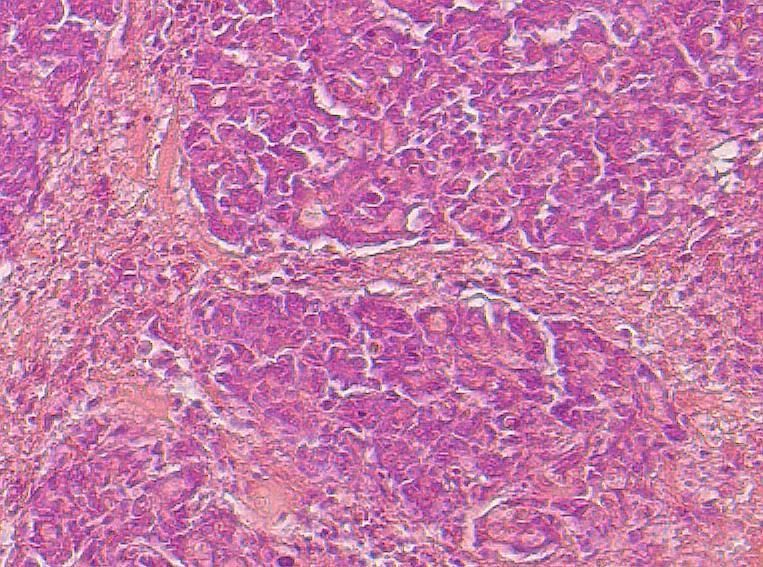
«Colloquium-journal» #9(96), 2021 / BIOLOGICAL SCIENCES 5
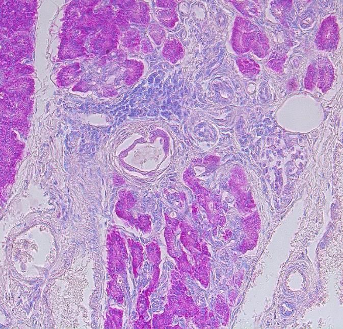
gan is tape-shaped, pale pink in wet form, wet in sec- stroma of the organ as well as in the walls of blood ves-
tion, the capsule is tense. In dry form, the pancreas is sels indicated a deep disorganization of connective tis-
light purple, slightly compacted. The most striking sue fibers (Fig. 1). In the deformed, considerably thick-
changes were detected by microscopic examination, ened walls of arterioles all layers are retouched, homo-
which differed significantly in the nature of the devel- geneous weight prevailed. The basement membrane of
opment of pathological processes in the wet form from arterioles is impregnated with PAS-positive com-
those in the dry form. pounds, endothelial exfoliation was noted. The lumen
Histologically, in the pancreas of cats at the wet of the arterioles is sharply narrowed, filled with plasma
form more pronounced changes were found in the and desquamated endothelium. Destructive-necrobiotic
stroma, in the structure of the arterio-venular system, processes in the smooth muscle and elastic fibers of the
receptors, excretory ducts. The stroma of the body is walls of the afferent arterioles have progressed. The vi-
loose. The connective tissue fibers of the stroma, the olation of the structural organization of arterioles con-
walls of the excretory duct are impregnated with fuch- tributed to a sharp increase in the permeability and yield
sinophilic compounds on preparations stained accord- of plasma proteins and led to the development of fi-
ing to McManus. The lumen of small venous vessels is brinoid necrosis. The revealed structural changes in the
filled with adhesive forms of erythrocytes, dissemi- arterio-venular system were functionally reflected by a
nated thrombosis and stasis in capillaries were noted. sharp disturbance of blood supply, oxygenation, devel-
The increase in the content of glycoproteins in the opment of significant metabolic disorders of the organ
tissue (Fig. 2).
Fig. 1. The pancreas of a cat in the wet form. Fuchsin- Fig. 2.
ophilicity of the stroma and walls of the duct and ve- The cat's pancreas in the wet form. Fibrinoid necrosis
nous vessels. McManus. m.e.10, vol. 20 of the arteriole wall. McManus. m.e. 10, vol. 20
The histological feature of the pancreas of cats is The cytoplasm of the basal parts of the exocrine
the presence in the interstitium of well-structured re- cells of the acinus is mainly pyroninophilic, nuclei with
ceptors of nerve fibers - Fater-Pacini bodies. The inner a high content of chromatin, which is a sign of the syn-
axon of the receptor is surrounded by perineural la- thesis phase (Fig. 4) in the parenchyma of the organ of
mellae, in which the stratification of annular lamellae cats with the wet form, on preparations stained by
was noted, with the formation of vesicles and the ac- Brache. A group of endocrine cells with an RNA-poor
cumulation of fuchsinophilic compounds in the lu- cytoplasm, the islets of Langenhars, stood out among
mens between them. The receptor is slightly enlarged the pyroninophilic exocrine cells of the acinuses. The
due to the expansion of the lumen between the lamel- dilated lumens of capillaries are well visible (Fig. 5)
lae. Histostructural changes in the receptors were among the weakly stained endocritocytes surrounded
functionally reflected by a weakening of the sensitiv- by fibrous fibers.
ity and neural conduction of the organ (Fig. 3).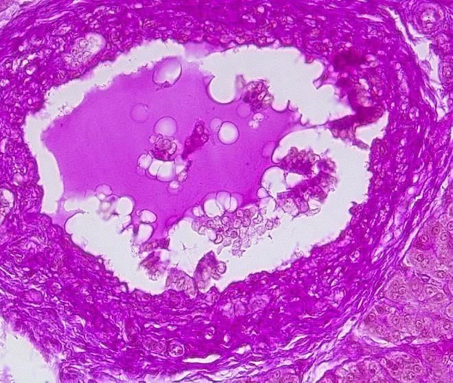
6                                  BIOLOGICAL SCIENCES / «Colloquium-journal» #9(96), 2021

 Fig. 3. The cat's pancreas in the         Fig. 4. Acinus of the cat's pan-       Fig. 5. The cat's pancreas in the
  wet form. Fater-Pacini's body.         creas in the wet form. Interacinar     wet form. Pyroninophilia of the cy-
Fuchsinophilicity of the lumen and        lumens are expanded. The cyto-         toplasm of exocrine cells. The en-
structures of perineural lamellae.        plasm of pyrinophilic exocrino-       docrinocytes of the islet of Langen-
    McManus. m.e. 10, vol. 40              cytes. Brachet. m.e.10, vol. 10       hars are weakly stained. Brachet.
                                                                                           m.e.10, vol. 40

     The intercinary stromal structures and the base-             It should be noted, while analyzing the histochem-
ment membrane of the intraparticle ducts are impreg-        ical changes (Brachet) in the pancreas of the cat at the
nated with glycoproteins on McManus-stained prepara-        wet form, that in all lobes pyroninophilia of the cyto-
tions. The lumen of the intraparticle ducts is markedly     plasm of the basal exocrine glands was expressed, indi-
expanded, their epithelial lining is preserved, and the     cating the phase of synthesis and accumulation of se-
basement membrane is thickened (Fig. 6). It should be       cretion granules in cells and a sharp decrease in secre-
noted that the interparticle duct system of the pancreas    tion in the duodenum. Prolonged decrease in secretion
underwent significant destructive changes. The base-        from cells causes metabolic disorders in them. How-
ment membrane, connective tissue fibers are inten-          ever, stagnation of secretion with a high content of pro-
sively impregnated with fuchsinophilic compounds,           teolytic enzymes in the interparticle parts of the excre-
which indicates the disorganization of structures. The      tory duct of the pancreas had a negative effect on the
available magenta content is mixed with the desqua-         epithelial lining, as it caused the digestion of its own
mated epithelium in the lumen of the dilated, signifi-      cells of the inner membrane.
cantly deformed duct. (Fig. 7).

Fig. 6. The pancreas of a cat in the wet form. The aci-     Fig. 7. The interparticle duct of the pancreas of a cat
nus and intraparticle excretory duct are lined with cu-     in the wet form. Fuchsinophilicity of the wall and the
      bic epithelium. McManus. m.e. 10, vol. 40             contents in the lumen of the duct. McManus. m.e. 10,
                                                                                    vol. 40

     In addition, it is necessary to focus on generalized   disorganization, destruction of connective tissue necro-
damage to the vascular system. Structural changes in        sis, fibril and was accompanied by a violation of the
the arteriovenular system (hyperemia, disseminated          structure of the walls and patency of the interparticle
thrombosis in venules, stasis in capillaries) were ac-      excretory ducts of the gland.
companied by increased vascular permeability with the             In the pancreas of cats in a less long course of the
release of not only water, electrolytes but also plasma     dry form of the disease productive pancreatitis devel-
proteins, which led to the development of irreversible      oped, which was characterized by round-cell infiltra-
                                                            tion of the stroma, especially in the perivascular and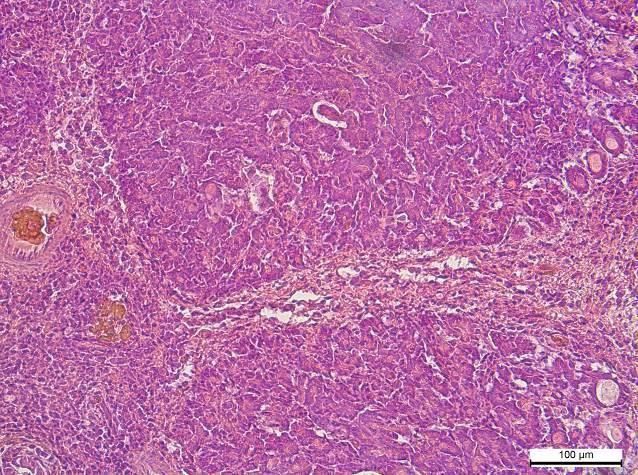
«Colloquium-journal» #9(96), 2021 / BIOLOGICAL SCIENCES 7
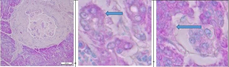
periductal areas. Brachet-stained preparations showed desquamated. The walls of venules and veins of aver-
accumulations of lymphoid elements around the ves- age caliber underwent the most significant changes.
sels, the interparticle duct and in the intercinary con- The lumen of the veins is sharply dilated, without
nective tissue (Fig. 8). Simultaneously, the histostruc- formed elements of blood, and in the lumen of the ven-
ture of arterioles, venules, capillaries is broken: the ba- ules - hypertrophied macrophages and single lympho-
sal layer of membranes is loosened, in places cytes. The peculiar feature for macrophages was the
homogenized. The endothelium of the vessel walls is presence of weakly pyroninophilic cytoplasmic inclu-
sions (Fig. 9).
Fig. 8. The cat's pancreas in dry form. T lymphocytes Fig. 9. The pancreas of a cat in dry form. The base-
in the stroma of the body. Brachet. m.e. 10, vol. 20 ment membrane of the venule is broken. Macrophage
elements in the lumen of the venule. m.e. 10, vol. 100
The ductal system of the pancreas underwent sig- Dystrophic-necrobiotic processes were developed
nificant destructive changes as well. The intercinary in the parenchyma organ, and the clarity of the structure
and interparticle excretory ducts of the gland were in- of acinuses and exocrinocytes was erased. Polygonal
volved in the inflammatory process. The contours of exocrine cells with altered outlines, cytoplasm was
the connective tissue fibers of the interstitial duct wall weakly stained with pyronine, nuclei enlarged with sig-
are retouched, almost do not absorb pyronin, and single nificantly reduced chromatin content. The contours of
nuclei of fibers with low DNA content on Brachet- swollen, deformed exocrinocytes were blurred, some of
stained preparations. The lumen of the interparticle ex- them acquired a bubble shape, others lysed (Fig. 10 b).
cretory duct is filled with secretion, which contains ex- There are cells with vacuolated cytoplasm and lysed
foliated epithelial fragments from the basement mem- nuclei. The islets of Langenhars have undergone
brane. (Fig. 10 a). It is probable that due to the change changes as well. The fibers surrounding the islands are
in the structure of the ducts of the pancreas there was a lysed in places. The structure of most endocrinocytes is
thickening of the secretion from the formation of clots disturbed: the contours of the cells were not deter-
together with the desquamated epithelium and blockage mined. The cytoplasm is swollen (Fig. 10 c).
of their lumen.
Fig. 10 a. Obstruction of the lumen Fig. 10 b. Acinus. Exocrinocytes Fig. 10 c. The islet of Langenhars.
of the interparticle duct by con- are weakly stained with pyronine, The fibers surrounding the islands
densed secretion and exfoliated ep- the nuclei are enlarged with re- are sometimes lysed. m.e. 10, vol.
ithelium of the lining. m.e. 10, vol. duced chromatin content. m.e. 10, 100
20 vol. 100
Fig. 10. The pancreas of a cat in dry form. Brachet.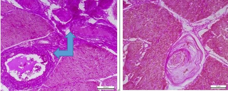
8 BIOLOGICAL SCIENCES / «Colloquium-journal» #9(96), 2021
The detected changes in the pancreas of cats in a acinuses were more often exposed to vacuolar dystro-
shorter course of the dry form of the disease indicated phy and atrophy (Fig. 11). The walls of the arterioles
the development of exocrine and endocrine insuffi- significantly thickened and deformed, and the lumen
ciency, which was expressed in a decrease in the pro- narrowed, productive-necrotic vasculitis progressed. A
duction of digestive enzymes and insulin by the gland. similar thickening due to intense roundcell infiltration
The formation of clots of secretion with desquamated occurred in the walls of the interparticle excretory
epithelium in the lumen of the excretory ducts has led ducts, which clearly led to a sharp narrowing and oc-
to obstruction and impaired patency of the ducts. clusion of their lumen.
In cats, atrophy of the lobes, significant thickening Productive-necrotic processes in the arterio-venu-
of the stroma due to intraparticle, interparticle, periduc- lar system, interstitium and interparticle excretory
tal and perivascular growth of connective tissue fibers, ducts of the pancreas in the dry form of infectious per-
among which cellular infiltrates of lymphocytes and itonitis of cats has led to the development of severe dys-
histiocytic elements are clearly visible, were noted for trophic-necrobiotic changes, parenchymal atrophy,
a longer course. At the same time, exocrinocytes in the sclerosis of the stroma, which had an irreversible organ
function.
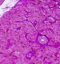
Fig. 11. Fig.12. The pancreas of a cat in a dry form. Interpar-
The pancreas of a cat in a dry form. Particle atrophy. ticle growth of connective tissue. Hematoxylin and
The growth of intraparticle and interparticle connec- eosin. m.e. 10, vol. 20
tive tissue. Hematoxylin and eosin. m.e. 10, vol. 40
Conclusions. 20, № 6. P. 961–965
1. It was found the development of pancreatosis, 2.Pedersen N. C., Allen C. E., Lyons L. A. Patho-
which was characterized by disorganization of connec- genesis of feline enteric coronavirus infection. J Feline
tive tissue, the development of fibrinoid necrosis of the Med Surg. 2008. Vol. 10. P. 529–541.
walls of arterioles, disseminated venous thrombosis, 3. Pedersen, N. C. An update on feline infectious
stasis in capillaries, obstruction with impaired duct pa- peritonitis: virology and immunopathogenesis. The
tency and functional hemorrhage tissue in the pancreas Veterinary Journal. 2014. Vol. 201, № 2. P. 123-132.
of cats in the wet form. Morphological changes have 4. Natural resistance to experimental feline in-
indicated the development of irreversible vascular-stro- fectious peritonitis virus infection is decreased rather
mal dystrophies in the pancreas of cats in the wet form. than increased by positive genetic selection. / Pedersen
2. Productive pancreatitis developed in the pan- N. C. et. al. Veterinary Immunology and Immunopatho
creas of cats in dry form. Meanwhile in cats for a 5. Morphologic features and development of
shorter course of the disease it was characterized by granulomatous vasculitis in feline infectious peritonitis
roundcell infiltration of the stroma, dystrophic-necrobi- / Kipar A. et. al. Vet Pathol. 2005. Vol. 42, № 3. P. 321-
otic processes of parenchymal cells, obstruction of the 330.
interparticle ducts. Fibrosis progressed over a longer 6.Merkulov, G. A. (1969). Kurs patologogystolo-
course in cats, which was expressed in the growth of hycheskoj tekhnyky [The course of pathohistological
interstitial connective tissue fibers, atrophy of the technique]. L.: Medycyna [in Russian].
parenchymal lobes, productive necrotic vasculitis, and 7. Goral's'kyj, L.P., Homych, V.T., Konons'kyj,
occlusion of the interparticle excretory ducts. O.I. (2005). Osnovy gistologichnoi' tehniky i mor-
References fofunkcional'ni metody doslidzhennja u normi ta pry
1.Baydar E., Eröksüx Y., Timurkan M. O. Feline patologii':[navchal'nyj posibnyk]. Zhytomyr: Polissja
infectious peritonitis with distinct ocular involvement (in Ukrainian).
in a cat in Turkey. Kafkas Univ Vet Fak Derg. 2014.
Vol.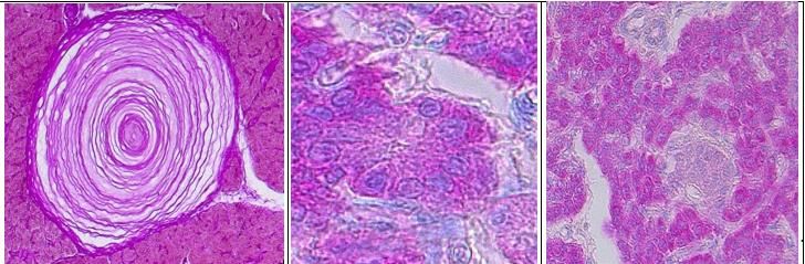
You can also read