Maxillary protraction using miniscrews in a Class III patient: case report
←
→
Page content transcription
If your browser does not render page correctly, please read the page content below
Research, Society and Development, v. 10, n. 2, e51910212759, 2021
           (CC BY 4.0) | ISSN 2525-3409 | DOI: http://dx.doi.org/10.33448/rsd-v10i2.12759

Maxillary protraction using miniscrews in a Class III patient: case report
Protração maxilar usando mini-implantes em paciente Classe III: relato de caso
Protracción maxilar mediante miniimplantes en un paciente Clase III: reporte de caso

                                                   Received: 02/08/2021 | Reviewed: 02/14/2021 | Accept: 02/17/2021 | Published: 02/27/2021

                                                                                                             Ricardo Alves de Souza
                                                                                             ORCID: https://orcid.org/0000-0003-2113-6663
                                                                                                  Southwest Bahia State University, Brazil
                                                                                                            E-mail: ricardoorto@gmail.com
                                                                                                         Gregório Bonfim Dourado
                                                                                            ORCID: https://orcid.org/0000-0002-2204-8860
                                                                                                                   College UniFTC, Brazil
                                                                                                E-mail: drgregoriodourado@outlook.com
                                                                                                  Ivanderson Santana de Almeida
                                                                                            ORCID: https://orcid.org/0000-0002-4542-1832
                                                                                                                          Innovare, Brazil
                                                                                                       E-mail: van-santana@hotmail.com
                                                                                                               João Batista de Paiva
                                                                                            ORCID: https://orcid.org/0000-0002-3178-4843
                                                                                                            University of Sao Paulo, Brazil
                                                                                                                   E-mail: jbpaiva@usp.br

      Abstract
      An already established alternative to conventional orthopedic treatment with palatal expansion and face mask for
      growing patients with skeletal Class III due to maxillary deficiency is the use of skeletal anchorage for protraction of
      the maxilla. Its use eliminates the adverse dental effects of conventional therapy, such as extrusion and mesialization
      of upper molars, projection of upper incisors and retroinclination of lower incisors. In addition, it eliminates the need
      to use an extra-oral device, which contributes to patients' motivation. Mini-plates are the most commonly used
      skeletal anchorage for this type of therapy, however the need for a surgical step for installation and another for
      removing plaques represents a disadvantage for this technique, and thus the use of miniscrews was proposed to
      perform maxillary protraction eliminating the surgical step. This work aims to describe the technique of maxillary
      prostration supported by orthodontic miniscrews, illustrated through a clinical case of a patient treated with this
      therapy, showing the positive facial and occlusal results achieved.
      Keywords: Angle class III malocclusion; Orthodontic anchorage procedures; Orthodontics interceptive.

      Resumo
      Uma alternativa já consolidada ao tratamento ortopédico convencional com disjunção palatina e máscara facial para
      pacientes em crescimento com Classe III esquelética por deficiência maxilar é a utilização de ancoragem esquelética
      para protração da maxila. Seu uso elimina os efeitos dentários adversos da terapia convencional, como extrusão e
      mesialização de molares superiores, projeção de incisivos superiores e retroinclinação de incisivos inferiores. Além
      disso elimina a necessidade de uso de um dispositivo extra-oral, o que contribui para a motivação dos pacientes. As
      miniplacas são a forma de ancoragem esquelética mais usada para esse tipo de terapia, entretanto a necessidade de um
      passo cirúrgico para instalação e outro para remoção das placas representa um ponto desvantajoso para essa técnica, e
      dessa forma o uso de mini implantes foi proposto com a finalidade de promover protração maxilar eliminando o passo
      cirúrgico. Este trabalho se propõe a descrever a técnica de prostração maxilar apoiada em mini implantes
      ortodônticos, ilustrada através de um caso clínico de um paciente tratado com esta terapia, mostrando os resultados
      faciais e oclusais positivos alcançados.
      Palavras-chave: Má oclusão de angle classe III; Procedimentos de ancoragem ortodôntica; Ortodontia interceptora.

      Resumen
      Una alternativa ya establecida al tratamiento ortopédico convencional con disyunción palatina y mascarilla facial para
      pacientes con crecimiento de clase III esquelética debido a deficiencia maxilar es el uso de anclaje esquelético para la
      prolongación del maxilar. Su uso elimina los efectos dentales adversos de la terapia convencional, como extrusión y
      mesialización de molares superiores, proyección de incisivos superiores y retroinclinación de incisivos inferiores.
      Además, elimina la necesidad de utilizar un dispositivo extraoral, lo que contribuye a la motivación de los pacientes.
      Las miniplacas son el anclaje esquelético más utilizado para este tipo de terapias, sin embargo la necesidad de un paso
      quirúrgico para la instalación y otro para la remoción de las placas representa un inconveniente para esta técnica, por
      lo que se propuso el uso de mini implantes con el propósito de promoviendo la prolongación maxilar eliminando el
                                                             1
Research, Society and Development, v. 10, n. 2, e51910212759, 2021
(CC BY 4.0) | ISSN 2525-3409 | DOI: http://dx.doi.org/10.33448/rsd-v10i2.12759
paso quirúrgico. Este trabajo tiene como objetivo describir la técnica de postración maxilar soportada por mini
implantes de ortodoncia, ilustrada a través de un caso clínico de un paciente tratado con esta terapia, mostrando los
resultados faciales y oclusales positivos obtenidos.
Palabras clave: Maloclusión de angle classe III; Métodos de anclaje em ortodoncia; Ontodoncia interceptiva.
1. Introduction
The treatment effectiveness of the of growing patients with maxillary deficiency using rapid maxillary expansion and
protraction with face mask protocol is undeniable. (Pangrazio-Kullbersh, Berger & Kersten, 1998; Nartallo-Turley & Turley,
1998; Baccetti, Franchi & McNamara, 2000; De Toffol, Pavoni, Baccetti, Franchi & Cozza, 2008) However, even this
therapeutic option continues to be the gold standard for the treatment of these patients, it has limitations. Undeniable dental
effects, such as dental mesialization of the upper arch due to the anchorage used for protraction, and psychosocial effects,
which hinder the collaboration of the patient by using face mask. (Cevidanes, Baccetti, Franchi, McNamara & De Clerck,
2010; Hino et al., 2013)
The adverse efects around facemask treatment motivated the use of skeletal anchorage through mini-plates installed in
the maxilla and mandible to adapt class III elastics. De Clerck, Cornelis, Cevidanes, Heymann and Tulloch (2009) are the
pioneer of this technique and reported promising results in maxillary protraction, with reduced dental effects and more evident
collaboration from patients in addition to the better aesthetics provided by this device (De Clerk, Cevidanes & Baccetti, 2010).
By the way, the discomfort caused in children due to the surgical step required to install and remove the miniplates, has
become a relevant disadvantage of this technique.
Thus, in order to reduce patient morbidity and cost reduction, a clinical trial was conducted by Souza, Neto & Paiva
(2019), demonstrating the feasibility of using orthodontic miniscrews for skeletal anchorage during maxillary protraction
showing promising results and whose technique will be described below.
2. Case Report
2.1 Diagnosis and planning
Patients selected for this treatment have skeletal class III malocclusion due to maxillary deficiency. The diagnosis of
the patients must be confirmed through clinical parameters, manipulating the children's jaw in a centric relation to avoid the
deviation of the mandible to the anterior, and also in centric occlusion to verify the molar relationship in Class III and the
presence of negative or top overjet. In the facial analysis, the deficiency of the middle third of the face and the zygymatic bone
is noticeable, determining a straight or concave profile. In cephalometry these individuals have Witts less than or equal to - 2
mm and ANB less than 1. The therapeutic window comprises those patients who are in the prepubertal growth phase, in the
second transitional period of mixed dentition or the early permanent dentition. The installation of miniscrews is possible as
soon as the permanent lower canines are erupted.
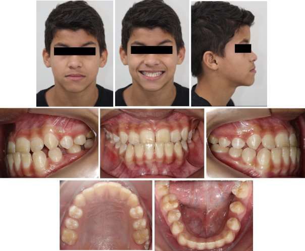
JVBS the patient, 12 years old, looked for orthodontic treatment because his “chin is too forward”. Facial exam
showed a concave profile, open nasolabial angle, a few zygomatic projection, characteristics of a Class III patient with
retraction of the maxilla. Intraoral exam showed an early permanent dentition, anterior crossbite with overjet of -3mm and a
molar relation of Angle Class III (Figure 1). Cephalometric measures showed wits -4 and ANB -2 (Figure 2).
2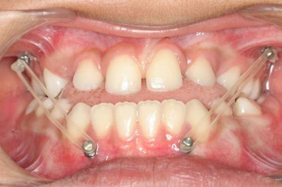
Research, Society and Development, v. 10, n. 2, e51910212759, 2021
(CC BY 4.0) | ISSN 2525-3409 | DOI: http://dx.doi.org/10.33448/rsd-v10i2.12759

                     Figure 1. Initial clinical aspect of the patient.

                                     Source: Authors.

                                            3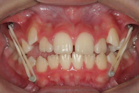
Research, Society and Development, v. 10, n. 2, e51910212759, 2021
(CC BY 4.0) | ISSN 2525-3409 | DOI: http://dx.doi.org/10.33448/rsd-v10i2.12759

                   Figure 2. Initial radiographic aspect of the patient.

                                     Source: Authors.

                                            4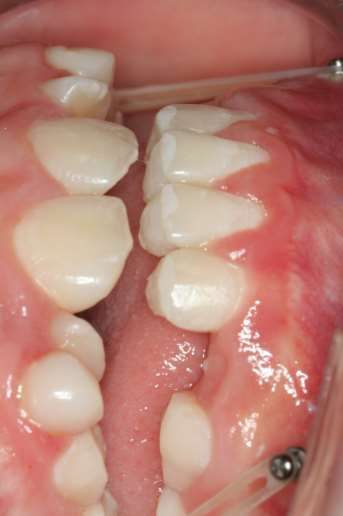
Research, Society and Development, v. 10, n. 2, e51910212759, 2021
              (CC BY 4.0) | ISSN 2525-3409 | DOI: http://dx.doi.org/10.33448/rsd-v10i2.12759

2.2 Technique description
         Four conventional titanium orthodontic miniscrews must be used. Long screws are recommended to reinforce the
anchorage through the greater insertion of the thread in the alveolar bone. The case described was treated using orthodontic
miniscrews from Morelli (Sorocaba, São Paulo, Brazil) 10mm long, 1.5mm in diameter and 2mm in transmucosal profile.
         After the patient has been carefully diagnosed and the prognosis and treatment plan determined with the aid of
complementary radiographic examinations, the installation of the devices begins. Preoperative procedures include the selection
and demarcation of the appropriate installation sites, mouthwash with 0.12% chlorhexidine digluconate for disinfection of the
mucosa and infiltrative anesthesia of the regions where the implants will be inserted.
         The site for installing miniscrews should be the mesial interradicular region of the first maxillary molars and the distal
interradicular region of lower canines, so that the elastic used between the screws is located in the lateral region of the arches,
without causing injury to the gingival tissues (Figure 3). However, because the therapeutic window for maxillary protraction
comprises the mixed dentition phase, and lower crowding is a common condition in growing patients with skeletal class III,
elective for this treatment, in some cases it would not be possible to install miniscrews in the distal region of the canines. In
these cases, it would be recommended to install in the mesial region of the lower canines, but being careful to observe if the
elastics are not compressing the patient's inserted gingiva, causing ischemia of the area, due to the curvature of the mandible in
that region (Figure 4).

        Figure 3. Miniscrews placed in mesial region of first maxilary molars and distal region of mandibular canines.

                                                         Source: Authors.

                                                                5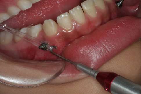
Research, Society and Development, v. 10, n. 2, e51910212759, 2021
              (CC BY 4.0) | ISSN 2525-3409 | DOI: http://dx.doi.org/10.33448/rsd-v10i2.12759

        Figure 4. Miniscrews placed in mesial region of first maxilary molars and mesial region of mandibular canines.

                                                         Source: Authors.

         After installing the miniscrews, they must be connected on each side by intermaxillary elastics. It is recommended
that the elastic exerts an average force between 180 and 200gr in occlusion to allow maxillary protraction. In the first month of
use, a lighter force of approximately 100gr should be used to adapt the patient. In general, ¼” elastic is used to obtain adequate
strength. In the case described, the Morelli brand latex elastic (Sorocaba, São Paulo, Brazil) was used, with light strength in the
first month, and medium strength from the second month of use (Figure 5).

                                                                6
Research, Society and Development, v. 10, n. 2, e51910212759, 2021
(CC BY 4.0) | ISSN 2525-3409 | DOI: http://dx.doi.org/10.33448/rsd-v10i2.12759
Figure 5. Measuring forces of the elastics. Usually ¼” latex elastics are used and 200gr are recommended to protraction of
maxilla.
Source: Authors.
It is recommended to bond a bite turbo on the lingual portion of the lower incisors. The ramp-shaped bite turbo must
be made of composite resin and has the objective of opening the bite, improving the forward movement of maxillary bone and
uncrossing the anterior bite (Figure 6).
Figure 6. Bite turbos in lingual of lower incisives. The disocclusion permit uncrossing the anterior bite.
Source: Authors.
7Research, Society and Development, v. 10, n. 2, e51910212759, 2021
(CC BY 4.0) | ISSN 2525-3409 | DOI: http://dx.doi.org/10.33448/rsd-v10i2.12759
Elastics must be used 24 hours by day, just removing for cleaning and feeding. Elastics must be changed daily, so that
there is no degradant of the latex by the saliva and the forces maintain balanced. The active phase of treatment lasts 12 months,
after this period retention phase begins, in which is recommended to use elastics just at night for more 6 months.
2.3 Results
After 12 months of active phase, the patient showed a harmonic face, with several improvement of the profile, now
straight. In intraoral exam, anterior crossbite was resolved, with an overjet of 2mm, and a molar relation of Angle Class I.
Cephalometric measures also improved to Wits 1 and ANB 2º. Retention phase lasted 6 months with maintenance of the
results.
Figure 7. Final clinical aspect of the patient.
Source: Authors.
8Research, Society and Development, v. 10, n. 2, e51910212759, 2021
(CC BY 4.0) | ISSN 2525-3409 | DOI: http://dx.doi.org/10.33448/rsd-v10i2.12759
Figure 8. Final radiographic aspect of the patient.
Source: Authors.
3. Discussion
Morphological differences, variability in facial growth and level of complexity of prognathism become essential for
the treatment plan and prognosis of Class III maloclusion (Ngan et al., 1998).
De Clerck et al. (2009) demonstrated the effectiveness of treatment in patients with Class III malocclusion due to
maxillary deficiency, through the use of orthodontic miniplates for skeletal anchorage during maxillary protraction. These
devices are installed through surgical access in the regions of infra-zygomatic ridges and between lower canines and lateral
9Research, Society and Development, v. 10, n. 2, e51910212759, 2021
(CC BY 4.0) | ISSN 2525-3409 | DOI: http://dx.doi.org/10.33448/rsd-v10i2.12759
incisors or between lower canines and first premolars, on the right and left side of the patient with general anesthesia in most
cases. At the end of the treatment, a new surgical access is required to remove the miniplates. The results in computed
tomography showed that the use of these anchorage methods allowed the forces to be transferred directly to the sites of the
maxillary sutures, improving the orthopedic effect, due to the force not being dissipated in the periodontal ligament (Kircelli &
Pektas, 2008). Therefore, this new protocol has advantages over conventional treatment with a face mask, as it avoids the
undesirable effects of tooth movement such as mesialization and extrusion of the upper molars, protrusion of the upper incisors
and retroinclination of the lower teeth (Ngan, Hägg, Yiu & Wei, 1997; Saadia & Torres, 2000; Franchi, Baccetti, &
McNamara, 2004; De Clerck et al., 2009; Cevidanes et al., 2010; De Clerck et al., 2010; Nguyen et al., 2011; Hino et al.,
2013), in addition to clockwise rotation of the mandible (Kapust, Sinclair & Turley, 1998). Another advantage would be that
by eliminating dental effects, such as molar extrusion, this new protocol can be used in patients with Class III malocclusion
associated with an increase in the lower facial height of the face (AFAI) and with a vertical growth pattern. In this case, REM
associated with traction with face mask is not indicated, but not the use of skeletal anchorage for maxillary protraction.
Although this new protocol has many advantages in relation to the classic treatment of Class III for maxillary
deficiency through REM and face mask, the major question and disadvantage would be the degree of invasion of the surgical
procedure to install the miniplates, often with the need for hospitalization. (De Clerck et al., 2009). Therefore, the possibility of
using miniscrews as a skeletal anchorage for the maxillary protraction procedure should be considered, as it considerably
minimizes the patient's discomfort, as shown by Souza et al. (2019)
Another point to highlight refers to the change in the aspect of collaboration on the part of the patient, due to the
possibility of not using the face mask. Thus, exchanging an extra-oral appliance supported on the face, which is extremely
unsightly and compromises social life, to use only intermaxillary elastics inside the mouth, significantly increases the patient's
collaboration when these two options are exposed. Thus, in general, the level of acceptance of this new protocol is much more
receptive on the part of patients when compared to the use of the face mask (Favero Winkler & Favero, 2012; Jeevarathan,
Koora, Sudhakar, Muthu & Prabhu, 2013).
In this treatment protocol, forces of 100g were initially used, up to a maximum of 230g, which are lower than the
forces generally used for face mask therapy. According to Souza et al. (2019), patients who were treated with miniscrews
showed improvements in facial aesthetics even using lower forces. The change in the amount of force for maxillary protraction
between the conventional protocols supported by dental units and the one with skeletal anchorage is an important point to be
discussed. Until then, it was essential and consolidated in the orthodontic literature, that heavy orthopedic forces over 400
grams (Delaire, 1971; Nanda, 1978; Ngan, Hägg, Yiu, Merwin D & Wei, 1996), acted in the period of 14 to 16 hours, to enable
ideal maxillary protraction. De Clerck et al. (2009, 2010, 2012); Cevidanes et al. (2010); Heymann, Cevidanes, Cornelis, De
Clerck & Tulloche, (2010) and Nguyen et al. (2011), used forces around 200 grams, and proved that they were sufficient to
protrude the maxilla efficiently. Most likely, the 24-hour use of intermaxillary elastics was decisive for this favorable response
in the protocol with skeletal anchorage, either through miniplates or miniscrews. In these cases, the constancy of the force,
even if of lesser intensity, seems to be more important than the amount of force. This factor reinforces the need for patient
collaboration, which becomes more tangible when using a totally intraoral device.
4. Conclusion
Maxillary traction using miniscrews were a positive option to treat a Class III with maxillary deficiency patient. The
treatment shown improvement in facial patterns and in profile of the patient, and the protocol were more receptive by the
patient due to the possibility of using intra-oral appliances, which is more aesthetic.
10Research, Society and Development, v. 10, n. 2, e51910212759, 2021
(CC BY 4.0) | ISSN 2525-3409 | DOI: http://dx.doi.org/10.33448/rsd-v10i2.12759
References
Baccetti, T., Franchi, L., McNamara, J. A. Jr. (2000) Treatment and posttreatment craniofacial changes after rapid maxillary expansion and facemask therapy.
Am J Orthod Dentofac Orthop, 118(4), 404–13.
Cevidanes, L., Baccetti, T., Franchi, L., McNamara, J. A. Jr, & De Clerck, H. (2010). Comparison of two protocols for maxillary protraction: bone anchors
versus face mask with rapid maxillary expansion. Angle Orthod, 80(5), 799–806.
De Clerck, H. J., Cornelis, M. A., Cevidanes, L. H., Heymann, G. C., & Tulloch, C. J. (2009). Orthopedic traction of the maxilla with miniplates: a new
perspective for treatment of midface deficiency. J Oral Maxillofac Surg, 67(10), 2123–9.
De Clerck, H., Cevidanes, L., & Baccetti, T. (2010). Dentofacial effects of bone anchored maxillary protraction: a controlled study of consecutively treated
Class III patients. Am J Orthod Dentofacial Orthop, 138(5), 577–81.
De Clerck, H., Nguyen, T., de Paula, L. K., & Cevidanes, L. (2012). Three-dimensional assessment of mandibular and glenoid fossa changes after bone-
anchored Class III intermaxillary traction. Am J Orthod Dentofacial Orthop, 142(1), 25-31.
Delaire, J. 1971Confection dur masque orthopedique. Rev Stomatol, 72: 579-84.
De Toffol, L., Pavoni, C., Baccetti, T., Franchl, L., & Cozza, P. (2008). Orthopedic treatment outcomes in Class III malocclusion. A systematic review. Angle
Orthod, 78(3), 561–73.
Favero, L., Winkler, A., & Favero, V. (2012). Non-compliant maxillary protraction by orthodontic micro-implants. Eur J Paediatr Dent, 13(3), 244-8.
Franchi, L., Baccetti, T., & McNamara, J. A. Jr. (2004). Postpubertal assessment of treatment timing for maxillary expansion and protraction therapy followed
by fixed appliances. Am J Orthod Dentofacial Orthop, 126(5), 555-68.
Heymann, G. C., Cevidanes, L., Cornelis, M., De Clerck, H. J., & Tulloche, J. F. C. (2010) Three-dimensional analysis of maxillary protraction with
intermaxillary elastics to Miniplates. Am J Orthod Dentofacial Orthop, 137(2), 274-84.
Hino, C. T., Cevidanes, L. H., Nguyen, T. T., De Clerck, H. J., Franchi, L, & McNamara, J. A. Jr. (2013). Three-dimensional analysis of maxillary changes
associated with facemask and rapid maxillary expansion compared with bone anchored maxillary protraction. Am J Orthod Dentofacial Orthop, 144(5), 705–
14.
Jeevarathan, J., Koora, K., Sudhakar, V., Muthu, M. S., & Prabhu, R. V. (2013). Correction of class III malocclusion using modified tandem appliance-two
case reports. J Indian Soc Pedod Prev Dent, 31(4), 286-91.
Kapust, A. J., Sinclair, P. M., & Turley, P. K. (1998). Cephalometric effects of face mask/expansion therapy in Class III children: a comparison of three age
groups. Am J Orthod Dentofacial Orthop, 113(2), 204-12.
Kircelli, B. H., & Pektas, Z. O. (2008). Midfacial protraction with skeletally anchored face mask therapy: a novel approach and preliminary results. Am J
Orthod Dentofacial Orthop, 133(3), 440-9.
Nanda, R. (1978). Protraction of maxilla in rhesus monkeys by controlled extraoral forces. Am J Orthod, 74(2), 121-41.
Nartallo-Turley, P. E., & Turley, P. K. (1998). Cephalometric effects of combined palatal expansion and facemask therapy on Class III malocclusion. Angle
Orthod, 68(3), 217–24.
Ngan, P., Hägg, U., You, C., Merwin, D., & Wei, S.H. (1996). Soft tissue and dentoskeletal profile changes associated with maxillary expansion and
protraction headgear treatment. Am J Orthod Dentofacial Orthop, 109(1), 38-49.
Ngan, P., Hägg, U., You, C., & Wei, S. H. (1997). Treatment response and long-term dentofacial adaptations to maxillary expansion and protraction. Semin
Orthod, 3(4), 255-64.
Ngan, P., You, C., Hu, A., Hagg, U., We, S. H., & Gunel, E. (1998). Cephalometric and occlusal changes following maxillary expansion and protraction. Eur J
Orthod, 20(3), 237-54.
Nguyen, T., Cevidanes, L., Cornelis, M. A., Heymann, G., de Paula, L. K., & De Clerck, H. (2011). Three-dimensional assessment of maxillary changes
associated with bone anchored maxillary protraction. Am J Orthod Dentofacial Orthop, 140(6), 790-8.
Pangrazio-Kulbersh, V., Berger, J., & Kersten, G. (1998). Effects of protraction mechanics on the midface. Am J Orthod Dentofacial Orthop, 114(5), 484–91.
Saadia, M., & Torres, E. (2000). Sagittal changes after maxillary protraction with expansion in Class III patients in the primary, mixed, and late mixed
dentitions: a longitudinal retrospective study. Am J Orthod Dentofacial Orthop, 117(6), 669-80.
Souza, R. A., Neto, J. R., & Paiva, J. B. (2019). Maxillary protraction with rapid maxillary expansion and facemask versus skeletal anchorage with mini-
implants in class III patients: a non-randomized clinical trial. Prog Orthod, 20(1),35. 10.1186/s40510-019-0288-7.
11You can also read