Characterization and Outcomes of Epileptic Seizures in Mexican Pediatric Patients With Anti-N-Methyl-D-Aspartate Receptor Encephalitis - Cureus
←
→
Page content transcription
If your browser does not render page correctly, please read the page content below
Open Access Original
                                          Article                                                  DOI: 10.7759/cureus.8211

                                          Characterization and Outcomes of Epileptic
                                          Seizures in Mexican Pediatric Patients With
                                          Anti-N-Methyl-D-Aspartate Receptor
                                          Encephalitis
                                          Melissa Chavez-Castillo 1 , Matilde Ruiz-Garcia 1 , Patricia Herrera-Mora 2

                                          1. Pediatric Neurology, Instituto Nacional de Pediatría, Mexico City, MEX 2. Pediatric Neurology,
                                          Instituto Nacional de Pediatría, Mexico CIty, MEX

                                          Corresponding author: Melissa Chavez-Castillo, melissa_chc@hotmail.com

                                          Abstract
                                          Introduction
                                          Anti-N-methyl-D-aspartate receptor (NMDAR) encephalitis is one of the most common
                                          autoimmune encephalitides. The frequency of anti-NMDAR encephalitis is known to exceed the
                                          frequency of any individual viral encephalitis in young subjects. Epileptic seizures are a
                                          cardinal symptom in anti-NMDAR encephalitis; a significant amount of pediatric patients
                                          exhibit seizures as the first symptom of the disease, and most of them will develop them during
                                          the acute phase. The use of antiepileptic drugs (AEDs) is a cornerstone of the treatment of
                                          these patients, but the choice of agent and duration of treatment is currently unknown.

                                          Materials and methods
                                          This was a single-center retrospective review case series of all pediatric patients with a
                                          confirmed diagnosis of anti-NMDAR encephalitis and epileptic seizures admitted to the
                                          National Institute of Pediatrics in Mexico City from January 2012 to July 2019.

                                          Results
                                          We included a total of 31 patients (males 64.5%, median age: 10 years). No patient showed
                                          evidence of teratoma; only 38% of cases had a viral prodrome. Most patients initially exhibited
                                          psychiatric symptoms (51%), but the leading cause in soliciting medical assistance was the
                                          presence of epileptic seizures (71%). About 85% of patients presented epileptic seizures during
                                          the course of the illness, predominantly focal onset seizures (42% focal to bilateral tonic-clonic
                                          seizures, 32% focal seizures with impaired awareness). Electroencephalogram (EEG) was
                                          abnormal in 97% of patients; the characteristic extreme delta brush pattern was found in 9% of
Received 04/15/2020
                                          patients. Two AEDs on average were required to control seizures during the acute stage. In six
Review began 04/29/2020
                                          (19%) patients, human herpesvirus (HHV) was detected in cerebrospinal fluid (CSF); all of them
Review ended 05/11/2020
Published 05/20/2020                      had epileptic seizures, which were more resistant to pharmacological treatment during the
                                          acute phase, requiring a higher number of AED (median 2.5 vs. 2). The development of epilepsy
© Copyright 2020
                                          after acute encephalitis was uncommon; at 24 months, only one patient continued to have
Chavez-Castillo et al. This is an open
access article distributed under the      epileptic seizures. One of the factors most closely related to the persistence of epileptic seizures
terms of the Creative Commons             was the inadequate response to immunotherapy after four weeks. The functional prognosis was
Attribution License CC-BY 4.0., which     generally good; at a two-year follow-up, only two (10%) patients had a significant disability
permits unrestricted use, distribution,
                                          [modified Rankin Scale (mRS) score: 3-5]; both patients had seizures at a one-year follow-up.
and reproduction in any medium,
provided the original author and
source are credited.
                                          Conclusions

                                          How to cite this article
                                          Chavez-Castillo M, Ruiz-Garcia M, Herrera-Mora P (May 20, 2020) Characterization and Outcomes of
                                          Epileptic Seizures in Mexican Pediatric Patients With Anti-N-Methyl-D-Aspartate Receptor Encephalitis.
                                          Cureus 12(5): e8211. DOI 10.7759/cureus.8211
Sustained use of AEDs after the acute phase of anti-NMDAR encephalitis is controversial. We
found that the continuation of AEDs after the acute phase could be considered in the following
scenarios: status epilepticus (SE), inadequate response to immunotherapy at four weeks, and a
high mRS score at discharge and during follow-up. In other cases, discontinuation of AED may
be warranted. More studies are needed in our country to replicate these results.
Categories: Neurology, Pediatrics, Allergy/Immunology
Keywords: anti-nmdar encephalitis, autoimmune encephalitis, seizures, epilepsy, antiepileptic drugs,
outcome, children
Introduction
Anti-N-methyl-D-aspartate receptor (NMDAR) encephalitis is one of the most common
autoimmune encephalitides and is associated with a characteristic clinical syndrome, which
includes the presence of seizures and epilepsy [1]. This disease was first described in 2005 when
Vitaliani et al. reported a new syndrome characterized by memory dysfunction, psychiatric
symptoms, alterations in consciousness, and autonomic dysfunction in four women with
ovarian teratoma [2]. Two years later, specific anti-NMDAR antibodies were found in the
cerebrospinal fluid (CSF) of these four patients and eight more with similar neurological
symptoms [3]. Its discovery has changed the diagnostic approach to encephalitides in both
adults and children and has significantly expanded our knowledge regarding antibody-
mediated epilepsy [4].
Women are more commonly affected than men (3:1 ratio), and of all the reported cases of anti-
NMDAR encephalitis over a five-year period (September 2007 to February 2011), 65% occurred
in patients under the age of 18. Although we lack a specific prevalence study, the frequency of
anti-NMDAR encephalitis is known to exceed the frequency of any individual viral encephalitis
in young individuals [5].
Epileptic seizures are a cardinal symptom in anti-NMDAR encephalitis; 30% of pediatric
patients exhibit them as the first symptom of the disease. Within the first four weeks of disease
onset, both children and adults develop a similar clinical syndrome [6]. The pathophysiology of
epileptic seizures in anti-NMDAR encephalitis is still unclear, but it is believed to be due to an
alteration in the balance of the excitatory and inhibitory mechanisms present in the central
nervous system (CNS) [7]. The importance and prevalence of the anti-NMDAR in the developing
brain could explain why epileptic seizures are more prevalent in pediatric age [8].
During the acute phase, over 80% of patients will develop seizures [9-11]. Both focal and
generalized seizures have been reported. Different studies agree that the most common type of
seizure during the acute phase is the generalized tonic-clonic, followed by focal seizures
afterward [7,10,11]. Therefore, the use of antiepileptic drugs (AEDs) is a cornerstone in the
treatment of these patients, although the choice of agent and duration of AED treatment is
currently unknown. The incidence of status epilepticus (SE) in anti-NMDAR encephalitis is
approximately 25-50%; about 35% will develop refractory SE [10].
Electroencephalogram (EEG) is abnormal in over 90% of patients. The most common finding is
generalized background slowing with a frontotemporal predominance [8,9]. Although most
patients develop seizures during disease progression, epileptic activity is a rare finding. It is
found in 24-50% of cases, especially during the early stages [12]. The EEG pattern known as
extreme delta brush is a unique finding in anti-NMDAR encephalitis. This finding is
characterized by a 1-3 Hz rhythmic delta activity with superimposed outbreaks of beta activity
at 20-30 Hz. It is transiently found in 30% of adult patients and is associated with a more
2020 Chavez-Castillo et al. Cureus 12(5): e8211. DOI 10.7759/cureus.8211 2 of 9extended hospitalization [13]. The frequency is lower in pediatric patients (5%), and its
association with prognosis is unclear [14,15].
In Mexico, there is a scarcity of studies regarding anti-NMDAR encephalitis, and most of the
available information comes from case reports and small case series of adult patients
[16,17]. This study aimed to describe the characteristics and outcomes of epileptic seizures,
EEG findings, and treatment choices of pediatric patients with anti-NMDAR encephalitis and
epileptic seizures at the National Institute of Pediatrics in Mexico City.
Materials And Methods
This was a single-center retrospective study of pediatric patients (Characteristics Values (n=31)
Male gender, n (%) 20 (64.5%)
Age (years), median (range) 10 (1-16)
Associated tumor, n (%) 0 (0%)
Prodromal symptoms, n (%) 12 (38%)
Initial symptom, n (%)
Psychiatric features 16 (51%)
Epileptic seizures 13 (42%)
Non-epileptic paroxysmal events 2 (7%)
Reason for medical consultation, n (%)
Epileptic seizures 22 (71%)
Psychiatric features 6 (19%)
Non-epileptic paroxysmal events 3 (9%)
Abnormal EEG, n (%) 29 (97%)
Diffuse slow activity 13 (42%)
Epileptiform activity 11 (35%)
Extreme delta brush pattern 3 (9%)
Focal slow activity 1 (3%)
Continuous epileptiform discharges 1 (3%)
Abnormal CSF analysis, n (%) 17 (55%)
Presence of HHV 6 (19%)
TABLE 1: Initial clinical and paraclinical features of the enrolled subjects
EEG: electroencephalogram; CSF: cerebrospinal fluid: HHV: human herpesvirus
None of the patients was found to have any type of neoplasm during the diagnostic approach or
follow-up. Twelve patients (38%) had a history of febrile illness preceding the symptom onset.
The initial clinical manifestations in the subjects were as follows: 16 patients (51%) presented
with psychiatric symptoms, 13 (42%) with epileptic seizures, and two (7%) had with non-
epileptic paroxysmal events. In 22 patients (71%), the reason for consultation was the presence
of epileptic seizures, it was psychiatric and behavioral disorders in six (19%), and non-epileptic
paroxysmal events in three (9%).
The seizure semiology was as follows: 13 patients (42%) developed focal to bilateral tonic-clonic
2020 Chavez-Castillo et al. Cureus 12(5): e8211. DOI 10.7759/cureus.8211 4 of 9seizures; 10 (32%) developed focal with impaired awareness; six (19%) had generalized tonic-
clonic seizures. In two patients (6%), seizure onset was unknown. Sixteen patients (52%)
developed SE during the course of the illness; 11 (35%) had non-refractory SE, four (13%)
refractory SE, and one patient (3%) had super-refractory SE (Table 2).
Characteristics Values (n=31)
Seizure onset, n (%)
Focal to bilateral tonic-clonic 13 (42%)
Focal with impaired awareness 10 (32%)
Generalized tonic-clonic 6 (19%)
Unkown onset 2 (7%)
Presence of SE, n (%) 16 (52%)
Established SE 11 (35%)
Refractory SE 4 (13%)
Super-refractory SE 1 (3%)
Number of AED used, median (range) 2 (1-5)
AED treatment at discharge, n (%) 29 (94%)
AED treatment at 1-year follow-up, n (%) 26 (89%)
AED treatment at 2-year follow-up, n (%) 11 (55%)
Seizures at 1-year follow-up, n (%) 4 (13%)
Seizures at 2-year follow-up, n (%) 1 (3%)
TABLE 2: Seizure characteristics, treatment, and outcomes
AED: antiepileptic drug; SE: status epilepticus
EEG was performed during the acute phase in 30 patients (97%), and 29 (97%) had an abnormal
EEG. Extreme delta brush was present in three (9%), diffuse slow activity in 13 (42%), and
epileptiform activity was detected in 11 patients (35%). One patient (3%) presented focal slow
activity, and continuous epileptiform discharges were found in one patient (3%). AED treatment
was given to 30 patients (97%) during acute disease. The median number of AEDs given was
two, with a minimum of one AED and a maximum of five. Five patients (16%) were classified as
having drug-resistant epilepsy during the acute period of the disease. A total of 15 patients
(48%) were on valproic acid (VPA), making it the most frequently used AED. The most common
AED combination was VPA and levetiracetam (LEV), which was used in 18 patients (58%).
Phenytoin (PTH) was used in 13 patients (42%), and oxcarbazepine was used in
seven patients (23%). Other AEDs used included carbamazepine, clonazepam, lacosamide,
lamotrigine, and topiramate.
2020 Chavez-Castillo et al. Cureus 12(5): e8211. DOI 10.7759/cureus.8211 5 of 9All patients had a CSF analysis, which was reported as abnormal (due to pleocytosis and
monocytosis) in 17 patients (55%). In six patients (19%), HHV virus was detected in CSF using
polymerase chain reaction (PCR). In five of these patients (83%), human herpesvirus type 7
(HHV-7) was detected, and human herpesvirus type 6 (HHV-6) was detected in one (17%). In
50% of these patients, a prodromal febrile illness was identified as the initial manifestation of
the disease. All patients who had herpetic infection had seizures during the course of the
disease, and the median number of AED required by these patients was 2.5.
Thirty patients (97%) required hospitalization. The median number of days of hospitalization
was 48, with a minimum of 14 days and a maximum of 124 days. Twenty-six patients (84%)
received first-line treatment with methylprednisolone and intravenous immunoglobulin
G (IVIG). In five patients (16%), the first-line treatment was IVIG. Fifteen patients (48%)
required second-line treatment: 14 of them (93%) received rituximab, and one (3%) received
azathioprine. A significant clinical improvement was seen in 19 patients (61%) four weeks after
the onset of immunotherapy; none of these patients presented epileptic seizures during follow-
up. Out of the 12 patients (39%) with an inadequate clinical response to immunotherapy, five
(41%) persisted with epileptic seizures at the six-month follow-up.
Out of the total sample, 29 patients (94%) were available for follow-up in the year after
diagnosis; 20 patients (64%) had a follow-up for two years. Four patients (13%) persisted with
epileptic seizures after a year; only one patient (3%) continued to exhibit epileptic seizures at
the two-year follow-up. Of the five patients classified with drug-resistant epilepsy during the
acute illness, only one continued with the same diagnosis after a two-year follow-up. At
discharge, 29 patients (94%) continued with an AED. At a one-year follow-up, 26 out of 29
patients (89%) still had an AED. Out of the 20 patients with two years of follow-up, 11 (55%)
continued with AED treatment. Twenty-one patients (72%) had a new EEG at the one-year
follow-up. Findings were as follows: eight patients (38%) had generalized slowing; seven
patients (33%) had a normal EEG; five patients (24%) had epileptiform activity; focal slowing
was identified in one patient (5%).
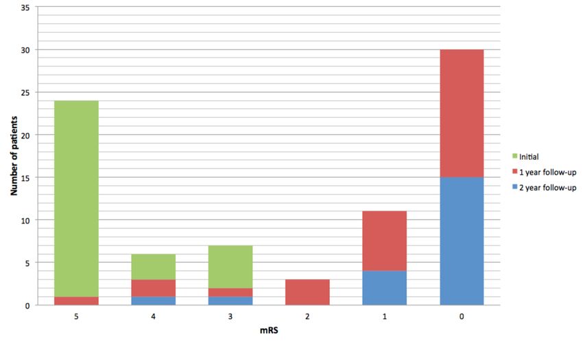
During the acute illness, 23 patients (74%) had a maximal mRS score of 5, three patients (10%)
had an mRS score of 4, and five patients (16%) had an mRS score of 3. All patients had an
improvement in the mRS score at one- and two-year follow-up after initial diagnosis. At one-
year follow-up, 15 out of 29 patients (52%) had an mRS score of 0; seven (24%) patients had an
mRS score of 1; three patients (10%) had an mRS score of 2, and four (14%) patients had an
unfavorable outcome (mRS score: 3-5). At the two-year follow-up, only two patients (10%)
presented an unfavorable outcome (Figure 1).
2020 Chavez-Castillo et al. Cureus 12(5): e8211. DOI 10.7759/cureus.8211 6 of 9FIGURE 1: Functional outcome as measured by mRS at
admission and follow-up
mRS: modified Rankin Scale
Discussion
Our study showed a predominance of affected males, which was in contrast with previous
reports [5]. To our knowledge, this demographic profile has not been previously highlighted and
could be relevant in the characterization of the disease in the pediatric population.
The presence of teratoma was intentionally sought, both in the acute phase and at follow-up,
but no cases were found. The paraneoplastic form of anti-NMDAR encephalitis is known to be
less common in the pediatric population [2,8,9]. Only 38% of cases had a viral prodrome, and
this supports the theory that there are other mechanisms not yet well studied that lead to the
development of anti-NMDAR encephalitis in pediatric patients apart from paraneoplastic and
post-infectious etiologies. HHV-6 and HHV-7 could play an essential role in this alternative
hypothesis.
Most patients started their illness with psychiatric symptoms, but the leading cause in
soliciting medical assistance was the presence of epileptic seizures. This finding suggests that
psychiatric disorders are the most common initial symptom in both children and adults but can
be minimized in the pediatric population. In this series, 85% of patients had epileptic seizures,
which was higher than what was previously reported (60-80%) [8,10,11]. Although there is no
specific type of epileptic seizure for this entity, focal seizures were predominantly found in this
study.
The mean number of AEDs required for the control of seizures during the acute stage was two,
which is consistent with previous studies [10,11]. The development of epilepsy after acute
encephalitis was uncommon; at 12 months, four patients persisted with seizures, and all of
them presented SE during the acute illness. At 24 months, only one patient continued to have
seizures. One of the factors most closely related to the persistence of post-acute encephalitis
seizures was a poor response to immunomodulatory treatment at four weeks.
2020 Chavez-Castillo et al. Cureus 12(5): e8211. DOI 10.7759/cureus.8211 7 of 9There is very little information available about the relationship between the presence of HHV
in CSF and anti-NMDAR encephalitis [19]. In this series, all patients with HHV infection had
epileptic seizures, which were more resistant to pharmacological treatment during the acute
phase, requiring a higher number of AED on average. Only 50% had a history of fever, and none
of them had a characteristic picture of herpetic encephalitis. Only one of these patients
persisted with seizures at 12 months, and none of them had seizures at 24 months. This finding
suggests that despite having more significant morbidity during the acute phase, patients with
HHV-6 and HHV-7 infection do not necessarily have a higher risk of epilepsy in the long term.
The characteristic extreme delta brush EEG pattern was found in 9% of patients; this pattern
was found in a lower percentage than in adult cohorts (30%). However, it was more prevalent
compared to results in other pediatric series (5%). A notable fact is that this series had a higher
number of pediatric patients included compared to previously published results [14,15]. None of
the patients with extreme delta brush EEG pattern persisted with long-term epilepsy.
As previously reported, the functional prognosis of these patients was generally good. At the
two-year follow-up, only two patients (10%) had a significant disability (mRS: 3-5); both
patients had seizures at the one-year follow-up. The median age of these patients was
3.5 years, which suggests that lower age is a risk factor for a poor functional outcome, which is
consistent with previously published data [20].
Very few studies have focussed on the semiology of epileptic seizures and the transition to
epilepsy in patients with anti-NMDAR encephalitis so far, and none of them are specific to the
pediatric population. To our knowledge, this is the first study describing the clinical
manifestations of seizures in pediatric patients with anti-NMDAR encephalitis. Sustained use
of AEDs after the acute phase of anti-NMDAR encephalitis is controversial. The low prevalence
of long-term epilepsy, as well as the absence of seizures in patients with an adequate response
to immunomodulatory treatment, support the theory that seizures present in patients with
anti-NMDAR encephalitis could be classified as symptomatic, and these patients do not have to
carry the burden of a lifelong diagnosis of epilepsy.
Conclusions
Based on our findings, we propose that the continuation of AEDs after the acute phase could be
considered in the following scenarios: SE, inadequate response to immunotherapy at four
weeks, and a high mRS score at discharge and during follow-up. In all other cases,
discontinuation of AED may be warranted. More studies are needed in our country to replicate
these results.
Additional Information
Disclosures
Human subjects: Consent was obtained by all participants in this study. Animal subjects: All
authors have confirmed that this study did not involve animal subjects or tissue. Conflicts of
interest: In compliance with the ICMJE uniform disclosure form, all authors declare the
following: Payment/services info: All authors have declared that no financial support was
received from any organization for the submitted work. Financial relationships: All authors
have declared that they have no financial relationships at present or within the previous three
years with any organizations that might have an interest in the submitted work. Other
relationships: All authors have declared that there are no other relationships or activities that
could appear to have influenced the submitted work.
References
1. Dalmau J, Lancaster E, Martinez-Hernandez E, Rosenfeld MR, Balice-Gordon R: Clinical
2020 Chavez-Castillo et al. Cureus 12(5): e8211. DOI 10.7759/cureus.8211 8 of 9experience and laboratory investigations in patients with anti-NMDAR encephalitis. Lancet
Neurol. 2011, 10:63-74. 10.1016/S1474-4422(10)70253-2
2. Vitaliani R, Mason W, Ances B, Zwerdling T, Jiang Z, Dalmau J: Paraneoplastic encephalitis,
psychiatric symptoms, and hypoventilation in ovarian teratoma. Ann Neurol. 2005, 58:594-
604. 10.1002/ana.20614
3. Dalmau J, Tüzün E, Wu HY, et al.: Paraneoplastic anti-N-methyl-D-aspartate receptor
encephalitis associated with ovarian teratoma. Ann Neurol. 2007, 61:25-36.
10.1002/ana.21050
4. Bien C, Holtkamp M: "Autoimmune epilepsy”: encephalitis with autoantibodies for
epileptologists. Epilepsy Curr. 2017, 17:134-141. 10.5698/1535-7511.17.3.134
5. Gable MS, Sheriff H, Dalmau J, Tilley DH, Glaser CA: The frequency of autoimmune N-
methyl-D-aspartate receptor encephalitis surpasses that of individual viral etiologies in
young individuals enrolled in the California Encephalitis Project. Clin Infect Dis. 2012,
54:899-904. 10.1093/cid/cir1038
6. Titulaer MJ, McCracken L, Gabilondo I, et al.: Treatment and prognostic factors for long-term
outcome in patients with anti-NMDA receptor encephalitis: an observational cohort study.
Lancet Neurol. 2013, 12:157-165. 10.1016/S1474-4422(12)70310-1
7. Cooray GK, Sengupta B, Douglas P, Englund M, Wickstrom R, Friston K: Characterising
seizures in anti-NMDA-receptor encephalitis with dynamic causal modelling. Neuroimage.
2015, 118:508-519. 10.1016/j.neuroimage.2015.05.064
8. Florance NR, Davis RL, Lam C, et al.: Anti-N-methyl-D-aspartate receptor (NMDAR)
encephalitis in children and adolescents. Ann Neurol. 2009, 66:11-18. 10.1002/ana.21756
9. Dalmau J, Gleichman AJ, Hughes EG, et al.: Anti-NMDA-receptor encephalitis: case series and
analysis of the effects of antibodies. Lancet Neurol. 2008, 7:1091-1098. 10.1016/S1474-
4422(08)70224-2
10. de Bruijn MAAM, van Sonderen A, van Coevorden-Hameete MH: Evaluation of seizure
treatment in anti-LGI1, anti-NMDAR, and anti-GABABR encephalitis. Neurology. 2019,
92:e2185-e2196. 10.1212/WNL.0000000000007475
11. Liu X, Yan B, Wang R, Li C, Chen C, Zhou D, Hong Z: Seizure outcomes in patients with anti-
NMDAR encephalitis: a follow-up study. Epilepsia. 2017, 58:2104-2111. 10.1111/epi.13929
12. Irani SR, Bera K, Waters P, et al.: N-methyl-d-aspartate antibody encephalitis: temporal
progression of clinical and paraclinical observations in a predominantly non-paraneoplastic
disorder of both sexes. Brain. 2010, 133:1655-1667. 10.1093/brain/awq113
13. Schmitt SE, Pargeon K, Frechette ES, Hirsch LJ, Dalmau J, Friedman D: Extreme delta brush: a
unique EEG pattern in adults with anti-NMDA receptor encephalitis. Neurology. 2012,
79:1094-1100. 10.1212/WNL.0b013e3182698cd8
14. Armangue T, Titulaer MJ, Málaga I, Bataller L, Gabilondo I, Graus F, Dalmau J: Pediatric anti-
N-methyl-D-aspartate receptor encephalitis-clinical analysis and novel findings in a series of
20 patients. J Pediatr. 2013, 162:850-856. 10.1016/j.jpeds.2012.10.011
15. Jeannin-Mayer S, André-Obadia N, Rosenberg S, Boutet C, Honnorat J, Antoine JC, Mazzola L:
EEG analysis in anti-NMDA receptor encephalitis: description of typical patterns . Clin
Neurophysiol. 2019, 130:289-296. 10.1016/j.clinph.2018.10.017
16. González-Latapi P, Rodríguez-Violante M, Cervantes-Arriaga A, Calleja-Castillo JM,
González-Aguilar A: Encefalitis por anticuerpos antirreceptor de N-metil-D-aspartato (anti-
NMDAR): reporte de un caso. (Article in Spanish). Gac Med Mex. 2014, 150:348-351.
17. Jiménez-Ruiz A, Cárdenas-Sáenz O, Ruiz-Sandoval JL: Encefalitis autoinmunitaria secundaria
a teratoma ovárico: un nuevo síndrome neuropsiquiátrico. Reporte de caso. (Article in
Spanish). Ginecol Obstet Mex. 2017, 85:472-479.
18. Scheffer IE, Berkovic S, Capovilla G, et al.: ILAE classification of the epilepsies: position paper
of the ILAE Commission for Classification and Terminology. Epilepsia. 2017, 58:512-521.
10.1111/epi.13709
19. Armangue T, Spatola M, Vlagea A, et al.: Frequency, symptoms, risk factors, and outcomes of
autoimmune encephalitis after herpes simplex encephalitis: a prospective observational study
and retrospective analysis. Lancet Neurol. 2018, 17:760-772. 10.1016/S1474-4422(18)30244-8
20. Zekeridou A, Karantoni E, Viaccoz A: Treatment and outcome of children and adolescents
with N-methyl-D-aspartate receptor encephalitis. J Neurol. 2015, 262:1859-1866.
10.1007/s00415-015-7781-9
2020 Chavez-Castillo et al. Cureus 12(5): e8211. DOI 10.7759/cureus.8211 9 of 9You can also read