Very Delayed Acute Hepatitis after Pembrolizumab Therapy for Advanced Malignancy: How Long Should We Watch? - MDPI
←
→
Page content transcription
If your browser does not render page correctly, please read the page content below
Case Report
Very Delayed Acute Hepatitis after Pembrolizumab
Therapy for Advanced Malignancy: How Long
Should We Watch?
Timothy Phan 1, *, Kurvi Patwala 1 , Lara Lipton 2 , Virginia Knight 3 , Ahmad Aga 3
and Stephen Pianko 1,4
 1   Monash Health, Clayton, Melbourne, VIC 3168, Australia; kurvi.patwala@monashhealth.org (K.P.);
     spianko@geds.com.au (S.P.)
 2   The Royal Melbourne Hospital, Parkville, VIC 3052, Australia; Lara.Lipton@mh.org.au
 3   Cabrini Medical Centre, Malvern, VIC 3144, Australia; Virginia.knight@monash.edu (V.K.);
     ahmad.aga@mps.com.au (A.A.)
 4   School of Clinical Sciences, Monash University, Melbourne, VIC 3168, Australia
 *   Correspondence: timothy.phan@monashhealth.org; Tel.: +61-467-070-312

 Received: 14 January 2021; Accepted: 9 February 2021; Published: 14 February 2021

 Abstract: Immune checkpoint inhibitors (ICIs) have led to major therapeutic advances in the
 management of malignancy. Despite promising outcomes for some cancers, ICIs are linked to unique
 side-effects known as immune-related adverse events (IrAEs). These may affect a wide array of
 organ systems. In particular, ICI-induced hepatitis is diagnostically challenging given its variable
 natural history and clinical manifestations. The onset of ICI-induced hepatitis often occurs between
 6 and 14 weeks after treatment initiation and rarely exhibits delayed presentations or manifests after
 treatment cessation. We present a case of very delayed-onset ICI-induced hepatitis, stressing the
 importance of long-term surveillance for immune-indued hepatitis in patients initiated on ICIs even
 long after treatment cessation.

 Keywords: autoimmune hepatitis; pembrolizumab; immune checkpoint inhibitor; immune-related
 adverse events; neoplasms

1. Introduction
     Immune checkpoint inhibitors (ICIs) have markedly improved the prognosis of patients with
some cancers. These novel agents augment the immune system by downregulating inhibitors of
the anti-cancer immune response, including: cytotoxic T-lymphocyte-associated antigen 4 (CTLA4),
program cell death receptor 1 (PD-1) and its ligand—programmed cell death ligand 1 (PD-L1) [1].
Despite promising clinical outcomes, ICIs are linked to unique side-effects known as immune-related
adverse events (IrAEs) due to the induction of autoimmunity. IrAEs have the potential to affect a wide
range of organ systems, commonly including: dermatological (skin rashes), endocrine, gastrointestinal
and hepatic. As ICI therapy becomes more widespread in cancer management, IrAEs are becoming
more common, including immune-induced hepatitis. We present a case of very delayed-onset
ICI-induced hepatitis that occurred 7 months post-treatment cessation, thereby highlighting the
importance of prolonged monitoring for an acute hepatitis with careful consideration of its diagnostic
and treatment nuances.

2. Case Report
      Written informed consent has been obtained from the patient to publish this paper.

Curr. Oncol. 2021, 28, 898–902; doi:10.3390/curroncol28010088                 www.mdpi.com/journal/curroncol
Curr. Oncol. 2021, 28 899
A 78-year-old female with stage IIIC breast cancer presented with subacute onset of jaundice.
Her breast cancer had been treated with pembrolizumab for 5 months and ceased 7 months prior to
presentation. She developed a prodrome of progressive fatigue and anorexia over 2 weeks. There were
no recent medication changes other than oral mesalazine, which was commenced 3 months prior
due to a new diagnosis of unspecified left-sided colitis confirmed during colonoscopy (possibly
immune mediated).
She was diagnosed with stage IIIB triple negative inflammatory breast cancer in 2015. This was
treated with neoadjuvant chemotherapy and surgery with subsequent adjuvant radiotherapy.
Approximately 12 months prior to her presentation, she developed a locally advanced right breast
recurrence requiring neoadjuvant chemotherapy and PD-1 inhibitor (pembrolizumab) prior to
double mastectomy.
Initial biochemistry revealed a severe transaminitis. Her bilirubin was 199 µmol/L (Curr. Oncol. 2021, 28 900
Figure 2. Graphical representation of bilirubin (µmol/L) and alanine transferase (ALT [U/L]) trends
over a 75-day follow-up period from time of presentation. Prednisolone was commenced on Day
5 post-presentation (*).
3. Discussion
The clinical manifestations and natural history of ICI-induced hepatitis are heterogenous.
Acute hepatitis occurs in 2–10% of patients on ICI therapy, which typically presents as an asymptomatic
transaminitis [2,3]. Occasionally, patients may develop a rapidly progressive acute hepatitis and even
fulminant liver failure associated with significant morbidity and mortality [4,5]. Contrary to the usual
timeframe for disease onset, our patient developed ICI-induced hepatitis 7 months post-treatment
cessation. Only one other author has reported a case of delayed ICI-induced hepatitis occurring
8 months post-nivolumab cessation [6]. Delayed-onset immune-induced hepatitis after treatment
cessation is rare and most reported cases occur during active treatment—between 6 and 14 weeks
after treatment initiation [7]. Hence, this case serves as a reminder to strictly monitor liver function
monitoring up to 12 months post-treatment cessation to detect an evolving hepatitis.
ICI-induced hepatitis can be pathologically difficult to differentiate from autoimmune hepatitis
(AIH) or drug-induced liver injury (DILI). Contrasting with AIH, auto-antibodies including anti-nuclear
and anti-smooth muscle antibodies may be negative in ICI-induced hepatitis. Similarly, serum IgG levels
are normal or slightly elevated in ICI-induced hepatitis as exemplified by our case [2]. Liver biopsy is
often not required in suspected ICI-induced hepatitis and is only indicated to exclude other causes
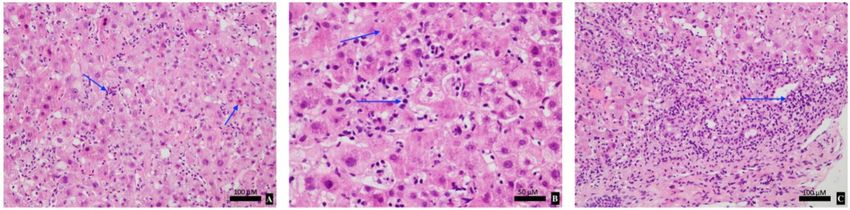
of acute hepatitis or quantify the degree of hepatocellular injury. Biopsy findings in ICI-induced
hepatitis are variable. Most commonly, panlobular hepatitis is observed in approximately 70%
of cases, typified by scattered focal necrosis and acidophilic bodies [8]. Histopathological factors
which differentiate AIH and DILI from ICP-hepatitis are summarised in Table 1 [2]. In our case,
liver biopsy demonstrated classical features of ICI-induced hepatitis without features of AIH or
DILI from mesalazine. The cellular infiltrate was suggestive of ICI-related pathology given a
lymphocytic predominance without plasmacytosis nor eosinophilia. Detailed knowledge around
the pathological and biochemical intricacies of ICI-induced hepatitis can greatly assist with prompt
diagnosis and treatment.Curr. Oncol. 2021, 28 901
Table 1. Comparison of histopathological features of ICI-induced hepatitis, autoimmune hepatitis
(AIH) and drug-induced liver injury (DILI). Adapted from Zen et al [2].
Histological Features ICI-Hepatitis AIH DILI
Confluent Necrosis Less common More common More common
Eosinophilic infiltration Uncommon Not specific More common
Bile plugs Uncommon Not specific More common
Plasmacytosis Uncommon Common Not specific
Hepatocellular rosettes Uncommon Common Not specific
Emperipolesis Uncommon Common Not specific
Management of ICI-induced hepatitis is based on the Common Terminology Criteria for Adverse
Events (CTCAE), which grades severity of disease from 1–4 [3]. Our patient presented with grade
4 hepatitis which was glucocorticoid responsive. Current guidelines advocate for prompt glucocorticoid
treatment with methylprednisolone or equivalent at 1–2 mg/kg/day for three days (up to 1 g/day),
followed by prednisolone 1–2 mg/kg/day over 4 weeks. In steroid-refractory cases, the introduction of
mycophenolate or azathioprine is prudent. Permanent discontinuation of ICI therapy is recommended
in grade 3–4 hepatitis [3,9]. Corticosteroid therapy in ICI-induced hepatitis is contentious. Spontaneous
improvement in LFTs can occur with observation and low doses of corticosteroids may be sufficient to
control disease [5]. Our patient was promptly commenced on prednisolone therapy at a relatively low
starting dose compared with pulsed methylprednisolone with rapid clinical response and excellent
efficacy. Our case supports that LFT resolution is achievable with a lower dose glucocorticoid,
thereby reducing glucocorticoid-related side effects. Further high-quality randomised-controlled
studies are required to assess optimal management strategies for ICI-induced hepatitis.
Delayed-onset IrAEs of any organ system are underrecognised and clinical vigilance is critical
even after treatment cessation [10]. The median interval to delayed-onset IrAE diagnosis is 6 months
post-immunotherapy cessation (IQR 3–28 months) [10]. Currently, there is a paucity of evidence
on delayed-onset IrAEs due to limited follow up periods and incompleteness of IrAE reporting in
immuno-oncology clinical trials [11]. Best practice guidelines allude to monitoring for delayed-onset
IrAEs up to 12 months after treatment discontinuation [9]. However, late-onset IrAEs are difficult
to predict with available investigations and are challenging to prevent. Surveillance for IrAEs
should be individualised based on a patient’s risk profile for developing IrAEs. A patient’s risk
for IrAEs is increased in the presence of patient and treatment factors including: pre-existing
connective tissue, vasculitic or auto-immune disease, and combination ICI usage [1]. The utility
of active surveillance strategies for delayed-onset IrAEs remains an area of active research with no
evidence-based algorithms available.
4. Conclusions
ICIs are becoming an integral part of cancer therapy, and monitoring for adverse events requires
awareness from all physicians and not just oncologists. This case pertinently emphasises the
importance of long-term surveillance for immune-induced hepatitis in patients initiated on ICI
therapy. Attentive appraisal of symptoms, signs and investigations is required for prompt diagnosis of
ICI-induced hepatitis allowing for the timely introduction of therapy. Systemic treatment is nuanced
and further high-quality evidence is required to guide optimal therapy. We advocate for individualised
surveillance strategies for delayed-onset IrAEs up to 12-months after treatment cessation. However,
stringent evidence-based surveillance protocols remains an area of ongoing development.
5. Clinical Practice Points
• Delayed-onset ICI-induced hepatitis can occur up to 12 months post-treatment cessation;
• Autoimmune biomarkers are often negative in ICI-induced hepatitis and plasmacytosis or
eosinophilia is uncommon in the biopsy cellular infiltrate;Curr. Oncol. 2021, 28 902
• Low-dose glucocorticoid therapy may be warranted in select ICI-induced hepatitis patients,
but further randomized control studies are required to assess optimal glucocorticoid dosing;
• Routine monitoring for delayed-onset IrAEs including liver function should be individualised
and can extend up to 12 months post-treatment cessation.
Author Contributions: T.P.—case analysis and writing. K.P., L.L., V.K., A.A. and S.P.—review and editing of
subsequent drafts. All authors have read and agreed to the published version of the manuscript.
Funding: This research received no external funding.
Conflicts of Interest: The authors declare no conflict of interest.
References
1. Martins, F.; Sofiya, L.; Sykiotis, G.P.; Lamine, F.; Maillard, M.; Fraga, M.; Shabafrouz, K.; Ribi, C.; Cairoli, A.;
Guex-Crosier, Y.; et al. Adverse effects of immune-checkpoint inhibitors: Epidemiology, management and
surveillance. Nat. Rev. Clin. Oncol. 2019, 16, 563–580. [CrossRef] [PubMed]
2. Zen, Y.; Yeh, M.M. Checkpoint inhibitor-induced liver injury: A novel form of liver disease emerging in the
era of cancer immunotherapy. Semin. Diagn. Pathol. 2019, 36, 434–440. [CrossRef] [PubMed]
3. Brahmer, J.R.; Lacchetti, C.; Thompson, J.A. Management of Immune-Related Adverse Events in Patients
Treated with Immune Checkpoint Inhibitor Therapy: American Society of Clinical Oncology Clinical Practice
Guideline Summary. J. Oncol. Pract. 2018, 14, 247–249. [CrossRef] [PubMed]
4. Kim, K.W.; Ramaiya, N.H.; Krajewski, K.M.; Jagannathan, J.P.; Tirumani, S.H.; Srivastava, A.; Ibrahim, N.
Ipilimumab associated hepatitis: Imaging and clinicopathologic findings. Investig. New Drugs 2013, 31,
1071–1077. [CrossRef] [PubMed]
5. De Martin, E.; Michot, J.M.; Papouin, B.; Champiat, S.; Mateus, C.; Lambotte, O.; Roche, B.; Antonini, T.M.;
Coilly, A.; Laghouati, S.; et al. Characterization of liver injury induced by cancer immunotherapy using
immune checkpoint inhibitors. J. Hepatol. 2018, 68, 1181–1190. [CrossRef] [PubMed]
6. Parakh, S.; Cebon, J.; Klein, O. Delayed Autoimmune Toxicity Occurring Several Months after Cessation of
Anti-PD-1 Therapy. Oncologist 2018, 23, 849–851. [CrossRef] [PubMed]
7. Weber, J.S.; Dummer, R.; de Pril, V.; Lebbé, C.; Hodi, F.S. Patterns of onset and resolution of immune-related
adverse events of special interest with ipilimumab: Detailed safety analysis from a phase 3 trial in patients
with advanced melanoma. Cancer 2013, 119, 1675–1682. [CrossRef] [PubMed]
8. LoPiccolo, J.; Brener, M.I.; Oshima, K.; Lipson, E.J.; Hamilton, J.P. Nodular Regenerative Hyperplasia
Associated with Immune Checkpoint Blockade. Hepatology 2018, 68, 2431–2433. [CrossRef] [PubMed]
9. Haanen, J.; Carbonnel, F.; Robert, C.; Kerr, K.; Peters, S.; Larkin, J.; Jordan, K. Management of toxicities from
immunotherapy: ESMO Clinical Practice Guidelines for diagnosis, treatment and follow-up. Ann. Oncol.
2017, 28 (Suppl. S4), iv119–iv142. [CrossRef]
10. Couey, M.A.; Bell, R.B.; Patel, A.A.; Romba, M.C.; Crittenden, M.R.; Curti, B.D.; Urba, W.J.; Leidner, R.S.
Delayed immune-related events (DIRE) after discontinuation of immunotherapy: Diagnostic hazard of
autoimmunity at a distance. J. Immunother. Cancer 2019, 7, 165. [CrossRef] [PubMed]
11. Chen, T.W.; Razak, A.R.; Bedard, P.L.; Siu, L.L.; Hansen, A.R. A systematic review of immune-related adverse
event reporting in clinical trials of immune checkpoint inhibitors. Ann. Oncol. Sep. 2015, 26, 1824–1829.
[CrossRef] [PubMed]
Publisher’s Note: MDPI stays neutral with regard to jurisdictional claims in published maps and institutional
affiliations.
© 2021 by the authors. Licensee MDPI, Basel, Switzerland. This article is an open access
article distributed under the terms and conditions of the Creative Commons Attribution
(CC BY) license (http://creativecommons.org/licenses/by/4.0/).You can also read