Clinical pattern of failure after a durable response to immune checkpoint inhibitors in non small cell lung cancer patients - Nature
←
→
Page content transcription
If your browser does not render page correctly, please read the page content below
www.nature.com/scientificreports
OPEN Clinical pattern of failure
after a durable response to immune
checkpoint inhibitors in non‑small
cell lung cancer patients
Ja Yoon Heo1,4, Shin Hye Yoo1, Koung Jin Suh2, Se Hyun Kim2*, Yu Jung Kim2,
Chan‑Young Ock1, Miso Kim1, Bhumsuk Keam1,3, Tae Min Kim1,3, Dong‑Wan Kim1,3,
Dae Seog Heo1,3 & Jong Seok Lee2
Although immune checkpoint inhibitors (ICIs) can induce durable responses in non-small-cell lung
cancer (NSCLC) patients, a significant proportion of responders still experience progressive disease
after a period of response. Limited data are available on the clinical patterns of acquired resistance
(AR) to ICIs. Clinical and radiologic data from 125 NSCLC patients treated with anti-PD-1 or PD-L1
antibodies between 2011 and 2018 at two tertiary academic institutions were retrospectively
reviewed. Overall, 63 (50.4%) patients experienced AR after ICI treatment in a median of 10.7 months.
Among the 13 patients with a partial response with ICI, 12 (32.4%) had only lymph node progression.
Most patients (n = 52, 82.5%) had one or two sites with progression (oligo-progression). The median
overall survival (OS) after progression was significantly longer in the extrathoracic group than in the
thoracic and liver progression groups (30.2 months [95% confidence interval (CI), 13.4 to not reached
(NR)], 11.7 months [95% CI, 9.5–21.1], and 5.4 months [95% CI, 2.6-NR], respectively, P < 0.001).
Patients with oligo-progression had significantly longer OS after AR than did the multi-progression
patients (18.9 months [95% CI, 10.6-NR] vs. 8.8 months [95% CI, 5.7-NR], P = 0.04). No significant
difference in progression-free survival was observed between the subsequent chemotherapy and the
ICI after AR groups (P = 0.723). Patients with AR after ICI treatment had a unique progression pattern
with oligo-progression and high rates of progression only in the lymph nodes. Local treatment and/or
continuation of ICIs beyond AR might be an effective option.
Immune checkpoint inhibitors (ICIs), including anti- programmed death 1 (PD-1) or programmed death ligand
1 (PD-L1) antibodies, represent a breakthrough in the treatment of non-small-cell lung cancer (NSCLC). Previ-
ous studies have shown that high levels of PD-L1 expression on tumors and/or immune cells by immunohisto-
chemistry are associated with improved clinical efficacy when compared with low levels or negative PD-L11–6.
In patients with pretreated advanced NSCLC, ICI monotherapy resulted in durable clinical responses lasting
more than 6 months and enabled patients to achieve 5-year overall survival (OS) without subsequent therapy and
disease progression6–8. However, a significant proportion of long-term responders still experience progressive
disease after a period of response time.
Acquired resistance (AR) refers to a clinical scenario in which cancer initially responds to immunotherapy,
but it continues to grow and metastasize to other organs after a period of time. In the epidermal growth factor
receptor (EGFR) mutant NSCLC, it is well-known that acquired mutations, including T790M, confer AR to
tumors after EGFR tyrosine kinase inhibitor (TKI) t reatment9. However, among patients with ICI treatment,
more efforts are needed to elucidate which patients will experience AR and how tumor evolution or changes
in the immune system lead to AR development. Defects in interferon-receptor signaling and antigen presenta-
tion pathways, tumor-associated neoantigen loss, alternate immune checkpoints, and alterations in the tumor
microenvironment are believed to be involved in AR m echanisms10–12.
1
Department of Internal Medicine, Seoul National University Hospital, Seoul, Republic of Korea. 2Department of
Internal Medicine, Bundang Hospital, Seoul National University College of Medicine, 82, Gumi‑ro 173 beon‑gil,
Bundang‑Gu, Seongnam, Gyeonggi‑do 13620, Republic of Korea. 3Cancer Research Institute, Seoul National
University, Seoul, Republic of Korea. 4Present address: Department of Internal Medicine, National Health
Insurance Service Ilsan Hospital, Goyang, Republic of Korea. *email: sehyunkim@snubh.org
Scientific Reports | (2021) 11:2514 | https://doi.org/10.1038/s41598-021-81666-x 1
Vol.:(0123456789)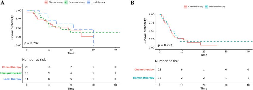
www.nature.com/scientificreports/
Recently, Gettinger et al.13 presented an impressive single-institution case series of 26 NSCLC patients who
experienced AR of the PD-1 blockade. The authors reported the lymph node (LN) as the most common site of
AR; their study included 11 cases with only LN progression. The majority of the included patients (88%) showed
progression in one or two sites of disease. Overall, 15 (58%) patients received local therapy to sites of AR, while
11 (42%) patients continued immunotherapy beyond progression14. To date, clinical data regarding subsequent
treatment strategies and survival according to progression patterns after AR to ICI in NSCLC patients is sparse,
especially for those with durable responses.
Therefore, in this study, we conducted a retrospective analysis to investigate the clinical patterns of AR after
a durable response to ICIs in NSCLC patients. Additionally, we determined the implications for subsequent
therapy in these patients.
Results
Patient characteristics. Of the 125 patients with a durable clinical benefit more than 6 months, 63 (50.4%)
patients with AR to ICIs were identified. The baseline clinicopathological characteristics of these patients are
summarized in Table 1. The majority (n = 47, 74.6%) of the patients were male, and just over half of the patients
were pathologically diagnosed with adenocarcinoma (n = 35, 55.5%). The majority of patients received ICI as
first or second-line treatment for metastatic NSCLC (n = 47, 74.6%); 21 (33.3%) and 26 (41.3%) patients received
it as first and second-line treatment, respectively. The study included 15 (23.8%) patients with driver muta-
tions. Fifty-two (82.5%) patients received monotherapy, while 11 (17.5%) patients received combination therapy,
including five (7.9%) patients with PD-1 and CTLA-4 inhibitors, five (7.9%) patients with PD-L1 and CTLA-4
inhibitors, and one (1.6%) patient with a PD-1 inhibitor and chemotherapy. The median duration of follow-up
was 18 months (range, 8–60) and 23 (36.5%) patients were alive at the time of data analysis in December 2018.
Clinical patterns of ICI failure. Among all the included patients, the median time to AR was 10.7 months.
The clinical patterns of AR after ICI treatment are summarized in Table 2 and shown based on their best objec-
tive response in Fig. 1. The most common site of progressive disease was the lungs (38/63, 60.3%), followed by
the LNs (31/63, 49.2%), and the brain (6/63, 9.5%). One of fifth (14/63, 22.2%) of patients had only LN progres-
sion at the time of AR. Twelve (85.7%) of the 14 patients with LN progression only had achieved PR as their best
response, while 2 (14.3%) of the 14 patients achieved SD during ICI treatment (P = 0.04).
Next, we evaluated the pattern of failure according to the type of ICI treatment. Less frequent lung progres-
sion was observed in patients treated with ICI as first- or second-line treatment than in patients treated with
ICI as at least third-line treatment (48.9% vs. 93.8%; P value = 0.039). Progressive disease only in the LNs was
observed among the patients who received first- or second line-treatment. There were no statistically significant
differences between the two groups regarding the number of progression sites, previous best response, and the
pre-existence of progression sites.
The clinical patterns of AR based on the number of progression sites are summarized in Table 3. Fifty-two
(82.5%) patients had progression in one or two lesions (oligo-progression group), while 11 (25.4%) patients had
progression in three or more lesions (multi-progression group). The lungs were the most frequent sites of progres-
sion in both groups. Progression of new lesions only was observed among 12 (23.1%) and 0 (0%) patients with
oligo-progression and multi-progression, respectively (P < 0.001). Progression in the LNs only was also more fre-
quent in patients with oligo-progression compared to patients with multi-progression (27.0% vs. 0%, P = 0.103).
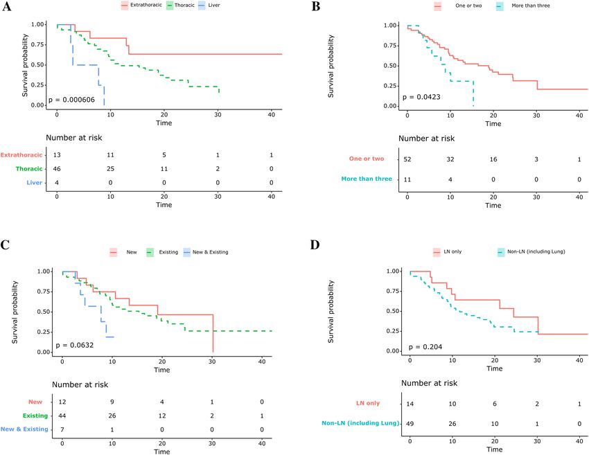
Survival differences based on the progression patterns. The median OS after AR was 10.9 months.
OS after AR was analyzed according to the pattern of progression. When the patients were classified into sub-
groups (i.e. those who had intrathoracic, extrathoracic [excluding the liver], and liver progression), the median
OS after progression was significantly longer in the extrathoracic subgroup than in the thoracic and liver pro-
gression subgroups (30.2 months [95% CI, 13.4 to not reached (NR)], 11.7 months [95% CI, 9.5–21.1], and
5.4 months [95% CI, 2.6-NR], respectively, P < 0.001, Fig. 2A). The median OS after progression was significantly
longer in the oligo-progression group compared to that in the multiple progression group (18.9 months [95%
CI, 10.6-NR] vs. 8.8 months [95% CI, 5.7-NR], P = 0.04, Fig. 2B). The progression patterns were not statistically
significant when classified by progression in new lesions (Fig. 2C) nor nodal progression (Fig. 2D).
Subsequent therapy after AR. Among the 63 patients with AR to ICIs, 52 (82.5%) patients received
subsequent therapy after progression. Eleven (21.2%) patients received local treatment, including radiotherapy
and surgical resection. Forty-one (78.8%) patients underwent systemic therapy after progression, including 25
(48.1%) and 16 (30.8%) patients who received cytotoxic chemotherapy and ICI after progression (Table 4). The
median OS of the local therapy, chemotherapy, and ICI groups was 27.4, 23.2, and 29.1 months, respectively;
there was no significant difference in survival between these groups (P = 0.787, Fig. 3A). Additionally, there was
no significant difference in the PFS between the chemotherapy and the ICI groups (P = 0.723, Fig. 3B).
Discussion
In this study, after a median time of 10.4 months, patients who had a durable clinical benefit experienced AR. The
majority of these patients (52/63, 82.5%) had progressive disease in one or two sites. Progression in the LNs only
was observed in 22.2% patients at the time of AR; such patients received ICI as first or second-line treatment.
Most (12/14, 85.7%) patients with progression in the LNs only had objective tumor shrinkage, partial response
according to RECIST, during ICI treatment. Among patients with oligo-progression (n = 52), progression of the
LNs only (27.0%) or appearance of new lesions without evident progression of pre-existing lesions (23.1%) was
observed. The patterns of progression observed in this cohort of patients with NSCLC at the time of AR are very
Scientific Reports | (2021) 11:2514 | https://doi.org/10.1038/s41598-021-81666-x 2
Vol:.(1234567890)www.nature.com/scientificreports/
N = 63
Age (years)
Median (range) 64 (34–84)
Sex
Male 47 (74.6)
Female 16 (25.4)
Ethnicity
Asian 63 (100.0)
Smoking
Never smoked 21 (33.3)
Current or ex-smoker 42 (66.7)
Pathology
Adenocarcinoma 35 (55.5)
Poorly differentiated 4 (6.3)
Sarcomatoid 3 (4.8)
Large cell neuroendocrine 2 (3.2)
Squamous 19 (30.2)
Driver mutation
EGFR 11 (17.4)
KRAS 2 (3.2)
ALK 1 (1.6)
ROS1 1 (1.6)
Not identified 48 (76.2)
ICI treatment line
1 21 (33.3)
2 26 (41.3)
≥3 16 (25.4)
ICI treatment
PD-1 inhibitor 36 (57.1)
PD-L1 inhibitor 16 (25.4)
PD-1 inhibitor + CTLA-4 inhibitor 5 (7.9)
PD-L1 inhibitor + CTLA-4 inhibitor 5 (7.9)
PD-1 inhibitor + chemotherapy 1 (1.6)
PD-L1 expression
≥ 50% 18 (28.6)
1–49% 10 (15.9)
0% 11 (17.5)
Not available 24 (38.1)
Best objective response
Stable disease 26 (41.3)
Partial response 37 (58.7)
Complete response 0 (0)
Table 1. Patient characteristics. EGFR epidermal growth factor receptor, ALK anaplastic lymphoma kinase,
PD-1 programmed death 1, PD-L1 programmed death ligand 1, CTLA-4 Cytotoxic T-Lymphocyte Associated
Protein 4. *Data are presented as the median (range) or number (%).
unique considering that most patients treated in the conventional chemotherapy era develop systemic progres-
sion including pre-existing diseases. Even after having AR, patients with oligo-progression could be treated with
local ablative therapy or continued ICI beyond AR. In the survival analysis, there was significantly longer OS
among the oligo-progressors compared to that among systemic or multiple site progressors. We observed no
significant difference in terms of PFS and OS between patients with subsequent chemotherapy and those with
continuation of ICI beyond progression.
A notable finding in our study was that a significant portion of patients with AR to ICIs had progression of the
LNs only, with sustained anti-cancer efficacy of ICI in pre-existing lesions. Among patients with a PR response
with ICI (n = 37, 58.7%), 12 (32.4%) patients had progression of the LNs only. In the previous study by Gettinger
et al. it was also reported that 42% of NSCLC patients who achieved PR after ICI treatment had AR limited to
the LNs11. The study also suggested that the malignant LN environment may lead to the immunosuppressive
evolution of cells within the node.
Scientific Reports | (2021) 11:2514 | https://doi.org/10.1038/s41598-021-81666-x 3
Vol.:(0123456789)www.nature.com/scientificreports/
All patients 1st/2nd line ICI ≥ 3rd line ICI
(n = 63) (n = 47) (n = 16) P-value
Previous best response
PR 37 (58.7) 16 (34.0) 6 (37.5) 0.089
SD (≥ 6 months) 26 (41.3) 31 (66.0) 10 (62.5)
Time to acquired resistance
Median (range) 10.7 (6.0–47.2) 10.8 (6.0–47.2) 10.2 (6.4–22.9) 0.052
Overall survival after progression
Median (range) 10.9 (0.0–43.7) 11.5 (0.0–43.7) 9.6 (0.0–30.4) 0.775
Number of progression sites
1 46 (73.0) 36 (76.6) 10 (62.5) 0.553
2 6 (9.5) 4 (8.5) 2 (12.5)
≥3 11 (17.5) 7 (14.9) 4 (25.0)
Progression sitea
Lung 38 (60.3) 23 (48.9) 15 (93.8) 0.004
Non-lung visceral organ
Liver 4 (6.3) 2 (4.3) 2 (12.5) 1
Colon 1 (1.6) 1 (2.1) 0 (0)
Adrenal gland 1 (1.6) 1 (2.1) 0 (0)
Heart (pericardium) 2 (3.2) 1 (2.1) 1 (6.2)
Brain 6 (9.5) 5 (10.6) 1 (6.2)
Bone 4 (6.3) 2 (4.3) 2 (12.5) 1
Lymph node 31 24 7 0.265
Neck 5 (7.9) 3 (6.4) 2 (12.5) 0.274
Axilla 2 (3.2) 1 (2.1) 1 (6.2)
Thoracic 20 (32.7) 16 (34.0) 4 (25.0)
Abdomen 4 (6.3) 4 (6.3) 0 (0)
Pre-existence of progression site
New only 13 (20.6) 11 (23.4) 1 (6.2) 0.201
Existing only 43 (68.3) 32 (68.1) 12 (75.0)
Both new and existing 7 (11.1) 4 (8.5) 3 (18.8)
Pattern of progression sites
Lymph node only 14 (22.2) 14 (30.0)) 0 (0) 0.013
Extranodal organs 49 (77.8) 33 (70.0) 16 (100)
Table 2. Clinical Patterns of the failure of immune checkpoint inhibitors. Bold values denote statistical
significance at the p < 0.05 level ICI immune checkpoint inhibitors, PR partial response, SD stable disease.
a
Patients were included in more than one group based on the site of progression.
Figure 1. Donut plot showing sites of acquired resistance by best objective response to immune checkpoint
inhibitors. LN lymph node, PR partial response, SD stable disease.
Scientific Reports | (2021) 11:2514 | https://doi.org/10.1038/s41598-021-81666-x 4
Vol:.(1234567890)www.nature.com/scientificreports/
Oligo-progression Multiple progression
(Progression site 1–2) (Progression site 3 ≤)
(n = 52) (n = 11) P-value
Age (years)
Median (range) 64 (34–84) 62 (49–74) 0.752
Sex
Male 38 (73.1) 9 (81.8) 0.714
Female 14 (26.9) 2 (18.2)
Smoking
Never smoked 19 (36.5) 2 (18.2) 0.31
Current or ex-smoker 33 (63.5) 9 (81.8)
Pathology
Squamous 16 (30.8) 3 (27.3) 0.822
Non-Squamous 36 (69.2) 8 (62.7)
ICI treatment line
01-Feb 40 (76.9) 7 (63.6) 0.449
3≤ 12 (23.1) 4 (36.4)
Monotherapy
PD-1 inhibitor 27 (51.9) 9 (81.8)
PD-L1 inhibitor 16 (30.8) 0 (0)
Combination therapy
PD-1 inhibitor + chemotherapy 1 (1.9) 0 (0)
PD-1 inhibitor + CTLA-4 inhibitor 3 (5.8) 2 (18.2)
PD-L1 inhibitor + CTLA-4 inhibitor 5 (9.6) 0 (0.0)
Previous response
SD 29 (55.8) 8 (72.7) 0.502
PR 23 (44.2) 3 (27.3)
Progression sitea
Lung 28 (53.8) 10 (90.9) 0.039
Non-lung visceral organ
Liver 2 (3.8) 2 (18.2) 0.275
Colon 1 (1.9) 0 (0)
Adrenal gland 1 (1.9) 0 (0)
Heart (pericardium) 1 (1.9) 1 (9.1)
Brain 5 (9.6) 1 (9.1) 1
Bone 1 (1.9) 3 (27.3) 0.015
Lymph node 14 (23.6) 11 (100) < 0.001
Neck 3 (5.8) 1 (9.1)
Axilla 1 (1.9) 1 (9.1)
Thoracic 10 (15.9) 10 (90.9)
Abdomen 4 (6.3) 0 (0)
Pre-existence of progression site
New only 12 (23.1) 0 (0) < 0.001
Existing only 39 (75.0) 5 (45.5)
Both new and existing 1 (1.9) 6 (54.5)
Pattern of progression sites
Lymph node only 14 (27.0) 0 (0) 0.103
Extranodal organs 38 (73.0) 11 (100.0)
Table 3. Baseline characteristics based on oligo-progression. Bold values denote statistical significance at the
p < 0.05 level PR partial response, SD stable disease, PD-1 programmed death 1, PD-L1 programmed death
ligand 1, CTLA-4 Cytotoxic T-Lymphocyte Associated Protein 4. a Patients can be included in more than one
group based on the site of progression13.
It should be also noted that that patients with oligo-progression showed prolonged OS compared to those with
multi-progression after AR in our study. Potential hypotheses for this finding might be the “the cancer-immunity
cycle”15. Tumors in still “immune surveillance active” hosts are not able to proliferate or metastasize to other
Scientific Reports | (2021) 11:2514 | https://doi.org/10.1038/s41598-021-81666-x 5
Vol.:(0123456789)www.nature.com/scientificreports/
Figure 2. Kaplan–Meier survival curves showing overall survival from the point of acquired resistance for (A)
the subgroup that had thoracic, extrathoracic, and liver progression only (B) the subgroup that had one or two
progression sites or more than three progression sites (C) the subgroup that had progression from new lesions,
existing lesions, and both new and existing lesions (D) the subgroup that had only lymph node and extranodal
progression (including the lung).
organs unless they obtain the novel phenotype of immune evasion. Therefore, it is easily conceivable that lung
cancer clones in the progressive site might have evolved to escape host immune surveillance with either loss of
heterozygosity in human leukocyte antigens or depletion of the expressed n eoantigens16.
In this study, physicians preferred local therapy and immunotherapy beyond progression for patients with
oligo-progression. For NSCLC, there was an interesting retrospective report from the phase III OAK study that
atezolizumab showed acceptable efficacy and safety after progression with patients with good performance17. To
date, ICI has been reported to show post-progression efficacies in a limited subset of patients including those with
metastatic urothelial carcinoma, renal cell carcinoma and melanoma, and N SCLC18–22. However, whether treat-
ment beyond progression will be effective is not yet elucidated in patients with AR. Gettinger et al. reported that
11 NSCLC patients continued immunotherapy beyond AR13. Adam LC et al. reported an AR case of successful
local control with cryotherapy combined with immunotherapy beyond progression23. Further analysis will help
to define the clinical benefit for NSCLC patients with a durable response treated with ICI beyond progression.
There are several limitations to this study. First, this was a retrospective analysis. As not many patients showed
a durable response to ICI, only a small number of patients were included in the study, resulting in limited sta-
tistical power. Therefore, the small study size prohibited us from performing comparisons between the different
subgroups. Second, the study population was very diverse, including not only patients with PD-1 inhibitor or
PD-L1 inhibitor monotherapy, but also patients with combination therapy including cytotoxic drugs or CTLA-4
inhibitors. Third, although the PD-L1 expression profile is a very important prognostic factor, our study included
the PD-L1 profile utilizing three different anti-PD-L1 antibodies (22C3, E1L3N, and SP263). As most of the
patients (81.0%) were involved in clinical trials, we were not able to ascertain the immunohistochemistry profiles
for some patients, due to patient information protection regulations. Besides, information on co-medication
such as antibiotics, corticosteroids, opioids, or proton pump inhibitors, which can affect ICI response, was not
fully available. Lastly, there may be bias in the selection of subsequent therapy, as each individual physician was
responsible for selecting the appropriate treatment.
Scientific Reports | (2021) 11:2514 | https://doi.org/10.1038/s41598-021-81666-x 6
Vol:.(1234567890)www.nature.com/scientificreports/
All patients 1st/2nd line ICI ≥ 3rd line ICI
(n = 52) (n = 40) (n = 12)
Local therapy (n = 11)
Surgery 2 (3.8) 2 (5.0) 0 (0)
Radiotherapy 7 (13.5) 6 (15.0) 1 (8.3)
Gamma-knife surgery 2 (3.8) 2 (5.0)) 0 (0)
Systemic therapy (n = 41)
Immunotherapy 16 (30.8) 16 (40.0) 0 (0.0)
Chemotherapy 25 (48.1) 16 (40.0) 9 (75.0)
Oligo-progression Multiple progression
All patients (Progression site 1–2) (Progression site 3 ≤)
(n = 52) (n = 43) (n = 9)
Local therapy (n = 11)
Surgery 2 (3.8) 2 (4.7) 0 (0)
Radiotherapy 7 (13.5) 6 (14.0) 1 (11.1)
Gamma-knife surgery 2 (3.8) 2 (4.7) 0 (0)
Systemic therapy (n = 41)
Immunotherapy 16 (30.8) 14 (32.6) 2 (22.2)
chemotherapy 25 (48.1) 19 (44.2) 6 (66.7)
Colon mass excision (1)
Local therapy (n = 11) Surgery
Axilla LN excision (1)
Abdomen LN (2)
Neck & SCN LN (1)
Radiotherapy
Lung mass (2)
Bone (2)
Gamma-knife surgery Brain (2)
Table 4. Subsequent therapy after progression. ICI immune checkpoint inhibitors.
Figure 3. Kaplan–Meier survival curves showing (A) overall survival from the point of acquired resistance
based on the subgroups of subsequent treatment (B) progression free survival based on the subsequent
treatment.
Despite these limitations, our study described the clinical features and patterns of AR in detail. To our knowl-
edge, this is the first study to show survival differences based on progression patterns. We believe that this report
may provide useful information to clinicians regarding advanced NSCLC patients; additionally, it highlights the
implications of subsequent therapy after AR.
In conclusion, advanced NSCLC patients who had durable responses (> 6 months) showed unique patterns
of progression with high rates of progression among the LN only, in addition to oligo-progression. Local treat-
ment and/or continuation of ICIs beyond AR might be an effective option for those patients. Further large-scale
prospective studies are required to confirm these findings.
Scientific Reports | (2021) 11:2514 | https://doi.org/10.1038/s41598-021-81666-x 7
Vol.:(0123456789)www.nature.com/scientificreports/
Methods
Design, patients, and data collection. We retrospectively reviewed the records of patients diagnosed
with NSCLC and treated with inhibitors of PD-1 and PD-L1 at the Seoul National University Hospital (SNUH)
and Seoul National University Bundang Hospital (SNUBH) between January 1, 2011 and November 31, 2018.
Adults without a previous history of severe systemic disease or autoimmune diseases who received PD-1/
PD-L1 blockade alone or with cytotoxic T-lymphocyte associated protein 4 (CTLA-4) inhibitor or chemotherapy
over the course of disease were included.
Patient classification and outcome measures. The patients were classified into the following sub-
groups: those who had intrathoracic, extrathoracic [excluding the liver], and liver progression. AR after durable
response was defined as disease progression after 6 months with the best response of the stable disease, partial
response, or complete response according to the Response Evaluation Criteria in Solid Tumors (RECIST) v1.1
criterion. Imaging studies included Computed tomography scan and Magnetic resonance imaging and were
independently reviewed by two observers. Progression-free survival (PFS) was calculated from the ICI treat-
ment initiation date to the date of disease progression using the RECIST v1.1 criteria24, as confirmed by imaging,
death, or the last follow-up date, if censored. OS was measured from the initiation of ICI treatment until death
or the last follow-up date, if censored.
Statistical analyses. Continuous variables were compared using the Students t-test or the Mann–Whitney
U-test, whereas categorical variables were compared using the chi-squared or Fisher exact tests. Survival analy-
ses were conducted according to the Kaplan–Meier method using the log-rank test. All tests were two-sided and
p-values < 0.05 were considered to be statistically significant. For survival analysis, R version 3.4.3 software (R
Development Core Team, https://www.r-project.org/) was used for computation.
Ethics. All the included patients provided written, informed consent for data collection before treatment.
The research team conducted retrospective analyses on cohort data. The institutional review boards of SNUH
and SNUBH approved the study protocol (IRB number: B-2004-606-105). All procedures were carried out in
accordance with the ethical standards of the institutional research ethics committee and the Helsinki declaration
revised in 2013 by the World Medical Association.
Received: 12 July 2020; Accepted: 11 January 2021
References
1. Garon, E. B. et al. Pembrolizumab for the treatment of non-small-cell lung cancer. N. Engl. J. Med. 372, 2018–2028. https://doi.
org/10.1056/NEJMoa1501824 (2015).
2. Gettinger, S. N. et al. Overall survival and long-term safety of nivolumab (anti-programmed death 1 antibody, BMS-936558,
ONO-4538) in patients with previously treated advanced non-small-cell lung cancer. J. Clin. Oncol. 33, 2004–2012. https://doi.
org/10.1200/jco.2014.58.3708 (2015).
3. Borghaei, H. et al. Nivolumab versus docetaxel in advanced nonsquamous non-small-cell lung cancer. N. Engl. J. Med. 373,
1627–1639. https://doi.org/10.1056/NEJMoa1507643 (2015).
4. Brahmer, J. et al. Nivolumab versus docetaxel in advanced squamous-cell non–small-cell lung cancer. N. Engl. J. Med. 373, 123–135.
https://doi.org/10.1056/NEJMoa1504627 (2015).
5. Rittmeyer, A. et al. Atezolizumab versus docetaxel in patients with previously treated non-small-cell lung cancer (OAK): a phase
3, open-label, multicentre randomised controlled trial. Lancet (London, England) 389, 255–265. https://doi.org/10.1016/s0140-
6736(16)32517-x (2017).
6. Gettinger, S. et al. Five-year followup of nivolumab in previously treated advanced non–small-cell lung cancer: results from the
CA209-003 study. J. Clin. Oncol. 36, 1675–1684. https://doi.org/10.1200/jco.2017.77.0412 (2018).
7. Hellmann, M. D. et al. Estimating long-term survival of PD-L1-expressing, previously treated, non-small cell lung cancer patients
who received pembrolizumab in KEYNOTE-001 and -010. J. Clin. Oncol. 35, 77–77. https://d oi.o
rg/1 0.1 200/J CO.2 017.3 5.7_s uppl.
77 (2017).
8. Gettinger, S. et al. Five-year follow-up of nivolumab in previously treated advanced non–small-cell lung cancer: results from the
CA209-003 study. J. Clin. Oncol. 36, 1675–1684. https://doi.org/10.1200/jco.2017.77.0412 (2018).
9. Yu, H. A. et al. Analysis of tumor specimens at the time of acquired resistance to EGFR-TKI therapy in 155 patients with EGFR-
mutant lung cancers. Clin. Cancer Res. Off. J. Am. Assoc. Cancer Res. 19, 2240–2247. https://d oi.o
rg/1 0.1 158/1 078-0 432.C cr-1 2-2 246
(2013).
10. Anagnostou, V. et al. Evolution of neoantigen landscape during immune checkpoint blockade in non-small cell lung cancer. Cancer
Discov. 7, 264–276. https://doi.org/10.1158/2159-8290.Cd-16-0828 (2017).
11. Gettinger, S. et al. Impaired hla class i antigen processing and presentation as a mechanism of acquired resistance to immune
checkpoint inhibitors in lung cancer. Cancer Discov. 7, 1420–1435. https://doi.org/10.1158/2159-8290.cd-17-0593 (2017).
12. Zaretsky, J. M. et al. Mutations associated with acquired resistance to PD-1 blockade in melanoma. N. Engl. J. Med. 375, 819–829.
https://doi.org/10.1056/NEJMoa1604958 (2016).
13. Gettinger, S. N. et al. Clinical features and management of acquired resistance to PD-1 axis inhibitors in 26 patients with advanced
non-small cell lung cancer. J. Thorac. Oncol. 13, 831–839. https://doi.org/10.1016/j.jtho.2018.03.008 (2018).
14. Shah, S. et al. Clinical and molecular features of innate and acquired resistance to anti-PD-1/PD-L1 therapy in lung cancer. Onco-
target 9, 4375–4384. https://doi.org/10.18632/oncotarget.23315 (2018).
15. Chen, D. S. & Mellman, I. Oncology meets immunology: the cancer-immunity cycle. Immunity 39, 1–10. https://d oi.o
rg/1 0.1 016/j.
immuni.2013.07.012 (2013).
16. Friedrich, M. et al. Tumor-induced escape mechanisms and their association with resistance to checkpoint inhibitor therapy.
Cancer Immunol. Immunother. 68, 1689–1700. https://doi.org/10.1007/s00262-019-02373-1 (2019).
Scientific Reports | (2021) 11:2514 | https://doi.org/10.1038/s41598-021-81666-x 8
Vol:.(1234567890)www.nature.com/scientificreports/
17. Gandara, D. R. et al. Atezolizumab treatment beyond progression in advanced NSCLC: results from the randomized, phase III
OAK study. J. Thorac. Oncol. 13, 1906–1918. https://doi.org/10.1016/j.jtho.2018.08.2027 (2018).
18. Long, G. V. et al. Nivolumab for patients with advanced melanoma treated beyond progression: analysis of 2 phase 3 clinical trials.
JAMA Oncol. 3, 1511–1519. https://doi.org/10.1001/jamaoncol.2017.1588%JJAMAOncology (2017).
19. Beaver, J. A. et al. Patients with melanoma treated with an anti-PD-1 antibody beyond RECIST progression: a US Food and Drug
Administration pooled analysis. Lancet Oncol. 19, 229–239. https://doi.org/10.1016/S1470-2045(17)30846-X (2018).
20. Kazandjian, D., Keegan, P., Suzman, D. L., Pazdur, R. & Blumenthal, G. M. Characterization of outcomes in patients with metastatic
non-small cell lung cancer treated with programmed cell death protein 1 inhibitors past RECIST version 1.1—defined disease
progression in clinical trials. Semin. Oncol. 44, 3–7. https://doi.org/10.1053/j.seminoncol.2017.01.001 (2017).
21. Necchi, A. et al. Atezolizumab in platinum-treated locally advanced or metastatic urothelial carcinoma: post-progression outcomes
from the phase II IMvigor210 study. Ann. Oncol. 28, 3044–3050. https://doi.org/10.1093/annonc/mdx518 (2017).
22. George, S. et al. Safety and efficacy of nivolumab in patients with metastatic renal cell carcinoma treated beyond progression: a
subgroup analysis of a randomized clinical trial. JAMA Oncol. 2, 1179–1186. https://d oi.o
rg/1 0.1 001/j amaon
col.2 016.0 775%J JAMA
Oncology (2016).
23. Adam, L. C. et al. Cryotherapy for nodal metastasis in NSCLC with acquired resistance to immunotherapy. J. Immunother. Cancer
6, 147. https://doi.org/10.1186/s40425-018-0468-x (2018).
24. Eisenhauer, E. A. et al. New response evaluation criteria in solid tumours: revised RECIST guideline (version 1.1). Eur. J. Cancer
(Oxford, England: 1990) 45, 228–247. https://doi.org/10.1016/j.ejca.2008.10.026 (2009).
Acknowledgements
This study was supported by the Grants from the SNUBH Research Fund (Grant Number: 13-2017-002 to SH
Kim).
Author contributions
Writing: J.Y.H. Data acquisition: J.Y.H and S.H.Y, Supervision and review: C.O, M.K., B.K., T.M.K., D.K., D.S.H
, K.J.S., S.H.K., Y.J.K. and J.S.L. Editing: J.Y.H. and S.H.K.
Competing interests
The authors declare no competing interests.
Additional information
Supplementary Information The online version contains supplementary material availlable at https://doi.org/
10.1038/s41598-021-81666-x.
Correspondence and requests for materials should be addressed to S.H.K.
Reprints and permissions information is available at www.nature.com/reprints.
Publisher’s note Springer Nature remains neutral with regard to jurisdictional claims in published maps and
institutional affiliations.
Open Access This article is licensed under a Creative Commons Attribution 4.0 International
License, which permits use, sharing, adaptation, distribution and reproduction in any medium or
format, as long as you give appropriate credit to the original author(s) and the source, provide a link to the
Creative Commons licence, and indicate if changes were made. The images or other third party material in this
article are included in the article’s Creative Commons licence, unless indicated otherwise in a credit line to the
material. If material is not included in the article’s Creative Commons licence and your intended use is not
permitted by statutory regulation or exceeds the permitted use, you will need to obtain permission directly from
the copyright holder. To view a copy of this licence, visit http://creativecommons.org/licenses/by/4.0/.
© The Author(s) 2021
Scientific Reports | (2021) 11:2514 | https://doi.org/10.1038/s41598-021-81666-x 9
Vol.:(0123456789)You can also read