Association of HSS score and mechanical alignment after primary TKA of patients suffering from constitutional varus knee that caused by combined ...
←
→
Page content transcription
If your browser does not render page correctly, please read the page content below
www.nature.com/scientificreports
OPEN Association of HSS score
and mechanical alignment
after primary TKA of patients
suffering from constitutional varus
knee that caused by combined
deformities: a retrospective study
Zhifeng Zhang1,3,9, Wei Chai2,9, Guanghui Zhao4, Qida Zhang5, Zhenxian Chen6,
Xinyao Wang5, Pingping Wei5, Yanwei Zhang5, Zhongmin Jin5,7,8* & Yusheng Qiu1*
For pre-operative osteoarthritis (OA) patients with varus knee, previous studies showed inconsistent
results. Therefore, we conducted this study to better identify the association of Hospital for Special
Surgery (HSS) score and mechanical alignment. 44 patients (51 knees) with constitutional varus knee
caused by combined deformities (LDFA (lateral distal femoral angle) > 90°and MPTA (medial proximal
tibial angle) < 85°)) were selected and analyzed with a mean follow-up period of 14 months after total
knee arthroplasty (TKA). From January 2015 to December 2016, patients were collected consecutively
after primary TKA. After filtering, fifty-one knees (44patients) were analyzed with a mean follow-up
period of 14 months. All patients were divided into two groups based on post-operative hip-
knee-ankle (HKA) acute angle: varus mechanical alignment (VMA) group (HKA < − 3°) and neutral
mechanical axis (NMA) group (− 3° ≤ HKA ≤ 3°). 30 knees were included in the NMA group, and 21
knees in the VMA group. Comparisons of HSS between NMA group and VMA group were performed.
After adjusting for age and Body Mass Index (BMI) confounders, Compared with NMA group, the HSS
score in VMA group decreased by 0.81 units (95% CI, − 3.37 to 1.75) p = 0.5370). For pre-operative
constitutional varus knee caused by combined deformities in chinese populations, no significant
association between post-operative lower limb mechanical alignment and HSS score was found.
Generally, achieving a post-operative neutral lower limb mechanical alignment within 3° is regarded as a golden
standard in total knee arthroplasty (TKA) for a long time, which is widely regarded being associated with optimal
clinical function. Mechanical alignment principle demands that tibial and femoral cuts are perpendicular to the
mechanical axis of the tibia and femur in the coronal plane, r espectively1. Based on this principle, the neutral
mechanical alignment in the coronal plane is defined as or characterized by hip-knee-ankle (HKA) acute angle
of 0° ± 3°(mean and standard deviation)2–4.
The Hospital for Special Surgery (HSS)score 5 which was proposed by American Hospital in 1976 consists
of the following six parts: pain (30 points), function (22 points), range of motion (18points), muscle strength
(10points), knee flexion deformity(10points), and stability (10points). When the patients walk with the help of
1
Department of Orthopaedics, The First Affiliated Hospital, Xi’an Jiaotong University, Xi’an 710061, Shaanxi,
China. 2Department of Orthopaedics, First Medical Center of PLA General Hospital, Beijing, China. 3Department of
Joint Surgery, The Second Affiliated Hospital, Inner Mongolia Medical University, Hohhot 010030, Inner Mongolia,
China. 4Hong-Hui Hospital, Xi’an Jiaotong University College of Medicine, Xi’an 710054, Shaanxi, China. 5State Key
Laboratory for Manufacturing System Engineering, School of Mechanical Engineering, Xi’an Jiaotong University,
Xi’an 710054, Shaanxi, China. 6Key Laboratory of Road Construction Technology and Equipment of MOE, Chang’an
University, Xi’an 710064, Shaanxi, China. 7School of Mechanical Engineering, Tribology Research Institute,
Southwest Jiaotong University, Chengdu 610031, Sichuan, China. 8Institute of Medical and Biological Engineering,
School of Mechanical Engineering, University of Leeds, Leeds LS2 9JT, UK. 9These authors contributed equally:
Zhifeng Zhang and Wei Chai. *email: zmjin@xjtu.edu.cn; yushengqiuxian@163.com
Scientific Reports | (2021) 11:3130 | https://doi.org/10.1038/s41598-021-81285-6 1
Vol.:(0123456789)www.nature.com/scientificreports/
Figure 1. Flowchart of patients included in the study.
the crutches or there are varus deformity or valgus deformity in their knee-joints, these points will be reduced
accordingly. HSS scoring s ystem6 emphasizes pain, function and range of motion, which is well known for its
high degree of inter-observer correlation7. The clinical function score is a reflection of the satisfaction score.
Dissatisfaction is the patients’ unsatisfied state or attitude; which consist of discontent; displeased, or a particu-
lar reason or feeling of displeasure or disappointment. It is estimated that 20% of elderly osteoarthritis patients
(about 60 million people) in the United States will need surgery by 20208 . However, about 20% of patients are
not satisfied with clinical outcome of TKA 9–11. Therefore, even a small promotion of satisfaction could have a
substantial impact on patients who are performed with TKA.
Constitutional varus knee was characterized by the physiologic mechanical axis is 3° varus or m ore12, which
takes a big proportion in medial OA p atients13. Although many studies have explored the association between
post-operative mechanical alignment and clinical outcomes in pre-operative varus knee populations, it is worth
noting that previous studies showed inconsistent results14–16. Some s tudies14,16 found that TKA performed in
patients with pre-operative varus deformity had superior clinical outcomes when the lower limb mechanical
alignment was left in residual varus, while another s tudies17–19 found that post-operative varus mechanical align-
ment has no significant effect on patient’s clinical outcomes compared with the neutral mechanical alignment.
And the majority of patients in above studies were Europeans or Americans who previously had varus knees,
and their results may not be generalized to other populations with different lifestyles or disease.
Therefore, we conducted a retrospective study in Chinese populations to better identify the association of
post-operative mechanical alignment and HSS score after primary TKA of patients suffering from constitutional
varus knee that caused by combined deformities.
Methods
Demographics. From January 2015 to December 2016, a doctor who was not involved in the surgery con-
secutively collected clinical data in this research. The target are patients suffering from constitutional varus
knee that caused by combined femoral and tibial deformities before surgery 12 was consecutively collected. To
get accurate experimental data, patients with bone on bone were excluded in this study. Each patient is selected
according to the following inclusion and exclusion criteria. The inclusion criteria encompasses following points:
(1) unilateral medial end-stage OA ((Kellgren-Lawrence grade) K/L grade III or IV) and contralateral knee joints
with cartilage loss less than 50% (K/L grade 0–II); (2) bilateral medial end-stage OA patients (K/L grade III). The
exclusion criteria includes five points: (1) rheumatoid or inflammatory arthritis; (2) external joint deformity;
(3) posttraumatic; (4) abnormal condition caused by diseases (such as Paget’s or rickets etc.); (5) inappropriate
preoperative or post-operative radiographs. 53 patients (78 knees) met above standard totally. After filtering,
44 patients (51 knees) who met inclusion criteria underwent follow-up (Fig. 1). 3 patients who met inclusion
criteria could not participate in the experiment because post-operative radiographs were not available (n = 3). 4
patients were lost during follow-up (n = 4); 2 patient’s radiographs did not meet Paley’s criteria (n = 2). This study
have got the informed consent of all patients. Ethical approval was granted by the Chinese PLA General Hos-
pital medical ethics committee (S-2018-018-01). All experiments were performed in accordance with relevant
guidelines and regulations.
Radiographic evaluation. Pre-operative and post-operative full-length standing radiographs were col-
lected and measured according to Paley’s criteria using the Mimics medical imaging programme (version 16.0;
Materialise, Leuven, Belgium) with an accuracy within 0.1 mm20. All patients stood barefoot with the patellae
pointed straight ahead20 during radiographic scan. The criteria of the femoral and tibial mechanical axis defined
by Cooke et al21 was adopted in this study.
Scientific Reports | (2021) 11:3130 | https://doi.org/10.1038/s41598-021-81285-6 2
Vol:.(1234567890)www.nature.com/scientificreports/
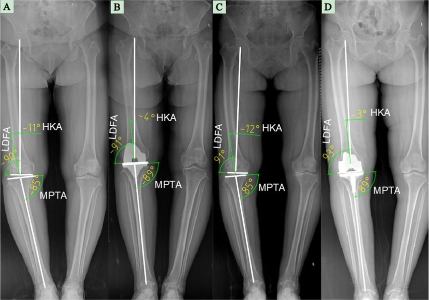
Figure 2. Radiographic measurements of HKA, LDFA and MPTA. Schematic diagrams of preoperative (A) and
postoperative (B) standing radiograph for VMA groups, and preoperative (C) and postoperative (D) standing
radiograph for NMA group.
LDFA. The LDFA is the lateral angle that formed by the mechanical axis of the femur and the knee joint line
of the femur in the coronal plane.
MPTA. The MPTA is the medial angle which formed by the mechanical axis of the tibia and the knee joint
line of the tibia in the coronal plane.
HKA. The HKA angle was defined as the included angle between the femoral mechanical axis and the tibial
mechanical axis22,23 (Fig. 2). This acute angle was expressed as a deviation from 0° with a negative value for varus
knee and positive value for valgus knee.
Clinical evaluation. Pre-operative and post-operative knee functional scores were calculated using the HSS
s cale24. The patients’ baseline characteristics included age, BMI and follow-up time. The maximum range of
motion of the knee was measured by a goniometer at 12 months after surgery.
Statistical analysis. Comparative statistical analyses between VMA group and NMA group after TKA
were performed in SPSS software 22.0 (SPSS, Chicago, IL) with the independent sample t test, a p value < 0.05
was adopted. These measured parameters were summarized by the mean and standard deviation.
Results
General observations. There was no significant difference in baseline characteristics regarding age, BMI
and Follow-up time, which are shown in Table 1. For all patients, there are significant differences between pre-
operative and post-operative HSS score, MPTA, LDFA, HKA angles, respectively. (Fig. 3A,B). In general, the
HSS score were improved from 48.23 ± 10.34 points before surgery to 86.71 ± 4.49 points at the final follow-up
examination (p < 0.05).
Pre‑operative and post‑operative comparison between two groups. Post-operative LDFA in
NMA group was larger than VMA group (90.87° ± 2.13° vs 94.06° ± 2.60°), there was a significant statistical
difference between the two groups (p = 0.001). Meanwhile, Post-operative MPTA in NMA group was larger
than VMA group (89.81° ± 1.54° vs 88.78° ± 1.79°), there was a significant difference between the two groups
(p = 0.038). For NMA group, the Mean and standard deviation of HKA was 1.80° ± 1.03°, for the VMA group,
the Mean and standard deviation (11.53° ± 4.15°) were more than three and two times larger than those of NMA
group. A significant difference was detected between the two groups (p < 0.05). No significant differences were
Scientific Reports | (2021) 11:3130 | https://doi.org/10.1038/s41598-021-81285-6 3
Vol.:(0123456789)www.nature.com/scientificreports/
Group NMA Group (n = 30) VMA Group(n = 21) p-value
Age 62.67 ± 5.72 64.13 ± 5.38 0.591
BMI 28.13 ± 3.19 27.42 ± 2.95 0.297
Pre-operative ROM 78.10 ± 4.02 78.83 ± 4.09 0.627
Pre-operative HSS 49.62 ± 10.07 47.27 ± 10.59 0.539
Table 1. Pre-operative baseline characteristics for all patients, grouped according to post-operative
mechanical alignment. The values are given as the mean and the standard deviation. NMA group neutral
mechanical axis, VMA group varus mechanical alignment, ROM range of motion, BMI body mass index, HSS
hospital for special surgery.
Figure 3. Statistic differences between preoperative and post-operative HSS score, MPTA, LDFA, HKA angles,
respectively.
NMA Group
VMA Group
100
80
60
Score
40
20
0
SS
H
Figure 4. The difference of HSS score between the two groups after TKA.
observed in HSS score between NMA group and VMA group (87.29 ± 3.41 vs 86.30 ± 5.13) after operation
(p < 0.05) (Fig. 4).
Discussion
Previous studies14,15 showed inconsistent results, a retrospective study was conducted to better identify the
association between mechanical alignment and post-operative HSS score after primary TKA in this study. The
most important finding of the current study was that we found no significant association between post-operative
lower limb mechanical alignment and HSS score for patients in Chinese with pre-operative constitutional varus
knee caused by combined deformities. .
Experimental results on the association between mechanical alignment and HSS score in this research is
consistent with previous studies 15,22,25,26. Michael et al22 proved that leaving varus knees in residual varus will
not improve clinical outcomes in a retrospective review of 361 primary TKAs. Dominique et al. 27 also found
Scientific Reports | (2021) 11:3130 | https://doi.org/10.1038/s41598-021-81285-6 4
Vol:.(1234567890)www.nature.com/scientificreports/
there were similar clinical outcomes for neutral or varus alignment. Richard et al.26 concluded that there was
no correlation between post-operative lower limb mechanical alignment and clinical outcomes for patients
with pre-operative varus deformity. However, the previous research objects were Europeans or Americans who
previously had varus knees. Here, the patients in Chinese with pre-operative constitutional varus knee caused
by combined deformities were selected, and a new evidence was provided in this study.
For these OA patients with constitutional varus knee, there are many intraoperative difficulties and post-
operative complications if applying neutral mechanical axis method. Firstly, it widely needs to release medial
soft tissue, including socket stripping insertion of medial soft tissue of the t ibia17, stripping popliteal ligament,
superficial medial collateral ligament and so o n18. It is widely known that excessive release of the soft tissue may
result in bleeding, knee instability. Secondly, by applying reduction osteotomy of tibial plateau, Dixon et al.19
corrected severe varus knee deformity and improved patients’ Knee Society Clinical Rating System (KSS) score
and activity level. However, along with the narrowing of the tibial plateau, the contact area of the tibial plateau
is decreased, meanwhile the wear of artificial joints will be increased under the same loading circumstance.
Moreover, tibial prosthesis cannot cover the lateral tibial margin after reduction osteotomy, which will increase
the sinking risk of the tibial component, because spongy bone of lateral-medial tibial plateau resulted in a rela-
tive ingression to adapt tracking of femoral component. When there exists residual varus deformity during the
operation, the above complications can be reduced. In addition, obtaining neutral mechanical axis among these
patients is more difficult and time-consuming procedure, which requires more complex bone cuts and larger
soft tissue releases. Sampath et al.28 also described the need for increased operative time to obtain neutral axis
of the lower limb in severe varus knees. In this study, slight varus mechanical alignment of the lower limb after
TKA can reduce the difficulty and save operative time for surgeon.
This study has several limitations. Firstly, this study was a retrospective. Second, in the case of proximal
tibia and distal femur bone on bone, the parameters of normal knee joint was measured. However, according
to Dargel’s r esult29, there was a positive correlation in morphometric data between two knees for one subject.
At last, static lower limb mechanical axis was not associated with clinical results while dynamic axis may be a
predictor of clinical performances after TKA, because Miller et al.30 found the standing mechanical axis after
TKA was not enough to predict dynamic behavior of the lower limb during gait. Thus, a prospective study with
large samples is required in the future.
Conclusion
For patients suffering from constitutional varus knee that caused by combined deformities among Chinese
populations, we found no significant association between post-operative lower limb mechanical alignment and
HSS score in our study. Slight varus mechanical alignment of the lower limb after TKA can reduce the difficulty
and save operative time for surgeon.
Received: 9 March 2020; Accepted: 31 December 2020
References
1. Insall, J., Scott, W. N. & Ranawat, C. S. The total condylar knee prosthesis. A report of two hundred and twenty cases. J. Bone Jt.
Surg. 61, 173–180 (1979).
2. Jeffery, R. S., Morris, R. W. & Denham, R. A. Coronal alignment after total knee replacement. J. Bone Jt. Surg. 73, 709–714 (1991).
3. Abdel, M. P. et al. Coronal alignment in total knee replacement: historical review, contemporary analysis, and future direction.
Bone Jt. J. 96-B, 857 (2014).
4. Thienpont, E., Schwab, P. E. & Fennema, P. J. B. J. J. A systematic review and meta-analysis of patient-specific instrumentation for
improving alignment of the components in total knee replacement. Bone Jt. J. 410, 1052–1061 (2014).
5. Insall, J. J. C. O. & Research, R. Rationale of the Knee Society clinical rating system. 248 (1989).
6. Davies, A. P. J. K. Rating systems for total knee replacement. Orthopedics 9, 261–266 (2002).
7. Bach, C. M. et al. Scoring systems in total knee arthroplasty. Bone Jt. J. 399, 184 (2002).
8. Walcott, B. P., Hanak, B. W., Caracci, J. R., Redjal, N. & Coumans, J. V. C. E. J. S. N. I. Trends in inpatient setting laminectomy for
excision of herniated intervertebral disc: Population-based estimates from the US nationwide inpatient sample. Surg. Neurol. Int.
2, 7 (2011).
9. Bourne, R. B., Chesworth, B. M., Davis, A. M., Mahomed, N. N. & Charron, K. D. J. Patient satisfaction after total knee arthroplasty:
Who is satisfied and who is not? 468, 57–63.
10. Robertsson, O., Dunbar, M., Pehrsson, T. r., Knutson, K. & Lidgren, L. J. A. O. Patient satisfaction after knee arthroplasty: A report
on 27,372 knees operated on between 1981 and 1995 in Sweden. 71, 262–267.
11. Scott, C. E. H., Howie, C. R., MacDonald, D., Biant, L. C. J. J. o. B. & Volume, J. S. B. Predicting dissatisfaction following total knee
replacement: A prospective study of 1217 patients. 92-B, 1253–1258.
12. Bellemans, J., Colyn, W., Vandenneucker, H. & Victor, J. The Chitranjan Ranawat award: Is neutral mechanical alignment normal
for all patients? The concept of constitutional varus. Clin. Orthop. Relat. Res. 470, 45–53. https: //doi.org/10.1007/s11999 -011-1936-5
(2012).
13. Vandekerckhove, P. et al. The relationship between constitutional alignment and varus osteoarthritis of the knee. Knee Surg. Sports
Traumatol. Arthrosc. 25, 2873–2879. https://doi.org/10.1007/s00167-016-3994-4 (2017).
14. Vanlommel, L., Vanlommel, J., Claes, S. & Bellemans, J. Slight undercorrection following total knee arthroplasty results in superior
clinical outcomes in varus knees. Knee Surg. Sports Traumatol. Arthrosc. 21, 2325–2330. https: //doi.org/10.1007/s00167 -013-2481-4
(2013).
15. Rames, R. D., Mathison, M., Meyer, Z., Barrack, R. L. & Nam, D. No impact of under-correction and joint line obliquity on clini-
cal outcomes of total knee arthroplasty for the varus knee. Knee Surg. Sports Traumatol. Arthrosc. 26, 1506–1514. https://doi.
org/10.1007/s00167-017-4507-9 (2018).
16. Nishida, K. et al. Remaining mild varus limb alignment leads to better clinical outcome in total knee arthroplasty for varus osteo-
arthritis. (2016).
17. Thienpont, E., Cornu, O., Bellemans, J. & Victor, J. Current opinions about coronal plane alignment in total knee arthroplasty: A
survey article. Acta Orthop. Belg. 81, 471–477 (2015).
Scientific Reports | (2021) 11:3130 | https://doi.org/10.1038/s41598-021-81285-6 5
Vol.:(0123456789)www.nature.com/scientificreports/
18. Morgan, S. S. et al. The influence of postoperative coronal alignment on revision surgery in total knee arthroplasty. Int. Orthop 32,
639–642. https://doi.org/10.1007/s00264-007-0391-0 (2008).
19. Shelton, T. J., Nedopil, A. J., Howell, S. M. & Hull, M. L. Do varus or valgus outliers have higher forces in the medial or lateral
compartments than those which are in-range after a kinematically aligned total knee arthroplasty? Limb and joint line alignment
after kinematically aligned total knee arthroplasty. Bone Jt. J. 99, 1319–1328. https://doi.org/10.1302/0301-620x.99b10.Bjj-2017-
0066.R1 (2017).
20. Dror Paley, F. Principles of Deformity Correction (Springer, Berlin, 2002).
21. Derek, T., Cooke, V., Sled, E. A. & Scudamore, R. A. Frontal plane knee alignment: A call for standardized measurement. J. Rheu-
matol. 34, 1796–1801 (2007).
22. Meneghini, R. M., Grant, T. W., Ishmael, M. K. & Ziemba-Davis, M. Leaving residual varus alignment after total knee arthroplasty
does not improve patient outcomes. J. Arthroplasty 32, S171–S176. https://doi.org/10.1016/j.arth.2017.02.064 (2017).
23. Nishida, K. et al. Remaining mild varus limb alignment leads to better clinical outcome in total knee arthroplasty for varus osteo-
arthritis. Knee Surg. Sports Traumatol. Arthrosc. 25, 3488–3494. https://doi.org/10.1007/s00167-016-4260-5 (2017).
24. Slupik, A. & Bialoszewski, D. Comparative analysis of clinical usefulness of the Staffelstein Score and the Hospital for Special Sur-
gery Knee Score (HSS) for evaluation of early results of total knee arthroplasties. Preliminary report. Ortop. Traumatol. Rehabil.
9, 627–635 (2007).
25. Stucinskas, J. et al. Moderate varus/valgus malalignment after total knee arthroplasty has little effect on knee function or muscle
strength. Acta Orthop. 86, 728–733 (2015).
26. Rames, R. D., Mathison, M., Meyer, Z., Barrack, R. L. & Nam, D. No impact of under-correction and joint line obliquity on clini-
cal outcomes of total knee arthroplasty for the varus knee. Knee Surg. Sports Traumatol. Arthrosc. https://doi.org/10.1007/s0016
7-017-4507-9 (2017).
27. Saragaglia, D. et al. Influence of the post-operative axis on the clinical results of total knee replacement for severe varus deformities:
Does a slight residual varus improve the results?. Int. Orthop. https://doi.org/10.1007/s00264-018-4092-7 (2018).
28. Sampath, S. A., Voon, S. H., Sangster, M. & Davies, H. The statistical relationship between varus deformity, surgeon’s expe-
rience, BMI and tourniquet time for computer assisted total knee replacements. Knee 16, 121–124. https://doi.org/10.1016/j.
knee.2008.09.008 (2009).
29. Dargel, J., Feiser, J., Gotter, M., Pennig, D. & Koebke, J. Side differences in the anatomy of human knee joints. Knee Surg. Sports
Traumatol. Arthrosc. 17, 1368–1376. https://doi.org/10.1007/s00167-009-0870-5 (2009).
30. Miller, E. J., Pagnano, M. W. & Kaufman, K. R. Tibiofemoral alignment in posterior stabilized total knee arthroplasty: Static align-
ment does not predict dynamic tibial plateau loading. J. Orthop. Res. 32, 1068–1074 (2014).
Author contributions
Z.Z. and W.C. contributed equally, and they are co-first author. Z.J. and Y.Q. are co-corresponding author. W.C.,
Y.Q. and Z.J. provided the ideas of this study. Z.Z. collected the data and wrote the manuscript. Y.Z., X.W. and
Z.C. analyzed the patientdata. G.Z., Q.Z. helped with the interpretation of data. P.W. made some contributions
to the revision.
Competing interests
The authors declare no competing interests.
Additional information
Correspondence and requests for materials should be addressed to Z.J. or Y.Q.
Reprints and permissions information is available at www.nature.com/reprints.
Publisher’s note Springer Nature remains neutral with regard to jurisdictional claims in published maps and
institutional affiliations.
Open Access This article is licensed under a Creative Commons Attribution 4.0 International
License, which permits use, sharing, adaptation, distribution and reproduction in any medium or
format, as long as you give appropriate credit to the original author(s) and the source, provide a link to the
Creative Commons licence, and indicate if changes were made. The images or other third party material in this
article are included in the article’s Creative Commons licence, unless indicated otherwise in a credit line to the
material. If material is not included in the article’s Creative Commons licence and your intended use is not
permitted by statutory regulation or exceeds the permitted use, you will need to obtain permission directly from
the copyright holder. To view a copy of this licence, visit http://creativecommons.org/licenses/by/4.0/.
© The Author(s) 2021
Scientific Reports | (2021) 11:3130 | https://doi.org/10.1038/s41598-021-81285-6 6
Vol:.(1234567890)You can also read