Can Clinical Presentation and Ultrasonography Predict the Risk of Malignancy in Thyroid Nodules?
←
→
Page content transcription
If your browser does not render page correctly, please read the page content below
Original Article
Middle East Journal of Cancer; July 2019; 10(3): 246-253
Can Clinical Presentation and
Ultrasonography Predict the Risk of
Malignancy in Thyroid Nodules?
Zahra Davoudi*, Arezoo Chouhdari**,***,
Hooman Bahrami-Motlagh****♦, Karim Bagheri*
*Endocrinology Department, Loghman Hakim Hospital, Shahid Beheshti University of
Medical Sciences, Tehran, Iran
**Skull Base Research Center, Loghman Hakim Medical Center, Shahid Beheshti
University of Medical Sciences, Tehran, Iran
***Clinical Research Development Center, Loghman Hakim Hospital, Shahid Beheshti
University of Medical Sciences, Tehran, Iran
****Radiology Department, Loghman Hakim Hospital, Shahid Beheshti University of
Medical Sciences, Tehran, Iran
Abstract
Background: Thyroid nodules are frequent occurrences. This study aims to evaluate
the risk of malignancy based on the 2015 American Thyroid Association Management:
Guidelines for Adult Patients with Thyroid Nodules and Differentiated Thyroid Cancer.
Methods: In this cross-sectional study, ultrasonography and clinical manifestations
were compared with pathology findings to predict the risk of malignancy in thyroid nodules.
Assessment of the ultrasound findings was based on the criteria recommended by The
2015 American Thyroid Association Management Guidelines for Thyroid Nodules. For
the evaluation of the association between clinical and ultrasound findings with
histopathology results, we used the chi-square and Fisher’s exact tests. The relative risk
and prediction of malignancy was assessed by multiple logistic regression analysis.
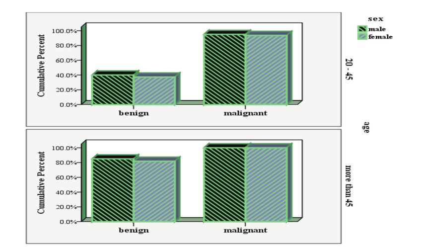
Data analysis was performed by the SPSS19. The significance level was set at PPrediction of Malignancy Risk in Thyroid Nodules Introduction 54.3%-74.2% for intranodular vascularity, and Thyroid nodules are a common medical 26.1%-59.1% for microcalcifications. Specificity problem in the general population with a ranges were 43.4%-94.3% for hypoechogenicity, prevalence of 10%-70%. Women are more 78.6%-80.8% for intranodular vascularity, and frequently diagnosed, in addition to individuals 85.5%-95% for microcalcifications.6,7 Recently, older than 40 years of age and those with a positive determination of tissue elasticity by ultrasound family history of thyroid diseases.1-4 Thyroid (elastography) has been proposed to detect cancer is found in 5%–15% of cases and depends malignancy in thyroid nodules.8 Considering the on sex, age, and exposure to other risk factors. The uncertainty of clinical findings and ultrasound in increased use of thyroid, head and neck ultrasound benign and malignant differentiation in detected has led to increased diagnosis of asymptomatic thyroid nodules, the aim in this study is to thyroid nodules and faster detection of thyroid investigate the relationship between clinical cancer in adults.5 Age, positive family history, manifestation and ultrasound characteristics with previous head and neck X-ray, and ultrasound pathologic confirmation of malignancy. parameters such as microcalcifications, absence of a halo, increased intranodal vascularity, hypoe- Materials and Methods chogenicity, tall shape of a nodule or irregular In this cross-sectional study, 130 cases of margins are traditionally associated with increased thyroid nodules larger than 1 cm were selected by risk of malignancy in thyroid nodules. However, convenient sampling method from patients who none of these parameters is sufficient and reliable presented to the Endocrinology Ward at Loghman to detect malignancy in these cases. Only Hakim Hospital, a tertiary university medical aspiration and pathology results are considered center in Tehran, Iran in 2017. Patients were definitive criteria for diagnosis by the physician. referred by endocrinologists, otolaryngologists, or Some studies have reported diagnostic sensitivity surgeons in the same hospital due to clinical ranges from 26.5%-87.1% for hypoechogenicity, suspicion of thyroid nodules. We used a data Figure 1. Type of thyroid nodules (pathology finding) based on age and sex. Middle East J Cancer 2019; 10(3): 246-253 247
Zahra Davoudi et al.

Table 1. The risk for malignancy according to the ultrasound criteria in the 2015 American Thyroid Association Management Guidelines.
Level of             Shape              Echogenic                         Margin                 Composition         Echogenicity
suspicion                                   foci
High suspicion       Taller than wide   Micro calcifications              Irregular, lobulated   Solid or            Hypoechoic
                                        rim calcifications                Extra thyroidal        partially cystic
                                                                          extension

Intermediate           Not taller      Without micro                 Smooth                    Solid                  Hypoechoic
suspicion              than wide       calcifications

Low suspicion          Not taller      Without micro                 Smooth                    Solid or partially    Iso/hyperechoic
                       than wide       calcifications                                          cystic with
                                                                                               eccentric solid areas
Very low               Not taller      Without micro                 Smooth
suspicion              than wide       calcifications

Benign                 Not taller      Without micro                 Smooth                    Spongiform or          Iso/hyperechoic
                       than wide       calcifications                                          Partially Cystic       Anechoic
                                                                                               Purely cystic

collection form to gather patients’ demographic                      Fisher’s exact tests were used to evaluate the
information such as age, sex, family history,                        association between variables. Finally, multivariate
previous thyroid diseases, clinical manifestations                   logistic regression was used to predict the possible
(pain, dysphonia, and other findings), and                           factors for malignancy in the thyroid nodules.
laboratory analyses (Thyroid Stimulating                             We used the Statistical Package for the Social
Hormone (TSH), Anti-thyroid peroxidase                               Sciences, version 19 for total analysis. P
Prediction of Malignancy Risk in Thyroid Nodules
Table 2. Association between demographic, clinical manifestation, ultrasound, and laboratory tests with pathology results of patients
with thyroid nodules (univariate analysis).
Variables Pathology P-value Variables Pathology P-value
Benign Malignant Benign Malignant
Number (%) Number (%)
Age (years) 0.09 Echogenicity 45 58 (54.2) 8 (34.8) Hyper/iso/mixed 86 (80.4) 0 (0)
Sex 0.7 CalcificationZahra Davoudi et al. Table 3. Prediction of malignancy in patients with thyroid nodules (multiple logistic regression). Variables OR (95% CI) P-value Size 12.1 (3.2-18) 0.04 >4 £4 (reference) Halo 28 (16.3-36)
Prediction of Malignancy Risk in Thyroid Nodules

of the patients had a history of radiotherapy. In       lowest OR in positive findings was related to
other studies, clinical manifestations suggestive of    internal vascularity, which supported the results
malignant nodules included age below 20 and             of a study by Moon HJ et al.31 In the current
over 60 years, male gender, radiotherapy history,       study, there was a significant association between
rapid growth of the nodule, changes in speaking         pathology findings and level of suspicion
and swallowing, as well as a family history of          according to sonography results. Hence, 95.7% of
thyroid cancer and related syndromes.17 We did          patients who had features of malignancy according
not observe any difference between gender and           to their pathology results had intermediate or
malignancy in the thyroid nodules as in the recent      high-level suspicions of cancer. Hypoechoic
study by Hegedüs et al.18 None of the cases were        appearance, microcalcifications, irregular margins,
less than 20 years of age. A comparison of patients     vascularity, absence of a halo, taller than wide,
less or equal to 45 years of age with those older       positive lymph nodes, and solid composition
than 45 years showed no significant difference in       according to sonography results were significant
the number of benign or malignant nodules, as in        for thyroid nodules associated with malignancy
a study by Abu-Ghanem et al. in Israel.19 which         (Univariate analysis). In addition to assessing the
is similar to our survey, evaluated the relationship    aggregated ultrasound features, our study predicted
between clinical manifestations and sonography          malignancy. We found that absence of halo and
for FNA in terms of malignant risk. They pointed        taller than wide had the most pronounced effect
clinical presentation and sonography findings           of any of the predictors examined in the course of
could not predict malignant or benign nodules.19        this study. The second solitary ultrasound predictor
In this study, size greater than 4 cm had a             of malignancy was irregular margins and solid or
significant association with thyroid malignancy,        predominantly solid composition, which supported
which was similar to other studies in this area.20,21   much of the literature.29-32 The absence of halo
Recently, Papini et al. conducted a study in Italy      facing has been considered in the older literature;
and reported that no significant relationship existed   whereas, taller than wide manifestation is known
between larger non-palpable nodules and increased       as a high-risk nodule pattern in the new ATA
risk of thyroid malignancy23 similar to another         criteria.33,34 In this study, different features of
study. 24 In some studies, higher TSH levels            echogenic foci, the radiologist's opinion, and
increased thyroid cancer risk; whereas, there was       quality of the ultrasound device might indicate a
not a significant association between malignant         lower OR for microcalcifications. Therefore, it is
nodules and TSH.25,26 There was a significant           better to exclude patients with inadequate imaging
association between negative anti-TPO and thyroid       from the study. The small sample size is a major
nodule malignancy; 91.3% malignant nodules              limitation of our analysis because of the low
were negative for anti-TPO. This indicated an           numbers of malignant cases.
association of benign thyroid disease with positive
anti-TPO levels. In the current study, all patients     Conclusion
with thyroid nodules greater than 1 cm underwent            Based on the results of this study, there is an
FNA. However, according to the ATA 2015                 increasing incidence of thyroid cancer in thyroid
guidelines, FNA should be performed on patients         nodules. Despite the small sample size, we have
who have intermediate and high-risk thyroid             found a significant association between clinical
nodules that are 1 cm and more in size. 27              and ultrasound findings with FNA of malignant
According to ultrasound results in other studies,       nodules, especially with the absence of a halo,
the highest specificity for thyroid cancer in the       taller than wide, the appearance and severity of the
nodules is the presence of microcalcifications,         solid composition, and irregular margins. We
hypoechoic appearances in the parenchyma, and           recommend more studies to be conducted
                                                        according to ATA2015 criteria and enroll larger
irregular margins.28-30 In the current study, the

Middle East J Cancer 2019; 10(3): 246-253                                                                            251
Zahra Davoudi et al.
sample sizes. These studies should perform FNA 10.1210/jc.2010-0901.
of the thyroid nodules and genetic studies on 8. Gharib H, Papini E, Garber JR, Duick DS, Harrell
RM, Hegedüs L, et al. American Association of Clinical
malignant nodules, especially in cases that have Endocrinologists, American College of Endocrinology,
positive family history, which could generalize and Associazione Medici Endocrinologi Medical
results of this study to the 2015 American Thyroid Guidelines for clinical practice for the diagnosis and
Association Management Guideline. management of thyroid nodules--2016 Update. Endocr
Pract. 2016;22(5):622-39. doi: 10.4158/EP161208.GL.
9. Haugen BR, Sawka AM, Alexander EK, Bible KC,
Acknowledgment Caturegli P, Doherty GM, et al. American Thyroid
The authors would like to thank the Clinical Association guidelines on the management of thyroid
Research Development Center (CRDC) of nodules and differentiated thyroid cancer task force
Loghman Hakim Hospital, Shahid Beheshti review and recommendation on the proposed renaming
University of Medical Sciences, Tehran, Iran for of Encapsulated Follicular Variant Papillary Thyroid
Carcinoma without invasion to Noninvasive Follicular
their support, cooperation, and assistance Thyroid Neoplasm with Papillary-Like Nuclear
throughout this study. Features. Thyroid. 2017;27(4):481-3. doi:
10.1089/thy.2016.0628.
Conflict of Interest 10. Castro MR, Gharib H. Thyroid fine-needle aspiration
biopsy: progress, practice, and pitfalls. Endocr Pract.
None declared. 2003;9(2):128-36.
11. Ferlay J, Steliarova-Foucher E, Lortet-Tieulent J,
References Rosso S, Coebergh JW, Comber H, et al. Cancer
1. Remonti LR, Kramer CK, Leitão CB, Pinto LC, Gross incidence and mortality patterns in Europe: estimates
JL. Thyroid ultrasound features and risk of carcinoma: for 40 countries in 2012. Eur J Cancer.
a systematic review and meta-analysis of observational 2013;49(6):1374-403. doi: 10.1016/j.ejca.2012.12.027.
studies. Thyroid. 2015;25(5):538-50. doi: 10.1089/thy. 12. Pellegriti G, Frasca F, Regalbuto C, Squatrito S, Vigneri
2014.0353. R. Worldwide increasing incidence of thyroid cancer:
2. Rezai-Delui H, Davachi B, Rahroh M. Incidence of update on epidemiology and risk factors. J Cancer
ultrasonographically-detected thyroid nodules in Epidemiol. 2013;2013:965212. doi: 10.1155/2013
persons between 10-70 years with no previous thyroid /965212.
disease. Iran J Otorhinolaryngol. 2004;16(3):7-13. 13. Davies L. How understanding thyroid cancer in
3. Wong KT, Ahuja AT. Ultrasound of thyroid cancer. Belgium can help us mitigate the problem of increasing
Cancer Imaging. 2005;5:157-66. incidence. J Clin Endocrinol Metab. 2013;98(10):3977-
4. Siadati S, Moazezi Z, Bayani MA, Mirzapour A, 9. doi: 10.1210/jc.2013-3505.
Nikbakhsh N, Ghaemian N, et al. The diagnostic value 14. Pathak KA, Leslie WD, Klonisch TC, Nason RW. The
of fine needle aspiration as compared to pathology changing face of thyroid cancer in a population-based
results in diagnosis of thyroid nodules: A 22-year cohort. Cancer Med. 2013;2(4):537-44. doi:
follow-up study. J Babol Univ Med Sci. 2015;17(9):39- 10.1002/cam4.103.
43. 15. Yassa L, Cibas ES, Benson CB, Frates MC, Doubilet
5. Smith-Bindman R, Lebda P, Feldstein VA, Sellami PM, Gawande AA, et al. Long-term assessment of a
D, Goldstein RB, Brasic N, et al. Risk of thyroid multidisciplinary approach to thyroid nodule diagnostic
cancer based on thyroid ultrasound imaging character- evaluation. Cancer. 2007;111(6):508-16.
istics: results of a population-based study. JAMA Intern 16. Wang CC, Friedman L, Kennedy GC, Wang H,
Med. 2013;173(19):1788-96. doi: 10.1001/ Kebebew E, Steward DL, et al. A large multicenter
jamainternmed.2013.9245. correlation study of thyroid nodule cytopathology and
6. Remonti LR, Kramer CK, Leitão CB, Pinto LC, Gross histopathology. Thyroid. 2011;21(3):243-51. doi:
JL. Thyroid ultrasound features and risk of carcinoma: 10.1089/thy.2010.0243.
a systematic review and meta-analysis of observational 17. Davies L, Welch HG. Current thyroid cancer trends in
studies. Thyroid. 2015;25(5):538-50. doi: the United States. JAMA Otolaryngol Head Neck Surg.
10.1089/thy.2014.0353. 2014;140(4):317-22. doi: 10.1001/ jamaoto.2014.1.
7. Rago T, Scutari M, Santini F, Loiacono V, Piaggi P, Di 18. Hegedüs L, Bonnema SJ, Bennedbaek FN.
Coscio G, et al. Real-time elastosonography: useful tool Management of simple nodular goiter: current status
for refining the presurgical diagnosis in thyroid nodules and future perspectives. Endocr Rev. 2003;24(1):102-
with indeterminate or nondiagnostic cytology. J Clin 32.
Endocrinol Metab. 2010;95(12):5274-80. doi: 19. Abu-Ghanem S, Cohen O, Lazutkin A, Abu-Ghanem
252 Middle East J Cancer 2019; 10(3): 246-253Prediction of Malignancy Risk in Thyroid Nodules
Y, Fliss DM, Yehuda M. Evaluation of clinical for US features of nodules: a step in establishing better
presentation and referral indications for ultrasound- stratification of cancer risk. Radiology.
guided fine-needle aspiration biopsy of the thyroid as 2011;260(3):892-9. doi: 10.1148/radiol.11110206.
possible predictors of thyroid cancer. Head Neck. 30. Moon WJ, Jung SL, Lee JH, Na DG, Baek JH, Lee YH,
2016;38 Suppl 1:E991-5. doi: 10.1002/hed.24143. et al. Benign and malignant thyroid nodules: US dif-
20. Espinosa De Ycaza AE, Lowe KM, Dean DS, Castro ferentiation--multicenter retrospective study. Radiology.
MR, Fatourechi V, Ryder M, et al. Risk of malignancy 2008;247(3):762-70. doi: 10.1148/radiol.2473070944.
in thyroid nodules with non-diagnostic fine-needle 31. Moon HJ, Kwak JY, Kim MJ, Son EJ, Kim EK. Can
aspiration: a retrospective cohort study. Thyroid. vascularity at power Doppler US help predict thyroid
2016;26(11):1598-604. malignancy? Radiology. 2010;255(1):260-9. doi:
21. McCoy KL, Jabbour N, Ogilvie JB, Ohori NP, Carty 10.1148/radiol.09091284.
SE, Yim JH. The incidence of cancer and rate of false- 32. Salmaslioğlu A, Erbil Y, Dural C, Işsever H, Kapran
negative cytology in thyroid nodules greater than or Y, Ozarmağan S, et al. Predictive value of sonographic
equal to 4 cm in size. Surgery. 2007;142(6):837-44; features in preoperative evaluation of malignant thyroid
discussion 844.e1-3. nodules in a multinodular goiter. World J Surg.
22. Campanella P, Ianni F, Rota CA, Corsello SM, 2008;32(9):1948-54. doi: 10.1007/s00268-008-9600-2.
Pontecorvi A. Quantification of cancer risk of each 33. Brito JP, Gionfriddo MR, Al Nofal A, Boehmer KR,
clinical and ultrasonographic suspicious feature of Leppin AL, Reading C, et al. The accuracy of thyroid
thyroid nodules: a systematic review and meta-analysis. nodule ultrasound to predict thyroid cancer: systematic
Eur J Endocrinol. 2014;170(5):R203-11. doi: review and meta-analysis. J Clin Endocrinol Metab.
10.1530/EJE-13-0995. 2014;99(4):1253-63. doi: 10.1210/jc.2013-2928.
23. Papini E, Guglielmi R, Bianchini A, Crescenzi A, 34. Reading CC, Charboneau JW, Hay ID, Sebo TJ.
Taccogna S, Nardi F, et al. Risk of malignancy in Sonography of thyroid nodules: a "classic pattern"
nonpalpable thyroid nodules: predictive value of diagnostic approach. Ultrasound Q. 2005;21(3):157-
ultrasound and color-Doppler features. J Clin 65.
Endocrinol Metab. 2002;87(5):1941-6.
24. Alexander EK, Hurwitz S, Heering JP, Benson CB,
Frates MC, Doubilet PM, et al. Natural history of
benign solid and cystic thyroid nodules. Ann Intern
Med. 2003;138(4):315-8.
25. Frates MC, Benson CB, Doubilet PM, Kunreuther E,
Contreras M, Cibas ES, et al. Prevalence and
distribution of carcinoma in patients with solitary and
multiple thyroid nodules on sonography. J Clin
Endocrinol Metab. 2006;91(9):3411-7.
26. Boelaert K, Horacek J, Holder RL, Watkinson JC,
Sheppard MC, Franklyn JA. Serum thyrotropin
concentration as a novel predictor of malignancy in
thyroid nodules investigated by fine-needle aspiration.
J Clin Endocrinol Metab. 2006;91(11):4295-301.
27. Haymart MR, Repplinger DJ, Leverson GE, Elson
DF, Sippel RS, Jaume JC, et al. Higher serum thyroid
stimulating hormone level in thyroid nodule patients
is associated with greater risks of differentiated thyroid
cancer and advanced tumor stage. J Clin Endocrinol
Metab. 2008;93(3):809-14.
28. Haugen BR, Alexander EK, Bible KC, Doherty GM,
Mandel SJ, Nikiforov YE, et al. 2015 American
Thyroid Association Management guidelines for adult
patients with thyroid nodules and differentiated thyroid
cancer: The American Thyroid Association guidelines
task force on thyroid nodules and differentiated thyroid
cancer. Thyroid. 2016;26(1):1-133. doi: 10.1089/thy.
2015.0020.
29. Kwak JY, Han KH, Yoon JH, Moon HJ, Son EJ, Park
SH, et al. Thyroid imaging reporting and data system
Middle East J Cancer 2019; 10(3): 246-253 253You can also read