Of human breast cancer - Role of TG2 and TGF β1 in the pathogenesis
←
→
Page content transcription
If your browser does not render page correctly, please read the page content below
ONCOLOGY LETTERS 20: 221, 2020

                        Role of TG2 and TGF‑β1 in the pathogenesis
                                  of human breast cancer
   HAI‑CHENG GAO1, YAN‑ZHI HUANG2, YU‑QI LIU2, YAN CHEN1, ZHE‑HUI WANG3 and GUANG‑HAO YIN4

       1
        Department of Clinical Pharmacy, School of Pharmaceutical Sciences, Jilin University, Changchun, Jilin 130021;
                    2
                      Department of Respiratory Medicine, Children's Hospital, Changchun, Jilin 130000;
                       3
                         Department of Surgery, China‑Japan Union Hospital, Changchun, Jilin 130031;
           4
             Department of Breast Surgery, Second Hospital, Jilin University, Changchun, Jilin 130041, P.R. China

                                          Received December 11, 2019; Accepted June 15, 2020

                                                      DOI: 10.3892/ol.2020.12057

Abstract. The present study analyzed the role of transforming       Introduction
growth factor-β1 (TGF‑β1) and tissue transglutaminase (TG2)
in breast cancer, as well as their protein levels in MCF‑7 cells    Breast cancer is the most commonly occurring type of cancer
treated with cisplatin. In addition, the present study investi-     among women (1). The Global Health Organization reported
gated the effects of TG2 and TGF‑β1 in MCF‑7 cells following        that 508,000 women succumbed to breast cancer in 2011 (2).
TGF‑β1 and TG2 inhibition or TGF‑β1 induction. The protein          There are a number of factors involved in the occurrence
levels of TG2 and TGF‑ β1 in breast cancer tissues and in           and development of breast cancer, such as tissue transgluta-
MCF‑7 cells treated with cisplatin, TG2 and TGF‑β1 inhibitors       minase (TG2) and transforming growth factor‑ β (TGF‑ β)
or 10 ng/ml TGF‑β1 were analyzed by immunohistochemical             family members (3,4).
staining, immunofluorescence and western blotting. The results          TGF‑β1 is a member of the transforming growth factor
revealed that the expression levels of TG2 and TGF‑β1 in breast     superfamily that is widely involved in various pathophysi-
cancer tissues were significantly higher compared with those        ological processes, such as inflammation, trauma and organ
in paracancerous tissues. The fluorescence intensity of TG2         fibrosis (5). TG2 is an enzyme that is upregulated in epithelial
and TGF‑β1 in MCF‑7 cells treated with cisplatin was lower          malignancies and participates in Ca 2+ ‑dependent protein
compared with that in untreated MCF‑7 cells. Using bioinfor-        post‑translational modifications and cross‑linking via the
matics analysis, the present study predicted that TGF‑β1 may        acyl‑transfer reaction between glutamine and lysine resi-
be associated with TG2. In addition, the expression levels of       dues (6). It has been reported that TG2 serves an important
TGF‑β1 and TG2 in MCF‑7 cells treated with inhibitors of            role in the epithelial‑to‑mesenchymal transition (EMT) (6). In
TGF‑β1 and TG2 were lower compared with those in untreated          addition, upregulation of TGF‑β is associated with metastasis,
MCF‑7 cells. By contrast, the expression levels of TGF‑β1           cell invasiveness and EMT in ovarian cancer (7,8). However,
and TG2 in MCF‑7 cells treated with TGF‑β1 were higher              to the best of our knowledge, the synergistic role of TG2 and
compared with those in untreated MCF‑7 cells. Therefore, the        TGF‑ β1 in regulating the occurrence and development of
present study demonstrated that TGF‑β1 and TG2 may serve            breast cancer has been reported.
an important role in breast cancer tissues and in MCF‑7 cells.          Cisplatin is a broad‑spectrum anticancer drug that is
In addition, it was revealed that TG2 and TGF‑β1 may have a         commonly used in ovarian, prostate, testicular and lung
synergistic role in MCF‑7 cells.                                    cancer, nasopharyngeal carcinoma, esophageal cancer, malig-
                                                                    nant lymphoma, head and neck squamous cell carcinoma and
                                                                    thyroid cancer (9). However, whether the expression of TG2
                                                                    and TGF‑β1 is regulated by cisplatin remains to be elucidated.
                                                                        The present study aimed to analyze the role of TG2 and
Correspondence to: Dr Zhe‑Hui Wang, Department of Surgery,
China‑Japan Union Hospital, 126 Xiantai Street, Changchun,          TGF‑β1 in breast cancer. In addition, the present study aimed
Jilin 130031, P.R. China                                            to investigate the protein levels of TGF‑β1 and TG2 in MCF‑7
E‑mail: wangzhehui999@163.com                                       cells treated with cisplatin and the effect of TG2 and TGF‑β1
                                                                    in MCF‑7 cells treated with TGF‑β1 and TG2 inhibitors or
                                                                    TGF‑β1.
Professor Guang‑Hao Yin, Department of Breast Surgery,
Second Hospital, Jilin University, 218 Ziqiang Street, Changchun,
Jilin 130041, P.R. China.                                           Materials and methods
E‑mail: yinguanghao98@163.com
                                                                    Tissue samples. A total of 30 pairs of breast cancer and para-
Key words: breast cancer, tissue transaminase, transforming         cancerous tissue samples were obtained from the China‑Japan
growth factor‑β1, cisplatin, cell death                             Union Hospital (Changchun, China) between March 2018 and
                                                                    March 2019. The median age is 38 years (range, 28‑45 years).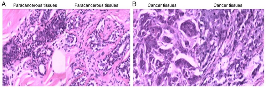
2 GAO et al: ROLES OF TG2 AND TGF-β1 IN BREAST CANCER
The study was approved by the Ethics Committee of the IgG antibody (cat. no. ZB‑2301; 1:2,000; Beijing Noble
China‑Japan Union Hospital. The samples were obtained with Technology Co., Ltd). TG2 and TGF‑β1 were detected using
signed informed consent from the patients or their family. ECL development solution (Pierce; Thermo Fisher Scientific,
Inc.). TG2 and TGF‑ β1 expression levels were determined
Cell culture. MCF‑7 cells were gifted from Jilin University using Quantity One v4.6.2 software (Bio‑Rad Laboratories,
School of Pharmacy. The cells were cultured in Dulbecco's Inc.).
modified Eagle's medium (DMEM) supplemented with
10% FBS, 100 U/ml penicillin G and 100 µg/ml streptomycin Immunofluorescence. The fluorescence intensity of TG2 and
in an incubator at 37˚C and 5% CO2. (all from Invitrogen; TGF‑β1 in MCF‑7 cells was assessed via immunofluorescence.
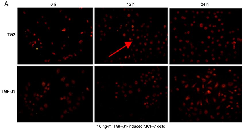
Thermo Fisher Scientific, Inc.). MCF‑7 cells (5x10⁴) were treated with cisplatin (1 mg/l), TGF‑β1
(150 µmol/l) and TG2 inhibitors (8.31 µmol/l), and incubated
Reagents. All materials for the SDS‑PAGE were purchased at 37˚C and 5% CO2 for 24 h. Rabbit monoclonal primary anti-
from Bio‑Rad Laboratories, Inc. The monoclonal antibody bodies against TG2 (1:300) and TGF‑β1 (1:300) were added to
against β‑actin (1,2000; cat. no. AAPR201‑100) was purchased the cells and incubated overnight at 4˚C. Following overnight
from Sigma‑Aldrich; Merck KGaA. Rabbit polyclonal anti- incubation with fluorescein‑conjugated IgG (cat. no. ZB‑2301;
bodies against TGF‑ β1 (1:2,000; cat. no. RAB‑0238) and Beijing Noble Technology Co., Ltd.) antibody at 4˚C. TG2
TG2 (1:2,000; cat. no. CTA‑DE056) were obtained from Cell and TGF‑β1 expression levels were determined using Quantity
Signaling Technology, Inc. The TG2 inhibitor (MDC) and the One v4.6.2 software (Bio‑Rad Laboratories, Inc.).
TGF‑β1 inhibitor (ITD) were purchased from Sigma‑Aldrich;
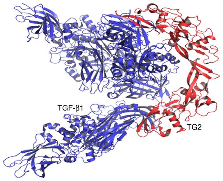
Merck KGaA. Molecular docking. The crystal structure of TG2 [in complex
with GTP; Protein Data Band (PDB) ID, 4PYG] and TGF‑β1
Hematoxylin and eosin (HE) staining. The sections (3 µm) (in complex with scFv GC1009; PDB ID, 4KV5) were
of breast cancer tissue were fixed in 4% paraformaldehyde obtained from the PDB (http://www.rcsb.org/pdb). The protein
followed by dehydration using a gradient ethanol series (80 files of TG2 and TGF‑β1 were prepared by removing water
and 95%). The section was subsequently stained with HE molecules and other ligands. Molecular docking studies and
at 25˚C (10 min). Images were captures on a fully automatic docking analysis were performed using the PatchDock server
photomicrography device (magnification x200; five field of (http://bioinfo3d.cs.tau.ac.il/PatchDock/). Analysis and visu-
views; Olympus PM‑10AO; Olympus Corporation). alization of interactions in the docked complexes obtained by
PatchDock server were analyzed by PyMOL (https://pymol.
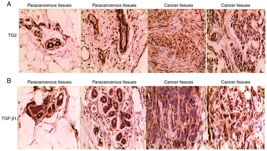
Immunohistochemistry Immunohistochemical staining was en.softonic.com/).
performed using a SABC kit (Biyuntian Biotechnology Co.,
Ltd.). 0.3% hydrogen peroxide formaldehyde solution was Statistical analysis. Quantitative data are presented as the
added to the paraffin sections and incubated at 37˚C. The mean ± standard deviation and were analyzed using SPSS 19.0
sections of breast cancer tissues and paracancerous tissues software (IBM Corp.). Student's t‑test was used to compare
were washed with PBS and incubated with 10% bovine serum two groups; one‑way ANOVA followed by Dunnett's test was
albumin (Thermo Fisher Scientific, Inc.) for 15 min. Rabbit used to compare all treatment groups against an untreated
anti‑human TGF‑β1 and TG2 polyclonal antibodies (1:300) control group. The χ2 test was used to analyze the associa-
were added and incubated at 4˚C for 12 h. The next day, color tions between protein expression and patient characteristics.
rendering was performed using 3,3'‑diaminobenzidine and P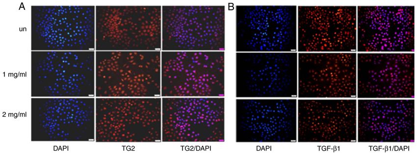
ONCOLOGY LETTERS 20: 221, 2020                                                                     3

  Figure 1. Hematoxylin and eosin staining of breast cancer and paracancerous tissues. (A) Paracancerous tissues. (B) Cancer tissues. Magnification x200.

Figure 2. Immunohistochemical staining of TG2 and TGF‑β1. (A and B) Expression of TG2 and TGF‑β1 in paracancerous and cancer tissues. (C) Expression
levels of TG2 and TGF‑β1 in paracancerous and breast cancer tissues. Magnification, x200. **P
4                                             GAO et al: ROLES OF TG2 AND TGF-β1 IN BREAST CANCER

Table I. Associations between patient clinicopathological characteristics and the expression of TG2 and TGF‑β1.
Characteristic                   TG2‑positive           TG2‑negative            P‑value          TGF‑β1‑positive            TGF‑β1‑negative             P‑value

Age, years, n
 35‑45                                   8                      6		                                       10                          5
 45‑55                                   9                      7		                                       10                          5
Stage, n
 II                                    30			                                                            30
Infiltration depth (%)             17 (56.7%) 13 (43.3%) 0.025                                      20 (66.7%)                  10 (33.3%)
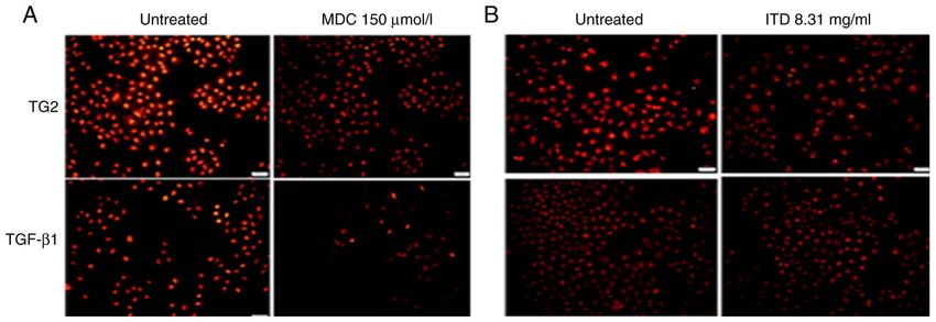
ONCOLOGY LETTERS 20: 221, 2020                                                               5

Figure 6. (A and B) Fluorescence of TG2 and TGF‑β1 in MCF‑7 cells treated with TG2 and TGF‑β1 inhibitors. (C and D) Fluorescence intensity of TG2 and
TGF‑β1 in MCF‑7 cells treated with TG2 and TGF‑β1 inhibitors. Magnification x200. *P
6                                            GAO et al: ROLES OF TG2 AND TGF-β1 IN BREAST CANCER

Table II. Binding scores of junction for TG2 with TGF‑β1.

Solution no.                         Score                        Area (Å)

 1                                  20,950                         3614.3
 2                                  20,368                         3214.5
 3                                  20,160                         3012.9
 4                                  19,952                         2918.5
 5                                  19,554                         3492.0
 6                                  19,460                         3311.2
 7                                  19,428                         2974.3
 8                                  19,288                         2760.4
 9                                  19,254                         2881.1
10                                  19,096                         2987.4

TGF‑β1, transforming growth factor-β1; TG2, tissue transglutaminase.

                                                                                Figure 9. (A) Protein expression of TG2 and TGF‑β1 in MCF‑7 cells treated
                                                                                with ITD, Pt and ITD+Pt. (B) Quantification of the expression levels of TG2
                                                                                and TGF‑β1 in untreated MCF‑7 and MCF‑7 cells treated with ITD, Pt and
                                                                                ITD+Pt. Magnification x200. *P
ONCOLOGY LETTERS 20: 221, 2020 7
fibrosis and patient mortality (16). The results of the present Patient consent for publication
study revealed that the expression levels of TG2 and TGF‑β1
in human breast cancer were higher compared with those in Not applicable.
paracancerous tissues. These results suggested that TGF‑ β1
and TG2 may serve an important role in the occurrence and Competing interests
development of breast cancer.
To further analyze the role of TGF‑β1 and TG2 in breast The authors declare that they have no competing interests.
cancer, the present study analyzed the expression levels
of TGF‑β1 and TG2 in MCF‑7 cells treated with cisplatin. References
Cisplatin is a selective drug used for the treatment of prostate 1. Zhu W, Harvey S, Macura KJ, Euhus DM and Aremov D:
cancer (17). A large proportion of patients develop resistance Invasive breast cancer preferably and predominantly occurs at
to cisplatin, inducing tumor relapse and limiting its clinical the interface between fibroglandular and adipose tissue. Clin
Breast Cancer 17: e11‑e18, 2017.
usefulness (17). In the present study, the results demonstrated 2. Alipour S, Jannat F and Hosseini L: Teaching breast cancer
that the fluorescence intensity of TG2 and TGF‑β1 in untreated screening via text messages as part of continuing education
MCF‑7 cells was higher compared with that in MCF‑7 cells for working nurses: A case‑control study. Asian Pac J Cancer
Prev15: 5607‑5609, 2014.
treated with cisplatin. These results suggested that cisplatin 3. Agnihotri N, Kumar S and Mehta K: Tissue transglutaminase as
may exert its chemotherapeutic effects in MCF‑7 cells by a central mediator in inflammation‑induced progression of breast
regulating the levels of TG2 and TGF‑β1 protein. cancer. Breast Cancer Res 15: 202, 2013.
4. Wang J, Xi C, Yang X, Lu X, Yu K, Zhang Y and Gao R: LncRNA
TG2 was associated with TGF‑β1 in the occurrence and WT1‑AS inhibits triple‑negative breast cancer cell migration
development of various diseases (8). To clarify the effect of and invasion by downregulating transforming growth factor-β1.
TG2 and TGF‑β1 in MCF‑7 cells, the present study investigated Cancer Biother Radiopharm 34: 671‑675, 2019.
5. Takai E, Tsukimoto M and Kojima S: TGF‑β1 downregulates
the effect of TG2 and TGF‑β1 in the MCF‑7 cells treated with COX‑2 expression leading to decrease of PGE2 production in
inhibitors of TGF‑β1 and TG2. The results demonstrated that human lung cancer A549 cells, which is involved in fibrotic
the fluorescence intensity of TG2 and TGF‑β1 in MCF‑7 cells response to TGF‑β1. PLoS One 8: e76346, 2013.
6. Shao M, Cao L, Shen C, Satpathy M, Chelladurai B, Bigsby RM,
treated with a TG2 or TGF‑β1 inhibitor was lower compared Nakshatri H and Matei D: Epithelial‑to‑mesenchymal transition
with that in untreated MCF‑7 cells. By contrast, the fluores- and ovarian tumor progression induced by tissue transgluta-
cence intensity of TG2 and TGF‑β1 in MCF‑7 cells treated minase. Cancer Res 69: 9192‑9120, 2009.
7. Humbert L, Ghozlan M, Canaff L, Tian J and Lebrun JJ: The leukemia
with TGF‑ β1 was higher compared with that in untreated inhibitory factor(LIF) and p21 mediate the TGFβ tumor suppressive
MCF‑7 cells. Therefore, TG2 and TGF‑β1 may serve a role in effects in human cutaneous melanoma. BMC Cancer 15: 200, 2015.
the occurrence and development of breast cancer. 8. Cao L, Shao M, Schilder J, Guise T, Mohammad KS and
Matei D: Tissue transglutaminase links TGF‑ β, epithelial to
mesenchymal transition and a stem cell phenotype in ovarian
Acknowledgements cancer. Oncogene 31: 2521‑2534, 2012.
9. Efferth T, Konkimalla VB, Wang YF, Sauerbrey A, Meinhardt S,
Zintl F, Mattern J and Volm M: Prediction of broad spectrum
Not applicable. resistanc‑e of tumors towards anticancer drugs. Clin Cancer
Res 14: 2405‑2412, 2008.
10. Segers‑Nolten IM, Wilhelmus MM, Veldhuis G, van Rooijen BD,
Funding Drukarch B and Subramaniam V: Tissue transglutaminase modulates
alpha‑synuclein oligomeri‑zation. Protein Sci 17: 1395‑1402, 2008.
The present study was supported by grants from the Science 11. Schmid AW, Chiappe D, Pignat V, Grimminger V, Hang I,
Moniatte M and Lashuel HA: Dissecting the mechanisms of
and Technology Department of Jilin Province Project tissue transglutaminase‑induced cross‑linkingof alpha‑synu-
(grant no. 20170204024YY) and the Health Department clein: Implications for the pathogenesis of parkinson disease.
Project of Jilin Province (grant no. 2016Q028). J BiolChem 284: 13128‑13142, 2009.
12. Assi J, Srivastava G, Matta A, Chang MC, Walfish PG and
Ralhan R: Transglutaminase 2 overexpression in tumor stroma
Availability of data and materials identifies invasive ductal carcinomas of breast at high risk of
recurrence. PLoS One 8: e74437, 2013.
13. Király R, Thangaraju K, Nagy Z, Collighan R, Nemes Z, Griffin M
The datasets used and/or analyzed during the current study are and Fésüs L: Isopeptidase activity of human transglutaminase
available from the corresponding author on reasonable request. 2: Disconnection from transamidation and characterization by
kinetic parameters. Amino Acids 48: 31‑40, 2016.
14. Zhao G, Zhang ZQ, Zhang B, Luo M, Sun YW and Wu ZY:
Authors' contributions Down‑Regulation of TG2 expression by RNAi inhibits HSC prolifera-
tion and attenuates liver fibrosis. Int J Clin Exp Pathol 4: 513‑520, 2011.
HCG conceived the study and drafted the manuscript. YC and 15. Alves MJ, Figuerêdo RG, Azevedo FF, Cavallaro DA,
Neto NI, Lima JD, Matos‑Neto E, Radloff K, Riccardi DM,
ZHW acquired the data. YZH, YQL and GHY analyzed the Camargo RG, et al: Adipose tissue fibrosis in human cancer
data and revised the manuscript. All authors read and approved cachexia: The role of TGF‑β pathway. BMC Cancer 17: 190, 2017.
the final manuscript. 16. Zhang YP, Cao SY, Ma JY, Wang LQ and Wang BF: The change
and significance of coagulation activity in bleomycin‑induced
lung fibrosis in rats. Zhonghua Jie He He Hu Xi Za Zhi 28:
Ethics approval and consent to participate 541‑544, 2005 (In Chinese).
17. Vesprini D, Narod SA, Trachtenberg J, Crook J, Jalali F, Preiner J,
Sridhar S and Bristow RG: The therapeutic ratio is preserved for
The study was approved by the Ethics Committee of China‑Japan radiotherapy or cisplatin treatment in BRCA2‑mutated prostate
Union Hospital (Changchun, China; approval no. 2018120506). cancers. Can Urol Assoc J 5: E31‑E35, 2011.
Signed informed consents were obtained from the patients This work is licensed under a Creative Commons
and/or the guardians. Attribution-NonCommercial-NoDerivatives 4.0
International (CC BY-NC-ND 4.0) License.You can also read