Systemic immunity markers associated with lymphocytes predict the survival benefit from paclitaxel plus bevacizumab in HER2 negative advanced ...
←
→
Page content transcription
If your browser does not render page correctly, please read the page content below
www.nature.com/scientificreports
OPEN Systemic immunity markers
associated with lymphocytes
predict the survival benefit
from paclitaxel plus bevacizumab
in HER2 negative advanced breast
cancer
Shogo Nakamoto 1*, Masahiko Ikeda1, Shinichiro Kubo1, Mari Yamamoto1,
Tetsumasa Yamashita1 & Akifumi Notsu2
Although paclitaxel plus bevacizumab (PB) therapy is an effective chemotherapeutic regimen
for HER2-negative advanced breast cancer (ABC), predictive markers for its effectiveness remain
undefined. We investigated the usefulness of systemic immunity markers associated with lymphocytes
as predictive markers for PB therapy in patients with HER2-negative ABC. We retrospectively reviewed
data from 114 patients with HER2-negative ABC who underwent PB therapy from November 2011
to December 2019. We calculated the absolute lymphocyte count (ALC), neutrophil-to-lymphocyte
ratio (NLR), platelet-to-lymphocyte ratio (PLR), and lymphocyte-to-monocyte ratio (LMR) as
representative systemic immunity markers. The time to treatment failure (TTF) and overall survival
(OS) of the patients with high ALC, low NLR, and high LMR were significantly longer compared with
those of the patients with low ALC, high NLR, and low LMR. A multivariable analysis revealed that
high ALC, low NLR, and low PLR were independent predictors for TTF and high ALC, low NLR, and high
LMR were independent predictors for OS. Systemic immunity markers were significantly associated
with longer TTF and OS in patients who underwent PB therapy and may represent predictive markers
for PB therapy in patients with HER2-negative ABC.
Paclitaxel plus bevacizumab (PB) therapy increases the progression-free survival (PFS) and overall response
rate (ORR) of patients with human epidermal growth factor receptor 2 (HER2)-negative advanced breast cancer
(ABC)1–4. The French Epidemiological Strategy and Medical Economics study reported a significant increase
in overall survival (OS) with PB therapy for HER2-negative A BC5. In an earlier study, we showed significant
increases in the time to treatment failure (TTF) and ORR for HER2-negative ABC and identified a patient sub-
group in which OS benefited from PB therapy through propensity score matching6. However, biomarkers that
identify patients who will experience a survival benefit from PB therapy remain u nclear3,7.
Inflammatory cells and mediators in the tumor microenvironment play an important role in cancer
progression8. The presence of an elevated peripheral neutrophil-to-lymphocyte ratio (NLR), a marker of sys-
temic immunity, has been recognized as a poor prognostic factor in various c ancers9,10. The absolute lymphocyte
count (ALC)11, platelet-to-lymphocyte ratio (PLR)12, and lymphocyte-to-monocyte ratio (LMR)13 are also useful
systemic immunity markers as a combined prognostic factor. The usefulness of these systemic immunity markers
in association with lymphocytes as a combined prognostic marker has been investigated in breast cancer14–17.
Studies have identified NLR and ALC as predictive markers for eribulin mesylate (eribulin) therapy for A BC18–20
21,22
and bevacizumab therapy for advanced non-small-cell lung cancer and metastatic colorectal cancer . How-
ever, it is unclear whether these systemic immunity markers are useful as predictive markers for PB therapy
in patients with ABC. Therefore, the aim of this retrospective study was to evaluate the effectiveness of these
1
Division of Breast and Thyroid Gland Surgery, Fukuyama City Hospital, 5‑23‑1 Zao, Fukuyama, Hiroshima,
Japan. 2Division of Clinical Research Center, Shizuoka Cancer Center, Shizuoka, Japan. *email: nakamoto0246@
fchp.jp
Scientific Reports | (2021) 11:6328 | https://doi.org/10.1038/s41598-021-85948-2 1
Vol.:(0123456789)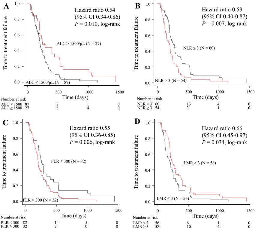
www.nature.com/scientificreports/

                                             Variables                                    Number of patients (%)
                                             Age, years, median (range)                   62.0 (32–89)
                                             ER status
                                             Positive                                      82 (71.9)
                                             Negative                                      32 (28.1)
                                             Diagnosis
                                             Advanced                                      44 (38.6)
                                             Recurrence                                    70 (61.4)
                                             Metastatic sites
                                             CNS                                           10 (8.8)
                                             Bone                                          61 (53.5)
                                             Lungs                                         55 (48.2)
                                             Pleura and/or lymphangiopathy                 46 (40.4)
                                             Lymph node                                    90 (78.9)
                                             Liver                                         53 (46.5)
                                             Soft tissue                                   73 (64.0)
                                             Type of metastases
                                             Visceral                                      93 (81.6)
                                             Non-visceral                                  21 (18.4)
                                             Number of metastatic sites, median (range)   3.5 (1–8)
                                             Number of metastatic sites
                                             ≥3                                            93 (81.6)
www.nature.com/scientificreports/
Figure 1. Time to treatment failure according to baseline levels of (A) ALC, (B) NLR, (C) PLR, and (D) LMR
in patients treated with paclitaxel plus bevacizumab. ALC absolute lymphocyte count, CI confidence interval,
LMR lymphocyte-to-monocyte ratio, NLR neutrophil-to-lymphocyte ratio, PLR platelet-to-lymphocyte ratio.
We performed univariable and multivariable analyses to evaluate independent predictors of PB therapy
(Tables 2 and 3). Each of the four multivariable analyses identified high ALC, low NLR, and low PLR as inde-
pendent predictive markers for TTF (P = 0.013, P = 0.023, and P = 0.004, respectively). The respective results of
the four multivariate Cox regression analyses are shown in Supplemental Table S2.
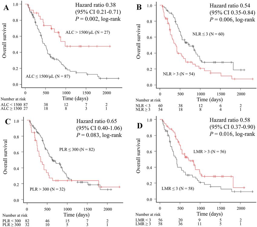
Correlation between systemic immunity markers and OS. We compared OS relative to the systemic
immunity markers (Fig. 2). The OS for patients with high ALC, low NLR, and high LMR was significantly longer
compared with that for patients with low ALC (988 days vs. 475 days, log-rank P = 0.002; Fig. 2A), high NLR
(722 days vs. 403 days, log-rank P = 0.006; Fig. 2B), and low LMR (722 days vs. 418 days, log-rank P = 0.016;
Fig. 2D), respectively. Although PLR showed a favorable tendency, no significant difference was observed
(639 days vs. 342 days, log-rank P = 0.083 for low PLR; Fig. 2C).
We performed univariable and multivariable analyses to evaluate independent predictors of PB therapy
(Tables 2 and 3). Each of the four multivariable analyses identified high ALC, low NLR, and high LMR as inde-
pendent predictive markers for OS (P = 0.010, P = 0.045, and P = 0.034, respectively). The respective results of
the four multivariate Cox regression analyses are shown in Supplemental Table S3.
Correlation between systemic immunity markers and ORR. We compared the ORR relative to
the systemic immunity markers and found no significant differences in ORR between the groups: 81.5% and
65.5% for the patients with high and low ALC, respectively (P = 0.15); 76.7% and 61.1% for the patients with
low and high NLR, respectively (P = 0.10); 73.2% and 59.4% for the patients with low and high PLR, respectively
(P = 0.18); and 74.1% and 64.3% for the patients with high and low LMR, respectively (P = 0.31).
Safety. Of the 114 patients, 81 (71.1%) discontinued PB therapy as a result of disease progression, 16 (14.0%)
discontinued because of adverse events, 12 (10.5%) discontinued due to “other reasons,” and 5 (4.4%) continued
the therapy to the data cut-off point. There were no new recorded therapy-related adverse events.
Scientific Reports | (2021) 11:6328 | https://doi.org/10.1038/s41598-021-85948-2 3
Vol.:(0123456789)www.nature.com/scientificreports/
TTF OS
Variables HR 95% CI P HR 95% CI P
Age (≥ 65 vs. < 65 years) 1.16 0.79–1.70 0.454 1.27 0.81–1.98 0.295
ER (negative vs. positive) 1.09 0.72–1.67 0.678 1.02 0.63–1.66 0.929
Diagnosis (recurrence vs. advanced) 1.36 0.92–2.00 0.127 1.35 0.86–2.14 0.195
Metastatic sites (yes vs. no)
CNS 1.28 0.66–2.47 0.463 1.39 0.67–2.89 0.380
Bone 0.96 0.65–1.40 0.818 0.98 0.63–1.53 0.945
Lung 0.58 0.39–0.86 0.007 0.65 0.42–1.02 0.060
Pleura and/or lymphangiopathy 0.97 0.66–1.43 0.876 0.83 0.53–1.30 0.425
Lymph node 0.97 0.61–1.54 0.895 0.77 0.46–1.31 0.334
Liver 1.37 0.93–2.00 0.110 1.44 0.92–2.24 0.109
Soft tissue 0.76 0.51–1.14 0.184 0.92 0.59–1.45 0.729
Visceral metastasis (yes vs. no) 0.81 0.50–1.31 0.392 0.96 0.55–1.68 0.880
Number of metastatic sites (≥ 3 vs. < 3) 0.70 0.43–1.14 0.153 0.79 0.45–1.38 0.403
Prior (neo) adjuvant chemotherapy* (yes vs. no) 1.54 1.02–2.32 0.039 1.28 0.80–2.05 0.304
Disease-free interval (< 24 months vs. ≥ 24 months) 1.16 0.79–1.70 0.438 1.23 0.79–1.91 0.367
Number of previous chemotherapies (< 2 vs. ≥ 2) 0.90 0.57–1.41 0.644 0.46 0.28–0.75 0.002
Marker of systemic immunity
ALC > 1500/μL versus ALC ≤ 1500/μL 0.54 0.34–0.86 0.010 0.38 0.21–0.71 0.002
NLR ≤ 3 versus NLR > 3 0.59 0.40–0.87 0.007 0.54 0.35–0.84 0.006
PLR ≤ 300 versus PLR > 300 0.55 0.36–0.85 0.006 0.65 0.40–1.06 0.083
LMR > 3 versus LMR ≤ 3 0.66 0.45–0.97 0.034 0.58 0.37–0.90 0.016
Table 2. Univariable analysis of time to treatment failure and overall survival (Cox hazard model). ALC
absolute lymphocyte count, CI confidence interval, CNS central nervous system, ER estrogen receptor, HR
hazard ratio, LMR lymphocyte-to-monocyte ratio, NLR neutrophil-to-lymphocyte ratio, OS overall survival,
PLR platelet-to-lymphocyte ratio, TTF time to treatment failure. *Chemotherapy included anthracycline and/
or taxane.
TTF OS
Variable HR 95% CI P HR 95% CI P
ALC > 1500/μL versus ALC ≤ 1500/μL 0.53 0.32–0.88 0.013 0.44 0.23–0.82 0.010
NLR ≤ 3 versus NLR > 3 0.63 0.43–0.94 0.023 0.62 0.39–0.99 0.045
PLR ≤ 300 versus PLR > 300 0.51 0.33–0.81 0.004 0.63 0.38–1.02 0.062
LMR > 3 versus LMR ≤ 3 0.70 0.47–1.03 0.069 0.60 0.38–0.96 0.034
Table 3. Multivariable analysis of time to treatment failure and overall survival (Cox hazard model). ALC
absolute lymphocyte count, CI confidence interval, HR hazard ratio, LMR lymphocyte-to-monocyte ratio, NLR
neutrophil-to-lymphocyte ratio, OS overall survival, PLR platelet-to-lymphocyte ratio, TTF time to treatment
failure.
Discussion
Our results identified that patients with HER2-negative ABC and high ALC, low NLR, and low PLR at baseline
had significantly improved TTF compared with those with low ALC, high NLR, and high PLR, respectively.
We also found that high ALC, low NLR, and high LMR were associated with longer OS. We demonstrated the
usefulness of these systemic immunity markers as predictive markers of PB therapy for patients with HER2-
negative ABC. These markers can be easily calculated from readily available blood tests routinely measured in
clinical practice.
Recent studies have demonstrated that systemic immunity markers (ALC, NLR, PLR, and LMR) are associ-
ated with better outcomes for patients with various carcinomas, including breast c ancer9–17. It has been reported
that a low ALC before the initiation of systemic therapy is associated with shorter OS and PFS in advanced
carcinomas, sarcomas, and lymphomas. In addition, a low baseline ALC is strongly associated with reduced
disease-free survival (DFS) (hazard ratio [HR] 5.24, 95% CI 2.23–12.30, P < 0.001) and OS (HR 6.13, 95% CI
2.21–17.0, P < 0.001) for patients with breast cancer who undergo primary c hemotherapy11,14. High ALC (≥ 1000/
μL or 1500/µL) was identified as an independent predictive marker for better OS in patients with ABC receiving
eribulin therapy19,20. In a previous meta-analysis of patients with unselected solid tumors, high NLR was associ-
ated with poor OS (HR 1.81, 95% CI 1.67–1.97, P < 0.001). In patients with breast cancer, high NLR (higher than
the cut-off value) was significantly associated with poor OS (HR 2.56, 95% CI 1.96–3.35, P < 0.001) and DFS
Scientific Reports | (2021) 11:6328 | https://doi.org/10.1038/s41598-021-85948-2 4
Vol:.(1234567890)www.nature.com/scientificreports/
Figure 2. Overall survival according to baseline levels (A) ALC, (B) NLR, (C) PLR, and (D) LMR in patients
treated with paclitaxel plus bevacizumab. ALC absolute lymphocyte count, CI confidence interval, LMR
lymphocyte-to-monocyte ratio, NLR neutrophil-to-lymphocyte ratio, PLR platelet-to-lymphocyte ratio.
(HR 1.74, 95% CI 1.47–2.07, P < 0.001)10,15. Further, low NLR (< 3) at baseline was significantly associated with
longer PFS18 and OS20 in patients treated with eribulin in ABC, thereby demonstrating that low NLR (< 3) was
an independent predictive marker for eribulin therapy. It has been reported that high PLR is associated with
shorter OS for various solid tumors and a meta-analysis showed that high PLR was associated with poor OS
(HR 1.55, 95% CI 1.07–2.25, P = 0.022) and DFS (HR 1.73, 95% CI 1.30–2.30, P < 0.001) in patients with breast
cancer12,16. Another systematic review and meta-analysis that included 11,197 patients from 29 studies showed
that low LMR (lower than the cut-off) was associated with poor OS (HR, 1.73 95% CI 1.55–1.93, P < 0.001) and
DFS (HR 1.56, 95% CI 1.31–1.86, P < 0.001) in non-hematologic solid t umors13. Ni et al. demonstrated that high
LMR predicted a favorable response and prognosis in A BC17. However, the utility of PLR and LMR as predictive
markers for patients with ABC remains unclear. Our results showed that high ALC (≥ 1500/µL) and low NLR (< 3)
were associated with longer TTF and OS compared with low ALC and high NLR, respectively. Furthermore, we
found that low PLR was associated with longer TTF and high LMR was associated with longer OS.
Our results support those of a previous study reporting that systemic immunity marker expression at the
beginning of PB therapy exhibited a significant association with improved prognosis and that these markers
were readily available as predictive markers of PB therapy in A BC23. In contrast, NLR and ALC showed no sig-
nificant association with increased PFS and OS in patients with ABC treated with nab-paclitaxel or a treatment
of the physician’s choice, such as taxanes18,20. In advanced non-small-cell lung cancer and metastatic colorectal
cancer, NLR was associated with survival outcome and was useful as a predictive marker in patients treated with
combination chemotherapy and bevacizumab, but not with chemotherapy a lone21,22. Therefore, we conclude that
systemic immunity markers can be useful as predictive markers by addition of bevacizumab to chemotherapy.
Recently, vascular endothelial growth factor (VEGF) has been recognized as an important mediator of
immune suppression, and VEGF blockade may be effective in the antitumor immune response in addition
to its direct effects on tumor vasculature. VEGF modulates the various processes of cancer immunity, includ-
ing promotion of T-regulatory cells, suppression of dendritic cell maturation, stimulation of tumor-associated
macrophages, and infiltration of myeloid-derived suppressor cells, leading to an immunosuppressive state24.
Given that bevacizumab modulates this immunosuppressive state through angiogenesis inhibition, a strategy
that combines bevacizumab and immune checkpoint inhibitors has been explored. Combined ipilimumab and
Scientific Reports | (2021) 11:6328 | https://doi.org/10.1038/s41598-021-85948-2 5
Vol.:(0123456789)www.nature.com/scientificreports/
bevacizumab therapy in patients with melanoma resulted in encouraging antitumor activity and had beneficial
effects on the host antitumor immune response25. In patients with metastatic non-squamous non-small-cell lung
cancer, the addition of atezolizumab to bevacizumab and chemotherapy significantly increased PFS and O S26.
Therefore, it is reasonable to suggest that systemic immunity markers can predict systemic antitumor activity
resulting from PB therapy in patients with ABC.
This study had several limitations. First, it was a retrospective single-center study with a small number of sub-
jects. Given that this was a single-center study, however, the patients were treated consistently and only two (1.8%)
patients were untraceable. Second, the optimal cut-off values for the systemic immunity markers are unclear.
Because the cut-off values for ALC at 1500/μL and NLR at 3 have often been reported as useful in ABC18,20,23, we
followed these parameters. Finally, which systemic immunity marker is the most effective at predicting survival
remains unclear. Therefore, further prospective studies are warranted to resolve this issue. Overall, our study
demonstrated the usefulness of systemic immunity markers associated with lymphocytes as predictive mark-
ers of PB therapy for patients with HER2-negative ABC. We also demonstrated that these systemic immunity
markers may play an important role in selecting candidates with HER2-negative ABC for bevacizumab therapy.
Methods
Study population and treatment. We reviewed the medical records of 114 patients with HER2-negative
ABC who underwent at least two cycles of PB therapy at the Fukuyama City Hospital (Japan) from November
2011 to December 2019. We excluded patients with missing systemic immunity marker data and patients who
had not undergone at least two cycles of PB therapy. The data cut-off was May 31, 2020. We defined the subtype
from the pathology reports at the time of surgery, initial biopsy, or at the time of recurrence. The subtype was
based on ER and HER2 expression, and ER-positivity was defined as ER ≥ 1% positive. HER2 overexpression was
defined according to the American Society of Clinical Oncology/College of American Pathologists guidelines27.
The treatment schedule was the same as that of the E2100 study1: Bevacizumab (10 mg/kg) was administered
on days 1 and 15 in combination with paclitaxel (90 mg/m2) on days 1, 8, and 15 of each 28-day cycle. Treatment
was continued until disease progression, unacceptable toxicity, or patient/physician decision. Tumor response
to treatment was assessed according to the Response Evaluation Criteria in Solid Tumors (RECIST) version 1.1,
and the physician determined the timing of the lesion assessment.
All procedures that involved human subjects were performed in accordance with the ethical standards of the
institutional and/or national research committees and with the 1964 Helsinki Declaration and its later amend-
ments or comparable ethical standards. This retrospective study was approved by the Fukuyama City Hospital’s
review board. Informed consent was obtained in the form of an opt-out on the website from all individual
participants included in the study.
Measurements of systemic immunity markers. Neutrophil, lymphocyte, platelet, and monocyte
counts were performed automatically using a Sysmex XE-2100 or XE-5000 automated hematology system (Sys-
mex Co., Kobe, Japan). ALC, NLR, PLR, and LMR were calculated from blood cell counts prior to administering
PB therapy, and the cut-off values for these markers were set in accordance with previous studies as follows:
1500/μL for ALC, 3 for NLR, 300 for PLR, and 3 for L MR12,13,15,20. All patients were divided into “low” and
“high” groups according to the cut-off values, respectively: low ALC (≤ 1500/μL, n = 87), high ALC (> 1500/μL,
n = 27); low NLR (≤ 3, n = 60), high NLR (> 3, n = 54); low PLR (≤ 300, n = 82), high PLR (> 300, n = 32); and low
LMR (≤ 3, n = 56), high LMR (> 3, n = 58). Neutrophil, lymphocyte, platelet, and monocyte counts are routinely
measured in clinical practice during treatment and the systemic immunity markers are easily calculated from
these blood cell counts. Therefore, these markers can be measured and analyzed easily and inexpensively without
additional equipment, software, or personnel specialized in analyzing the results.
Statistical analysis. The Wilcoxon rank sum test was used to compare continuous variables (such as
median age) and Fisher’s exact test was used to compare the proportions of categorical variables (such as metas-
tasis type) between groups. The distribution of TTF and OS was estimated by the Kaplan–Meier method. We
performed a univariate Cox regression analysis to determine the association between baseline patient character-
istics and TTF and OS. To evaluate the association between each systemic immunity marker (ALC, NLR, PLR,
and LMR) and TTF or OS, we conducted a multivariate Cox regression analysis. We considered the baseline
patient characteristics with P < 0.20 in the univariate Cox regression analysis as confounders and included them
in the multivariate analysis. Since the systemic immunity markers (ALC, NLR, PLR, and LMR) were correlated
with each other, we did not include these four markers simultaneously in the multivariate analysis. These mark-
ers were included independently in each multivariate analysis of TTF and OS. Finally, we performed this analysis
eight times. A P value < 0.05 was considered statistically significant, and all statistical analyses were performed
with EZR software (Saitama Medical Center, Jichi Medical University, Saitama, Japan), a graphical user interface
for R (The R Foundation for Statistical Computing, Vienna, Austria)28.
We defined TTF as the time from the administration of PB therapy to the discontinuation of treatment for
any reason, including disease progression, treatment toxicity, patient/physician decision, and death from any
cause. OS was defined as the time from the administration of PB therapy to the date of death from any cause.
ORR was defined as the percentage of patients who achieved a complete or partial response according to the
RECIST criteria.
Data availability
The datasets generated during and/or analyzed during the current study are available from the corresponding
author upon reasonable request.
Scientific Reports | (2021) 11:6328 | https://doi.org/10.1038/s41598-021-85948-2 6
Vol:.(1234567890)www.nature.com/scientificreports/
Received: 24 October 2020; Accepted: 2 March 2021
References
1. Miller, K. et al. Paclitaxel plus bevacizumab versus paclitaxel alone for metastatic breast cancer. N. Engl. J. Med. 357, 2666–2676.
https://doi.org/10.1056/NEJMoa072113 (2007).
2. Gray, R., Bhattacharya, S., Bowden, C., Miller, K. & Comis, R. L. Independent review of E2100: a phase III trial of bevacizumab
plus paclitaxel versus paclitaxel in women with metastatic breast cancer. J. Clin. Oncol. 27, 4966–4972. https://doi.org/10.1200/
JCO.2008.21.6630 (2009).
3. Miles, D. et al. Bevacizumab plus paclitaxel versus placebo plus paclitaxel as first-line therapy for HER2-negative metastatic breast
cancer (MERiDiAN): a double-blind placebo-controlled randomised phase III trial with prospective biomarker evaluation. Eur.
J. Cancer 70, 146–155. https://doi.org/10.1016/j.ejca.2016.09.024 (2017).
4. Miles, D., Cameron, D., Hilton, M., Garcia, J. & O’Shaughnessy, J. Overall survival in MERiDiAN, a double-blind placebo-controlled
randomised phase III trial evaluating first-line bevacizumab plus paclitaxel for HER2-negative metastatic breast cancer. Eur. J.
Cancer 90, 153–155. https://doi.org/10.1016/j.ejca.2017.10.018 (2018).
5. Delaloge, S. et al. Paclitaxel plus bevacizumab or paclitaxel as first-line treatment for HER2-negative metastatic breast cancer in a
multicenter national observational study. Ann. Oncol. 27, 1725–1732. https://doi.org/10.1093/annonc/mdw260 (2016).
6. Nakamoto, S., Watanabe, J., Ohtani, S., Morita, S. & Ikeda, M. Bevacizumab as first-line treatment for HER2-negative advanced
breast cancer: paclitaxel plus bevacizumab versus other chemotherapy. In Vivo 34, 1377–1386. https://doi.org/10.21873/invivo.
11917 (2020).
7. Miles, D. W. et al. Biomarker results from the AVADO phase 3 trial of first-line bevacizumab plus docetaxel for HER2-negative
metastatic breast cancer. Br. J. Cancer 108, 1052–1060. https://doi.org/10.1038/bjc.2013.69 (2013).
8. Hanahan, D. & Weinberg, R. A. Hallmarks of cancer: the next generation. Cell 144, 646–674. https://doi.org/10.1016/j.cell.2011.
02.013 (2011).
9. Guthrie, G. J. et al. The systemic inflammation-based neutrophil-lymphocyte ratio: experience in patients with cancer. Crit. Rev.
Oncol. Hematol. 88, 218–230. https://doi.org/10.1016/j.critrevonc.2013.03.010 (2013).
10. Templeton, A. J. et al. Prognostic role of neutrophil-to-lymphocyte ratio in solid tumors: a systematic review and meta-analysis.
J. Natl. Cancer Inst. 106, 124. https://doi.org/10.1093/jnci/dju124 (2014).
11. Ray-Coquard, I. et al. Lymphopenia as a prognostic factor for overall survival in advanced carcinomas, sarcomas, and lymphomas.
Cancer Res. 69, 5383–5391. https://doi.org/10.1158/0008-5472.CAN-08-3845 (2009).
12. Templeton, A. J. et al. Prognostic role of platelet to lymphocyte ratio in solid tumors: a systematic review and meta-analysis. Cancer
Epidemiol. Biomark. Prev. 23, 1204–1212. https://doi.org/10.1158/1055-9965.EPI-14-0146 (2014).
13. Nishijima, T. F., Muss, H. B., Shachar, S. S., Tamura, K. & Takamatsu, Y. Prognostic value of lymphocyte-to-monocyte ratio in
patients with solid tumors: a systematic review and meta-analysis. Cancer Treat. Rev. 41, 971–978. https://doi.org/10.1016/j.ctrv.
2015.10.003 (2015).
14. Vicente Conesa, M. A. et al. Predictive value of peripheral blood lymphocyte count in breast cancer patients treated with primary
chemotherapy. Breast 21, 468–474. https://doi.org/10.1016/j.breast.2011.11.002 (2012).
15. Ethier, J. L., Desautels, D., Templeton, A., Shah, P. S. & Amir, E. Prognostic role of neutrophil-to-lymphocyte ratio in breast cancer:
a systematic review and meta-analysis. Breast Cancer Res. 19, 2. https://doi.org/10.1186/s13058-016-0794-1 (2017).
16. Zhu, Y. et al. Platelet-lymphocyte ratio acts as an indicator of poor prognosis in patients with breast cancer. Oncotarget 8, 1023–
1030. https://doi.org/10.18632/oncotarget.13714 (2017).
17. Ni, X. J. et al. An elevated peripheral blood lymphocyte-to-monocyte ratio predicts favorable response and prognosis in locally
advanced breast cancer following neoadjuvant chemotherapy. PLoS ONE 9, e111886. https://doi.org/10.1371/journal.pone.01118
86 (2014).
18. Miyagawa, Y. et al. Significant association between low baseline neutrophil-to-lymphocyte ratio and improved progression-free
survival of patients with locally advanced or metastatic breast cancer treated with eribulin but not with nab-paclitaxel. Clin. Breast
Cancer 18, 400–409. https://doi.org/10.1016/j.clbc.2018.03.002 (2018).
19. Watanabe, J., Saito, M., Horimoto, Y. & Nakamoto, S. A maintained absolute lymphocyte count predicts the overall survival
benefit from eribulin therapy, including eribulin re-administration, in HER2-negative advanced breast cancer patients: a single-
institutional experience. Breast Cancer Res. Treat. 181, 211–220. https://doi.org/10.1007/s10549-020-05626-1 (2020).
20. Miyoshi, Y. et al. High absolute lymphocyte counts are associated with longer overall survival in patients with metastatic breast
cancer treated with eribulin-but not with treatment of physician’s choice-in the EMBRACE study. Breast Cancer 27, 706–715.
https://doi.org/10.1007/s12282-020-01067-2 (2020).
21. Botta, C. et al. Systemic inflammatory status at baseline predicts bevacizumab benefit in advanced non-small cell lung cancer
patients. Cancer Biol. Ther. 14, 469–475. https://doi.org/10.4161/cbt.24425 (2013).
22. Passardi, A. et al. Inflammatory indexes as predictors of prognosis and bevacizumab efficacy in patients with metastatic colorectal
cancer. Oncotarget 7, 33210–33219. https://doi.org/10.18632/oncotarget.8901 (2016).
23. Miyagawa, Y. et al. Baseline neutrophil-to-lymphocyte ratio and c-reactive protein predict efficacy of treatment with bevacizumab
plus paclitaxel for locally advanced or metastatic breast cancer. Oncotarget 11, 86–98. https://doi.org/10.18632/oncotarget.27423
(2020).
24. Ott, P. A., Hodi, F. S. & Buchbinder, E. I. Inhibition of immune checkpoints and vascular endothelial growth factor as combina-
tion therapy for metastatic melanoma: an overview of rationale, preclinical evidence, and initial clinical data. Front. Oncol. 5, 202.
https://doi.org/10.3389/fonc.2015.00202 (2015).
25. Hodi, F. S. et al. Bevacizumab plus ipilimumab in patients with metastatic melanoma. Cancer Immunol. Res. 2, 632–642. https://
doi.org/10.1158/2326-6066.CIR-14-0053 (2014).
26. Socinski, M. A. et al. Atezolizumab for first-line treatment of metastatic nonsquamous NSCLC. N. Engl. J. Med. 378, 2288–2301.
https://doi.org/10.1056/NEJMoa1716948 (2018).
27. Wolff, A. C. et al. Recommendations for human epidermal growth factor receptor 2 testing in breast cancer: American society of
clinical oncology/college of American pathologists clinical practice guideline update. J. Clin. Oncol. 31, 3997–4013. https://doi.
org/10.1200/jco.2013.50.9984 (2013).
28. Kanda, Y. Investigation of the freely available easy-to-use software “EZR” for medical statistics. Bone Marrow Transpl. 48, 452–458.
https://doi.org/10.1038/bmt.2012.244 (2013).
Acknowledgements
We would like to thank Enago for editing a draft of this manuscript.
Scientific Reports | (2021) 11:6328 | https://doi.org/10.1038/s41598-021-85948-2 7
Vol.:(0123456789)www.nature.com/scientificreports/
Author contributions
All authors contributed to the study conception and design. S.N. prepared the materials and performed the data
collection and analysis. S.N. wrote the first draft of the manuscript, and all authors commented on the previous
versions of the manuscript. All authors have read and approved the final manuscript.
Competing interests
Dr. Nakamoto reports having received personal fees from Chugai Pharmaceuticals, Eisai, and Taiho Pharmaceu-
ticals outside of the submitted work; Dr. Ikeda reports having received personal fees from AstraZeneca, Chugai
Pharmaceuticals, Daiichi-Sankyo, Eisai, Eli-Lilly, Kyowa Kirin, Pfizer, Nippon Kayaku, Novartis, Mundipharma,
Celltrion Healthcare, and Sawai Pharmaceuticals outside of the submitted work; Dr. Kubo has received lecture
fees from Eli-Lilly outside of the submitted work; Dr. Yamamoto has received lecture fees from Bayer outside of
the submitted work. The other authors have no conflicts of interest.
Additional information
Supplementary Information The online version contains supplementary material available at https://doi.org/
10.1038/s41598-021-85948-2.
Correspondence and requests for materials should be addressed to S.N.
Reprints and permissions information is available at www.nature.com/reprints.
Publisher’s note Springer Nature remains neutral with regard to jurisdictional claims in published maps and
institutional affiliations.
Open Access This article is licensed under a Creative Commons Attribution 4.0 International
License, which permits use, sharing, adaptation, distribution and reproduction in any medium or
format, as long as you give appropriate credit to the original author(s) and the source, provide a link to the
Creative Commons licence, and indicate if changes were made. The images or other third party material in this
article are included in the article’s Creative Commons licence, unless indicated otherwise in a credit line to the
material. If material is not included in the article’s Creative Commons licence and your intended use is not
permitted by statutory regulation or exceeds the permitted use, you will need to obtain permission directly from
the copyright holder. To view a copy of this licence, visit http://creativecommons.org/licenses/by/4.0/.
© The Author(s) 2021
Scientific Reports | (2021) 11:6328 | https://doi.org/10.1038/s41598-021-85948-2 8
Vol:.(1234567890)You can also read