IN VITRO ANTICANCER ACTIVITY OF PAPPEA CAPENSIS MEDICINAL PLANT FROM
←
→
Page content transcription
If your browser does not render page correctly, please read the page content below
Archives • 2021 • vol.2 • 234-241

IN VITRO ANTICANCER ACTIVITY OF PAPPEA CAPENSIS MEDICINAL PLANT FROM
                              MOKOPANE
                              Makhoahle Paksio*1 , Mashele Samson* 1
              1
             Department of Health Sciences, Faculty of Health and Environmental Sciences
     Central University of Technology, Free State, Private Bag X20539, Bloemfontein, South Africa

                           *smashele@cut.ac.za and pmakhoahle@cut.ac.za

   Abstract
    The present study reports on the anticancer activity of Pappea capensis, used by traditional healers
in the Limpopo province of South Africa for the treatment of most prominent diseases in humans
including cancer. More than 80% of the world population rely on natural products due the economic
state of not affording western medicine. Even though this medicinal plant is easily accessible, cheap
and found everywhere its efficacy is an issue that scientists should assess. MCF7 (breast cancer);
HCT116 (colon cancer), and PC30 (prostate cancer) cells used in-vitro Sulforhodamine B (SRB) assay.
The anticancer activities of Pappea capensis extracts dichloromethane/methanol (1:1) and aqueous
extracts were tested for their growth inhibitory effects in vitro against three human cancer cell lines.
Parthenolide was used as a standard. Data analysis was performed using GraphPad Prism software.
Fifty per cent (50%) of cell growth inhibition (IC 50) was determined by non-linear regression and
absorbance measured at 540nm. Extracts were classified into four categories based on their total
growth inhibition of the cell lines. Extracts which exhibited a total growth inhibition (TGI) of less than
6.25 μg/mL were regarded as potent. Dichloromethane/methanol extract of Pappea capensis exhibited
pronounced activity especially against the PC30 cell line. A relatively low Dichloromethane/methanol
extract concentration was necessary to reach 50% proliferation inhibition of the PC-3 cell line and
moderate for MCF-7 cell line and weak for HCT-116 cell line. The Dichloromethane/methanol extract was
classified as weakly active. So, Pappea capensis extracts are promising as possible drugs for the
treatment of prostate cancer. Further tests such as genotoxicity, antioxidant determination, phenolic
and active compounds should be done scientifically to establish compounds responsible for growth
inhibition towards prostate PC3 cell lines.

   Keywords: Anticancer activity, cancer, Limpopo

                                       http://pharmacologyonline.silae.it
                                                ISSN: 1827-8620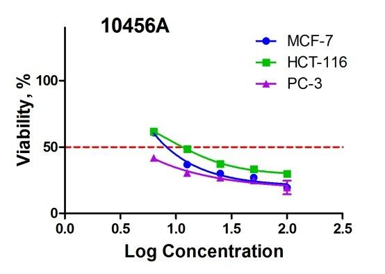
PhOL                                    Makhoahle, et al.                    235 (pag 234-241)

Introduction                                                 developed great interest towards understanding
                                                             the role of free radical reactions in biology,
Cancer is a major global public health problem (1).
                                                             suggesting that such reactions are important for
It is presented when abnormalities are observed
                                                             several metabolic reactions and could also be
within the cells of an individual’s body and can as
                                                             potentially harmful to health. Free radicals such
a result lead to death (2). It is possible that these
                                                             as reactive oxygen species (ROS) have been
cells grow due to imbalance in the body,
                                                             reported to play an important role in the
therefore by correcting this imbalance cancer
                                                             development of tissue damage in living
may be treated (2, 3). Cancer accounts for 3500
                                                             organisms. There is an increase in evidence
million deaths worldwide, contributing 2-3% of the
                                                             relating the occurrence of cancer due to the
world deaths recorded annually (4; 5). Cancer is             oxidative damage to DNA, proteins and lipids in
the second leading cause of death in America
                                                             the body caused by radicals and other
with breast cancer being the most common in
                                                             carcinogens (19). Although medicinal plants are
women and prostate cancer in men worldwide (2,
                                                             not used as antioxidants in traditional medicine,
6, 7, 8). Among South African women one out of
                                                             studies have reported plants containing several
every 31 is likely to develop breast cancer at some
                                                             phytochemicals        which      possess       strong
time in her life time (7). A lot of money has been
                                                             antioxidants. There is a possibility that these
spent on cancer research but it is still not fully
                                                             antioxidants may prevent and cure cancer, as well
understood what causes cancer (7). Despite the
                                                             as other diseases by protecting the cells from
progress of medicinal research in the past
                                                             damage caused by free radicals (20, 21).
decades cancer treatment remains vague.                      Considering the importance of this untapped
Several chemotherapeutic agents are used in the
                                                             research area of the medicinal plant Pappea
treatment of cancer, but they cause toxicity that
                                                             capensis, the aim of the study was to evaluate the
prevents their usage leading to the possible use
                                                             anticancer activities of this medicinal plant used in
of plants for cancer treatment (5). Chemotherapy
                                                             Limpopo for the treatment of cancer. The
being used for the control of advanced stage of
                                                             ultimate objective of this research is to reveal the
malignancies and a prophylactic against possible
                                                             anticancer inhibitory effect of this plant.
metastasis, exhibit severe toxicity to normal
tissues (6). Treatments like chemotherapy,
radiotherapy and surgery are less accessible in the
                                                             Methods
developing countries (1). According to World               Plant material
Health Organization, more that 80% of population           The plant was collected from the south of Limpopo
in these countries rely on traditional medicine for        province; 209 km from the town Naboomspruit
some aspects of primary health care (9, 10). Other         using R101 to Mokopane in April 2014. A voucher
studies have shown that more than 60% of cancer            specimen was supplied to the Free State Museum
patients use vitamins and herbs as therapy (11, 12).       and the Free State Botanical Gardens. The plant was
Plants have played an important role as an                 positively identified by Dr Zietsman at the Free State
effective anticancer agent, and recently over 60%          Museum and verified by the National Botanical
of the presently used anticancer agents are                Gardens in Pretoria.
derived from natural sources including plants (13,         Extraction of plant material
14, 15; 16). The increased incidence of cancer and         Finely ground plant mixture (100g) was separately
the lack of anticancer drugs have forced                   extracted           using           water          and
researchers to study the pharmacological and               dichloromethane/methanol          (1:1)    at    room
chemical investigations in the area of medicinal           temperature for 24 hours. Organic extracts were
plants to search for the discovery of new possible         filtered and concentrated by rotary vacuum at 50-
anticancer agents (8).                                     60ºC; the aqueous extracts were concentrated to
Recently there has been great scientific interest in       dryness using a freeze-dryer. The plant extracts
the discovery of new anti-cancer drugs from                generated was stored in a cold room at -20ºC until
natural products sources (2, 17; 18). Research has         further use. Five times two-fold serial dilution of the

                                      http://pharmacologyonline.silae.it
                                               ISSN: 1827-8620
PhOL Makhoahle, et al. 236 (pag 234-241)
plant extract and 10 times two fold serial dilutions of was extracted with 10mM Tris base for optical
the control (parthenolide) were used with density determination at the wavelength 540 nm
concentration of 100-6.25 ug/ml and 100-0.20 ug/ml using a multi well spectrophotometer.
respectively.
Determination of inhibitory effect on three cell lines Data analysis was performed using GraphPad
For the estimation of the growth inhibitory effects, Prism software. Fifty per cent (50%) of cell growth
the extract was tested in the 3-cell line panels inhibition (IC 50) was determined by non-linear
consisting of MCF7 (breast cancer), HCT116 (colon regression and absorbance measured at 540nm.
cancer) and PC3 (prostate cancer) cells by
Results
Sulforhodamine B (SRB) assay.The SRB assay was
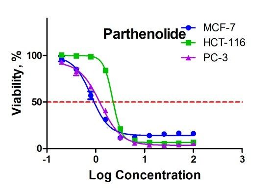
developed by Skehan and colleagues to measure The results (Table 1 and Figure 1) indicated that the
drug-induced cytotoxicity and cell proliferation. Its proliferation activities of PC-3, HTC-116 and MCF-7
principle is based on the ability of the protein dye cell lines were inversely related to the increased
sulforhodamine B (Acid Red 52) to bind levels of parthenolide concentrations. This
electrostatically in a pH-dependent manner to proliferation inhibition is acceptable for
protein basic amino acid residues of trichloroacetic parthenolide as it indicates the accuracy of the
acid-fixed cells. Under mild acidic conditions it binds assay, a significant dose response manner was
to the fixed cellular protein, while under mild basic shown (Z=0.9). A relatively low parthenolide
conditions it can be extracted from cells and concentration was necessary to reach 50%
solubilized for measurement. The SRB Assay was proliferation inhibition of the three cell lines.
performed at Council for Scientific and Industrial The results (Table 2 and Figure 2) indicated that the
Research (CSIR) of South Africa in accordance with proliferation activity of PC-3, HTC-116 and MCF-7cells
the protocol of the Drug Evaluation Branch, USA lines were inversely related to the increased levels
National Cancer Institute (NCI), and the assay has of the plant extract concentration. This proliferation
been adopted for this screen. inhibition effect is acceptable for plant extract, a
The human cancer cell lines MCF7 and PC3 were significant dose response manner was shown
obtained from NCI in a collaborative research (Z=0.9). A relatively low plant extract concentration
program between CSIR and NCI. The HCT-116 cell was necessary to reach 50% proliferation inhibition
line was obtained from European Collection of Cell of the PC-3 and moderate for MCF-7 and HCT-116 cell
Culture (ECACC). Cell lines were routinely lines.
maintained as a monolayer cell culture at 37ºC, 5% The CSIR interpretation criterion (Table 4) was
CO2, 95% air and 100% relative humidity in RPMI used since as it describes the sample is
containing 5% fetal bovine serum, 2 mM L-glutamine considered inactive if parameter IC 50 for two or
and 50µg/ml gentamicin. three cell lines is higher than 100 µg/ml. Table 3
For the screening experiment, the cells (3-19 reports the IC 50 values for the investigated in vitro
passages) were inoculated in a 96-well microtiter anticancer activity of the plant extract. The
plates at plating densities of 7-10 000 cells/well and extract showed a potent anti-cancer activity
were incubated for 24 hours. After 24 hours the cells against PC-3 and moderate activity against MCF-7
were treated with the experimental drug which and HTC-116.
were previously dissolved in DMSO and diluted in Discussion
medium to produce 5 concentrations. Cells without
Recently, relevant attention has been focused on
drug served as control. The blank contained
the screening for the best possible plants and
complete medium without cells and Parthenolide
plant’s parts that have anticancer activity for the
was used as a standard.
treatment of cancer. The results indeed indicated
The plates were incubated for 48 hours after
that the plant Pappea capensis have high
addition of the plant extract. Viable cells were fixed
anticancer activity against PC-3 and can be a
to the bottom of each well with cold 50%
suitable source for the treatment of prostate
trichloroacetic acid, washed, dried and dyed by SRB.
cancer. These results correlated to what have
Unbound dye was removed, and protein-bound dye
http://pharmacologyonline.silae.it
ISSN: 1827-8620PhOL Makhoahle, et al. 237 (pag 234-241)
been published by previous studies indicating the underexplored source of anticancer drug.
importance of traditional medicine and plants as Natural Products Resources, 6, 5:115-119.
natural sources for cancer treatment (15, 13, 14, 12, 6. American Cancer society. Facts and figures,
8, 16, 11). The present screen justifies the use of (1999).
this plant by the traditional healer as a prime 7. Estrogen and Cancer website., 2006.
source of the drugs for the treatment of cancers. www.womenshealth.com,ww.amazon.com
Based on the obtained data the results suggested 8. Koduru, S., Grierson, D.S., van de Venter, M.,
that this plant could be a new potential source of & Afolayan, A.J., (2006). Antitumor activity
anticancer agent. No data relative to cytotoxic of Solanum aculeastrum berries on three
components had been reported, however bio- carcinoma cells. International Journal of
guided fractionation and isolation to identify Cancer Research, 2, 397-406.
compounds responsible for the observed activity 9. Fransworth, N.R., Akelere, O., Bingel, A.S.,
is in progress Soejarto, D.D., & Guo, Z., (1985). Medicinal
plants in therapy. World Health Organization
Bulletin, 63, 965-981.
Acknowledgments
10. Akerel, O., (1988). Medicinal plants and
Mrs MR Matabane (the traditional healer) consulted primary health care: an agenda for action.
for plant use, collection for been the original source Fitoterapi, 59,355-363.
of the plant and helping with the collection. 11. Madhuri, S., & Pandy, G., (2008). Some
The Free State Museum (Dr Zeizzman) and National dietary agricultural plants with anticancer
Botanical Gardens for botanical identification of the properties. Plant Archives, 8,13-16.
plant. 12. Sivalokanathan, S., Ilayaraja, M., &
The Council for Scientific and Industrial Research Balasubramanium, M.P. (2005). Efficacy of
(CSIR) for performing the SRB assays on the sample. Terminalia arjuna (Roxb.) on N-
nitrosodiethylamine induced hepatocellular
carcinoma in rats. Indian Journal of
References Experimental Biology, 43, 264-267.
13. Kinghorn, A.D., Fransworth, N.R., Soejarto,
1. Sawadogo, W.R., Maciuk, A., Banzouzi, J.T., D., Cordell, G., Swanson, S., Pezzuto, J.,
Champy, P., Figadere, B., Guissou, I.P., & Wani, M., Wall, M., Oberlies, N., Kroll, D.,
Nacuolma, G.O. (2012). Mutagenic effect, Kramer, R., Rose, W., Vite, G., Fairchild, C.,
antioxidant and anticancer activities of six Peterson, R., & Wild , R. (2008). Novel
medicinal plants from Burkina Faso. Nature strategies for the discovery of plants derived
Product Research, 26, 575-579. anticancer agents. Pharmaceutical Biology,
2. Madhuri, S., & Pandey, G.(2009). Some 41(s1), 53-67.
anticancer medicinal plants of foreign origin. 14. Clardy, J., & Walsh, C. (204) Lessons from
Current Science, 96, 779-783. nature molecules. Nature, 432, 829-837.
3. Malo, M., Charriere-Bertrand, C., Chettaoui, 15. Cragg, G.M., & Newman, D.J. ( 2000).
C., Fabre-Guillevin, E., Maquerlot, F., Lackmy, Antineoplastic agents from natural sources:
A., Vallee, B., Delaplace, F., & Barlovatz- achievements and future directions. Experts
Meimon , G. (2006). Microenvironnement Opinion on Investigational Drugs, 9,1-15.
cellulaire, PAI-1 et migration cancereuse. 16. Rethy, B., Csupor-Loeffler, B., Zupko, I.,
Elsevier, 329:919-927. Hajdu, Z., Mathe, I., Hohmann, J., Redei, T.,
4. American Cancer Society (2006). A & Falkay, G. (2007). Antiproliferative activity
biotechnology company dedicated to cancer of Hungarian Asteraceae specie against
treatment.viewed on 25 January 2014; human cancer cell lines. Part I. Phytotherapy
ww.cancervax.com/infor/index.htm Resesearch, 21,1200-1208.
5. Kathiresan, K., Boopathy, N.S., & Kavitha, 17. Stevigny, C., Bailly, C., & Quetin-Leclercq, J.
S.(2006). Coastal vegetation- an (2005). Cytotoxic and antitumor
http://pharmacologyonline.silae.it
ISSN: 1827-8620PhOL Makhoahle, et al. 238 (pag 234-241)
potentialities of aporphinoid alkaloids. 19. Halliwell B. Antioxidants in human health
Current Medicinal Chemistry, Anti-Cancer and disease (1996). Annual Review of
Agents, 5, 173-182. Nutrition , 6,33-50
18. Mothana, R,A,A., Kriegisch, S., Harms, M., 20. Pandey, G., & Madhuri, S. (2006) Medicinal
Wende, K., & Lindequist, U. (2011). plants: better remedy for neoplasm. Indian
Assessment of selected Yemeni medicinal Drugs,43, 869-874.
plants for their in vitro antimicrobial, 21. Agrawala, S.K., Chatterjee, S., Misra,
anticancer, and antioxidant activities. S.K. (2001). Immunepotentiation activity of a
Pharmaceutical Biology, 49, 200-210s. polyherbal formulation ‘ Immu-21’ (research
name). Phytomedica, 2,1-22
http://pharmacologyonline.silae.it
ISSN: 1827-8620PhOL Makhoahle, et al. 239 (pag 234-241)
Table1: The survival rate of the three cell lines exposed to different parthenolide (control)
concentrations
Con Lo
c. g %Viability %Viabilit %Viability
SD SD SD
(μg/ Co MCF-7 y HCT-116 PC-3
ml) nc.
100 2.0 16.13 0.20 6.86 0.92 5.82 0.61
50 1.7 16.56 0.38 5.91 1.21 4.07 0.90
25 1.4 15.55 1.07 5.42 0.40 3.35 0.79
12.5 1.1 13.89 0.21 6.57 0.51 3.87 0.61
6.25 0.8 12.10 0.17 8.94 0.97 5.98 1.01
3.13 0.5 11.54 0.17 21.27 2.13 12.24 2.51
1.56 0.2 31.35 1.01 84.10 2.38 40.80 1.16
0.78 -0.1 57.15 4.13 98.87 0.98 66.60 0.19
0.39 - 85.28 1.99 99.63 0.80 82.52 4.09
0.20 0.4
- 96.70 2.90 100.59 0.27 93.16 2.29
0.7
Z’ factor: 0.9
Table2: The survival rate of the three cell lines exposed to different plant extract (Pappea capensis)
concentrations
Z’ factor: 0.9
Conc
Log %Via
. %Viabilit %Viabilit
Conc SD SD bility SD
(μg/ y MCF-7 y HCT-116
. PC-3
ml)
5.1
100 2.0 19.46 1.90 29.88 0.83 19.58 4
24.8 0.6
50 1.7 27.05 1.82 33.40 0.59 3 5
26.8 2.6
25 1.4 30.04 1.30 37.26 1.33 4 2
30.5 2.6
12.5 1.1 36.86 1.35 48.49 0.35 6 1
0.1
6.25 0.8 60.84 0.81 61.63 1.37 41.78 6
Table3. Results summary
No Compound IC50 for IC50 for IC50 for
MCF-7, µg/ml HCT-116,µg/ml PC-3,µg/ml
1 Pappea 8.528 12.52PhOL Makhoahle, et al. 240 (pag 234-241)
Table4: CSIR standard criteria for IC50 interpretation
IC50,µg/ml Status
> 100 µg/ml Inactive
< 100 µg/ml Weak Activity
>15 µg/ml
< 15 µg/ml Moderate Activity
> 6.25 µg/ml
< 6.25 µg/ml Potent Activity
Figure 1: A viability curve of the three cell lines exposed to Parthenolide (control)
http://pharmacologyonline.silae.it
ISSN: 1827-8620PhOL Makhoahle, et al. 241 (pag 234-241)
Figure 2: A viability curve of the three cell lines exposed to plant extract (Pappea capensis (CSIR Lab no 10456A)
http://pharmacologyonline.silae.it
ISSN: 1827-8620You can also read