1927 nm Thulium Laser Successfully Treats PostInflammatory Hyperpigmentation in Skin of Color
←
→
Page content transcription
If your browser does not render page correctly, please read the page content below
Hindawi
Dermatology Research and Practice
Volume 2021, Article ID 5560386, 5 pages
https://doi.org/10.1155/2021/5560386

Research Article
1927 nm Thulium Laser Successfully Treats PostInflammatory
Hyperpigmentation in Skin of Color

          Mana Abdullah Alharbi
          Department of Dermatology, Imam Mohammad Ibn Saud Islamic University, Riyadh, Saudi Arabia

          Correspondence should be addressed to Mana Abdullah Alharbi; mahharbi@imamu.edu.sa

          Received 3 February 2021; Revised 5 March 2021; Accepted 9 March 2021; Published 25 March 2021

          Academic Editor: E. Helen Kemp

          Copyright © 2021 Mana Abdullah Alharbi. This is an open access article distributed under the Creative Commons Attribution
          License, which permits unrestricted use, distribution, and reproduction in any medium, provided the original work is
          properly cited.
          Background. Treatment of postinflammatory hyperpigmentation (PIH) in patients with dark skin is challenging as the treatment
          itself might provoke paradoxical PIH. Only few studies examined the safety and efficacy of nonablative laser treatment in these
          patients. The objective was to examine efficacy and safety of nonablative 1927 nm wavelength laser followed by bleaching creams
          in the treatment of PIH. Methods. It was a prospective interventional pilot study that was conducted during 2019. All patients were
          of Fitzpatrick skin type IV who had unsatisfactory response to topical bleaching creams used for at least three months. Patients
          received one to four sessions of laser treatment (6 weeks apart) followed by topical hydroquinone 4% cream twice daily for 6
          weeks. Improvement was assessed by two blinded independent dermatologist evaluators. Results. A total of nine patients were
          enrolled and the outcome could not be assessed in one patient who was lost for follow-up. The affected sites were the abdomen,
          face, and other body parts. Three of the eight evaluated patients had excellent response (37.5%), four had satisfactory response
          (50.0%), and one had nonsatisfactory response (12.5%). The downtime was manifested as edema and erythema that disappeared
          after 5 to 7 days. Improvement was more evident in first session and it declined in subsequent sessions. None of the patients had
          paradoxical pigmentation after treatment. Conclusions. Low energy low density nonablative fractional 1927 nm wavelength laser
          treatment followed by topical hydroquinone 4% cream for 6 weeks is a safe and effective modality for improving PIH in patients
          with darker skin types.

1. Introduction                                                         after skin infections (such as impetigo, chickenpox, and
                                                                        herpes zoster), drug reactions, sunburn, trauma, and friction
Postinflammatory hyperpigmentation (PIH) is an acquired                  [3, 4]. Additionally, it occurs following a number of der-
pigmentary disorder characterized by reactive hyper-                    matologic procedures such as laser treatment and chemical
melanosis of the skin secondary to various endogenous and               peeling [3, 4]. The intensity of PIH is probably determined
exogenous conditions [1]. PIH results from the overpro-                 by the inherent skin color and degree and depth of in-
duction of melanin or abnormal distribution of melanin                  flammation [4]. The course of the disease is chronic with
pigment deposited in the epidermis and/or dermis [1, 2].                irregularly shaped lesions that vary in color from light-
PIH affects all ages and equally affects both genders [3]. It is          brown to bluish-grey [2].
frequently seen among dark-skinned racial/ethnic groups                     The management of PIH is largely dependent on pre-
such as those with African, Asian, and South American                   vention and treatment of the underlying inflammatory
ancestry [3]. It represents a common reason for visiting                conditions [1, 5]. Additionally, topical depigmenting creams,
dermatologic clinics in people with darker skin [1, 2]. PIH             such as hydroquinone, azelaic acid, kojic acid, and arbutin,
may develop secondary to several inflammatory dermatoses                 have been tried with limited success [1, 5]. More recently, a
such as acne, folliculitis, eczema, papulosquamous disorders,           number of nonablative fractional laser treatment modalities
and connective tissue diseases [3, 4]. PIH can also develop             have been successfully used in the treatment of PIH [6, 7].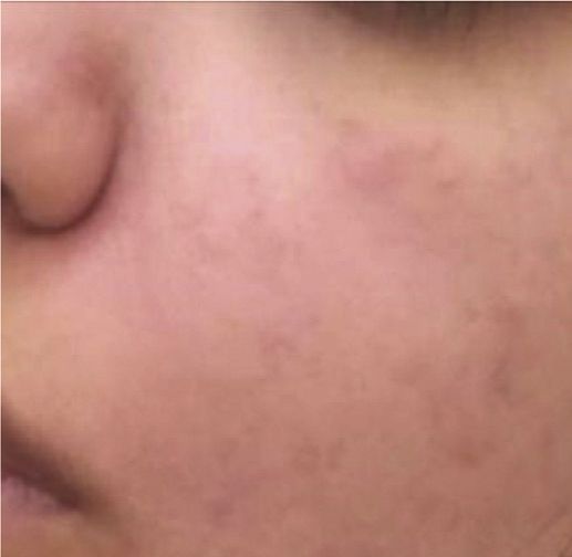
2                                                                                         Dermatology Research and Practice

They work by stimulating a robust wound healing after            classified as excellent, satisfactory, or nonsatisfactory. Ad-
creating zones of microscopic thermal injury surrounded by       ditionally, patients were asked to assess their satisfaction at
normal skin to help complete and rapid reepithelization          each visit and during follow-up using a quartile grading
[8, 9]. Treatment of PIH in patients with dark skin is           scale: grade 1, less than 25% clearance; grade 2, 26–50%
challenging as the treatment itself might provoke inflam-         clearance; grade 3, 51–75% clearance; grade 4, more than
matory response and end up exacerbating PIH [10–12].             75% clearance.
Additionally, only few studies examined the safety and ef-
ficacy of laser treatment in skin of color [13, 14]. The efficacy
and safety data of nonablative fractional laser in Saudi         2.5. Statistical Analysis. Categorical variables were presented
patients is limited [15, 16] with no data that focus on new      as frequencies and percentages. Continuous variables were
technologies such as 1550 nm/1927 nm dual wavelength             presented as means and standard deviations (SD). Statistical
laser. The objective of the current study was to examine         Package for the Social Sciences software (SPSS Version 25.0;
efficacy and safety of nonablative 1927 nm wavelength laser        Armonk, NY, IBM Corp) was used for all statistical analyses.
followed by a depigmenting cream in the treatment of Saudi
patients with PIH.                                               3. Results
2. Methods                                                       A total nine patients with Fitzpatrick skin type IV and
                                                                 unsatisfactory response to topical depigmenting creams used
2.1. Setting and Design. The current study was conducted in      for at least three months were enrolled in this study. Table 1
a private dermatology practice in Riyadh, Saudi Arabia. It       shows clinical data of the enrolled patients. The affected sites
was a prospective interventional pilot study that was con-       were the abdomen (three patients), face (two patients),
ducted during 2019.                                              forearm, breast, legs, and dorsum of foot (one patient each).
                                                                 The cause of pigmentation was variable and included
                                                                 postprocedure (abdominoplasty, mammoplasty, and lipo-
2.2. Subjects. Patients enrolled in this study were of Fitz-     suction), postlaser treatment (leg and abdomen), eczema,
patrick skin type IV who had PIH provoked by different            acne, chemical peeling, and burn scare with PIH. The
reasons including dermatitis, acne, and chemical peeling as      pretreatment duration of pigmentation ranged between 3
well as previous laser treatments. All included patients had     and 11 months, with an average of 6.7 ± 2.5 months. The
unsatisfactory response to topical bleaching creams used for     number of laser sessions received ranged between one and
at least three months before enrolling in this study. Pho-       four sessions, with an average of 1.7 ± 1.0 sessions. Five
tography was taken before each session and 6 weeks after the     patients (55.6%) received one session, three patients (33.3%)
last session. Photographs were taken using a digital camera      received two sessions, and one patient (11.1%) received four
under a constant light setting.                                  sessions.
                                                                     One patient was lost for follow-up after receiving one
2.3. Laser Treatments. Skin preparation was carefully per-       session and the downtime and response could not be
formed with a gentle cleanser to remove debris and makeup        evaluated. The downtime of the eight patients evaluated
before treatment. A topical anesthetic ointment was applied      ranged between 5 and 7 days, with an average of 5.9 ± 0.8
to the treatment area approximately 30 minutes before            days. The downtime was manifested as edema and erythema
treatment. The laser treatment used in this study was de-        in the first 24 hours followed by superficial crustations at
                                                                 sites of MTZ which slough over later on. According to
                  ®
livered by Fraxel DUAL 1550/1927 laser system (Solta
Medical, USA). All patients were treated with nonablative        independent dermatologist evaluation, three of the eight
1927 nm thulium fiber laser. Treatments were performed            patients had excellent response (37.5%), four patients had
with 30% surface area coverage at pulse energy of 20 mJ (per     satisfactory response (50.0%), and one patient had non-
microthermal zone (MTZ)). Only four passes were done in          satisfactory response (12.5%). According to the patient own
order to avoid overheating. Patients received one to four        evaluation, three of the eight patients had grade 4 clearance
sessions (6 weeks apart) according to the response. Patients     (37.5%), three had grade 3 clearance (37.5%), and two had
were given oral steroid 0.5 mg/kg after each session in ad-      grade 2 clearance (25.0%). Low patient satisfaction was
dition to topical clobetasol cream for 7 days to keep in-        observed in postabdominal liposuction and burn scar at the
flammatory response to minimum. Patients were also given          dorsum of foot. Dermatologist and patient evaluations were
topical hydroquinone 4% cream twice daily for 6 weeks            similar in all but one patient.
starting one week after the laser treatment and strict sun-          Improvement was more evident in first session and it
screen was advised.                                              declined in subsequent sessions. The three patients who had
                                                                 excellent response received only one session while the pa-
                                                                 tient who had nonsatisfactory response received four ses-
2.4. Outcome Evaluation. It was done by comparing the            sions. Facial lesions had excellent improvement (Figure 1).
before and after digital photographs taken at each visit.        The pretreatment duration of pigmentation was 5.7 ± 3.0
Improvement was assessed by two blinded independent              months in three patients who had excellent response and
dermatologist evaluators using a visual analog scale for the     7.4 ± 2.5 months in the other five patients. None of the
percentage of pigment clearance. The final response was           patients had paradoxical pigmentation after treatment.
Dermatology Research and Practice

                                 Table 1: Response to 1927 nm wavelength laser among patients with postinflammatory hyperpigmentation.
                                                             Duration of                                      Dermatologist
                  Affected         Cause of        Skin                         Number of       Downtime                           Patient evaluation of    Paradoxical
    Age Gender                                              pigmentation                                       evaluation of
                    site        pigmentation      type                        laser sessions    (days)                                improvement         pigmentation
                                                              (months)                                         improvement
1   22    F        Face       Chemical peeling    IV             3                  1             7              Excellent                 4                 None
2   18    F        Face             Acne          IV             5                  1             7              Excellent                 4                 None
3   28    F      Forearm          Eczema          IV             7                  1             5             Satisfactory               3                 None
4   39    F       Breast     Postmammoplasty      IV             4                  2             6             Satisfactory               3                 None
5   25    F        Legs      Laser hair removal   IV             8                  2             6             Satisfactory               3                 None
6   42    F      Abdomen       Postliposuction    IV             7                  4             5           Not satisfactory             2                 None
                             Postcarbondioxide
7   21    F      Abdomen                          IV             9                  1             6              Excellent                 4                 None
                                     laser
                 Dorsum of
8 30      F                  Burn scar with PIH   IV             11                 2             5             Satisfactory               2                 None
                   foot
9 44      F      Abdomen     Postabdominoplasty   IV             6                  1            NA                 NA                    NA                  NA
                                                                                                                                                                         3
4 Dermatology Research and Practice
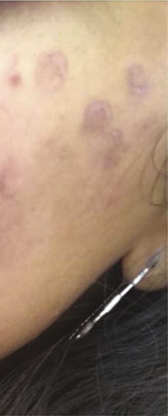
(a) (b)
Figure 1: Before and after photographs of a female patient with postinflammatory hyperpigmentation secondary to chemical peeling with
excellent response after one session of 1927 nm wavelength laser treatment combined with topical hydroquinone 4% cream for 6 weeks after
the laser treatment.
4. Discussion higher coefficient for absorption of water compared with the
1,550 nm wavelength emitted by the same DUAL machine,
We are reporting our successful experience in treating pa- which allows greater ability to target epidermal lesions such as
tients with PIH of different reasons with nonablative pigmentation and dyschromia [17]. This may also explain the
1927 nm wavelength laser followed by a depigmenting less satisfactory response among the two patients who had PIH
cream. The majority of the current patients had either ex- with scaring secondary to second degree burn and liposuction.
cellent or satisfactory improvement. Similar findings were Additionally, scaring may affect the fractional photo-
reported in the few reports that examined the same wave- thermolysis mechanism of laser and consequently obstruct the
length in dark-skinned patients with PIH [13, 14]. For ex- transepidermal elimination of dermal content [19].
ample, Wilson and colleagues reported 50% improvement During the current study, we did not encounter any
after 4 laser treatment sessions with or without topical paradoxical pigmentation which might be explained by
depigmenting cream among 40 patients with dark skin who using low energy low density wavelength in addition to the
had facial hyperpigmentation and/or melasma [13]. Simi- use of depigmenting cream and strict sun avoidance after
larly, Bae and colleagues reported 43% improvement after at treatment. Similarly, no scaring or paradoxical pigmentation
least 2 laser treatment sessions without topical depigmenting was reported in the studies that used nonablative 1927 nm
cream among 60 patients with dark skin who had PIH [14]. wavelength laser among dark-skinned [13, 14] and light-
Interestingly, nonablative 1927 nm wavelength laser was skinned [17, 20] patients with PIH. The modest downtime
reported to have better response in light-skinned patients experienced by the current patients was similar to what
with PIH [17, 18]. For example, Polder and colleagues re- reported before in the form of tolerable pain, moderate
ported >75% improvement after three laser treatment ses- erythema, and mild edema that resolve within 7–10 days
sions among 9 light-skinned patients with nonfacial [17, 20].
hyperpigmentation [17]. Similarly, Brauer and colleagues Improvement in the current study was more evident in
reported marked to very significant improvement in 55% of first session and it declined in subsequent sessions. Con-
the 23 patients with largely light skin after 4–6 laser treat- sistently, Wilson and colleagues showed that the improve-
ment sessions to treat facial PIH or melasma [18]. ment does not considerably change after the first two laser
The excellent or satisfactory improvement in the majority treatment sessions [13]. The improvement was 43.5% after 2
of our patients may be related to the use of topical hydro- sessions, 44.3% after 3 sessions, 40.6% after 4 sessions, and
quinone 4% cream for 6 weeks after the laser treatment. 43.8% after >5 sessions [13]. Improvement in the current
Consistent with this hypothesis, Wilson and colleagues re- study was better in facial lesions and lesions with shorter
ported a better response to nonablative laser treatment as pretreatment duration of pigmentation. This might be re-
assessed by Global Aesthetic Improvement Scale at week 12 lated to the severity of condition rather than response to
posttreatment among patients with facial hyperpigmentation laser itself. Lower MTZ density appears to be an important
who were concomitantly receiving topical hydroquinone factor to protect against PIH and severe downtime after laser
compared with those who were concomitantly receiving a treatment [11, 21, 22].
bland moisturizer [13]. Additionally, it may be also related to In conclusion, this pilot study showed that low energy
the appropriate choice of the patients with epidermal lesions. low density nonablative fractional 1927 nm wavelength laser
For example, nonablative 1927 nm wavelength laser has a treatment followed by topical hydroquinone 4% cream for 6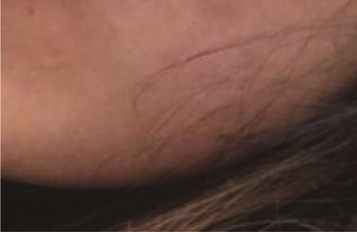
Dermatology Research and Practice                                                                                                            5

weeks after the laser treatment is a safe and effective mo-              [13] V. Wilson, I. T. Jones, J. Bolton, L. Larsen, and S. G. Fabi, “The
dality for improving PIH in patients with darker skin types.                 safety and efficacy of treatment with a 1,927-nm diode laser
                                                                             with and without topical hydroquinone for facial hyperpig-
                                                                             mentation and melasma in darker skin types,” Dermatol Surg,
Data Availability                                                            vol. 44, no. 10, pp. 1304–1310, 2018.
                                                                        [14] Y. S. C. Bae, S. Rettig, E. Weiss, L. Bernstein, and
All data used in this study are available upon request.                      R. Geronemus, “Treatment of post-inflammatory hyperpig-
                                                                             mentation in patients with darker skin types using a low
Conflicts of Interest                                                        energy 1,927 nm non-ablative fractional laser: a retrospective
                                                                             photographic review analysis,” Lasers in Surgery and Medi-
The author declares no conflicts of interest.                                 cine, vol. 52, no. 1, pp. 7–12, 2020.
                                                                        [15] S. Altalhab, M. Aljamal, T. Mubki et al., “Q-switched 532 nm
                                                                             Nd:YAG laser therapy for physiological lip hyperpigmenta-
References                                                                   tion: novel classification, efficacy, and safety,” Journal of
                                                                             Dermatological Treatment, pp. 1–5, 2020, inprint.
 [1] V. D. Callender, S. St.Surin-Lord, E. C. Davis, and M. Maclin,     [16] M. A. Alharbi, “Q-switched double-frequency Nd:YAG (532
     “Postinflammatory hyperpigmentation,” American Journal of                nm) laser is an effective treatment for racial lip pigmentation,”
     Clinical Dermatology, vol. 12, no. 2, pp. 87–99, 2011.                  Journal of Cosmetic Dermatology, vol. 18, no. 6, pp. 1672–1674,
 [2] E. C. Davis and V. D. Callender, “Postinflammatory hyper-                2019.
     pigmentation: a review of the epidemiology, clinical features,     [17] K. D. Polder, A. Harrison, L. E. Eubanks, and S. Bruce, “1,927-
     and treatment options in skin of color,” The Journal of Clinical        nm fractional thulium fiber laser for the treatment of non-
     and Aesthetic Dermatology, vol. 3, no. 7, pp. 20–31, 2010.              facial photodamage: a pilot study,” Dermatologic Surgery,
 [3] B. P. Kaufman, T. Aman, and A. F. Alexis, “Postinflammatory              vol. 37, no. 3, pp. 342–348, 2011.
     hyperpigmentation: epidemiology, clinical presentation,            [18] J. A. Brauer, H. Alabdulrazzaq, Y. S. Bae, and
     pathogenesis and treatment,” American Journal of Clinical               R. G. Geronemus, “Evaluation of a low energy, low density,
     Dermatology, vol. 19, no. 4, pp. 489–503, 2018.                         non-ablative fractional 1927 nm wavelength laser for facial
 [4] N. Silpa-archa, I. Kohli, S. Chaowattanapanit, H. W. Lim, and           skin resurfacing,” Journal of Drugs in Dermatology: JDD,
     I. Hamzavi, “Postinflammatory hyperpigmentation: A com-                  vol. 14, no. 11, pp. 1262–1267, 2015.
     prehensive overview: Epidemiology, pathogenesis, clinical          [19] B. M. Hantash, V. P. Bedi, V. Sudireddy, S. K. Struck,
     presentation, and noninvasive assessment technique,” Journal            G. S. Herron, and K. F. Chan, “Laser-induced transepidermal
     of the American Academy of Dermatology, vol. 77, no. 4,                 elimination of dermal content by fractional photo-
     pp. 591–605, 2017.                                                      thermolysis,” Journal of Biomedical Optics, vol. 11, no. 4,
 [5] S. Chaowattanapanit, N. Silpa-Archa, I. Kohli, H. W. Lim, and           Article ID 041115, 2006.
     I. Hamzavi, “Postinflammatory hyperpigmentation: A com-             [20] V. A. Narurkar, T. S. Alster, E. F. Bernstein, T. J. Lin, and
     prehensive overview: Treatment options and prevention,”                 A. Loncaric, “Safety and efficacy of a 1550nm/1927nm dual
     Journal of the American Academy of Dermatology, vol. 77,                wavelength laser for the treatment of photodamaged skin,”
     no. 4, pp. 607–621, 2017.                                               Journal of Drugs in Dermatology: JDD, vol. 17, no. 1, pp. 41–46,
 [6] O. Agbai, I. Hamzavi, and J. Jagdeo, “Laser treatments for              2018.
     postinflammatory hyperpigmentation,” JAMA Dermatology,              [21] L. Izikson and R. R. Anderson, “Resolution of blue mino-
     vol. 153, no. 2, pp. 199–206, 2017.                                     cycline pigmentation of the face after fractional photo-
 [7] T. Barrett and S. de Zwaan, “Picosecond alexandrite laser is            thermolysis,” Lasers in Surgery and Medicine, vol. 40, no. 6,
     superior to Q-switched Nd: YAG laser in treatment of                    pp. 399–401, 2008.
     minocycline-induced hyperpigmentation: a case study and            [22] M. H. Jih and A. Kimyai-Asadi, “Fractional photothermolysis:
     review of the literature,” Journal of Cosmetic and Laser                a review and update,” Seminars in Cutaneous Medicine and
     Therapy, vol. 20, no. 7-8, pp. 387–390, 2018.                           Surgery, vol. 27, no. 1, pp. 63–71, 2008.
 [8] E. P. Tierney, D. J. Kouba, and W. C. Hanke, “Review of
     fractional photothermolysis,” Dermatologic Surgery, vol. 35,
     no. 10, pp. 1445–1461, 2009.
 [9] D. Manstein, G. S. Herron, R. K. Sink, H. Tanner, and
     R. R. Anderson, “Fractional photothermolysis: a new concept
     for cutaneous remodeling using microscopic patterns of
     thermal injury,” Lasers in Surgery and Medicine, vol. 34, no. 5,
     pp. 426–438, 2004.
[10] A. Alajlan, “Crescent-shaped hyperpigmentation following
     laser hair removal: case series of fifteen patients,” Lasers in
     Surgery and Medicine, vol. 53, no. 3, 2020.
[11] H. H. L. Chan, D. Manstein, C. S. Yu, S. Shek, T. Kono, and
     W. I. Wei, “The prevalence and risk factors of post-inflam-
     matory hyperpigmentation after fractional resurfacing in
     Asians,” Lasers in Surgery and Medicine, vol. 39, no. 5,
     pp. 381–385, 2007.
[12] S. B. Kaushik and A. F. Alexis, “Nonablative fractional laser
     resurfacing in skin of color: evidence-based review,” The
     Journal of Clinical and Aesthetic Dermatology, vol. 10, no. 6,
     pp. 51–67, 2017.
You can also read