Case Report Contrast-Enhanced Mammography in the Diagnosis of Breast Angiosarcoma
←
→
Page content transcription
If your browser does not render page correctly, please read the page content below
Hindawi
Case Reports in Radiology
Volume 2021, Article ID 5542786, 7 pages
https://doi.org/10.1155/2021/5542786

Case Report
Contrast-Enhanced Mammography in the Diagnosis of
Breast Angiosarcoma

          Maya Grisaru Kacen ,1 Nikhil Sangle ,2 and Anat Kornecki                                    1

          1
              Department of Medical Imaging, Breast Division, St. Joseph’s Health Care, Western University, London Ontario, Canada
          2
              Department of Pathology and Laboratory Medicine, London Health Sciences Center & St. Joseph’s Health Care,
              London Ontario, Canada

          Correspondence should be addressed to Anat Kornecki; anat.kornecki@sjhc.london.on.ca

          Received 17 March 2021; Revised 18 April 2021; Accepted 20 July 2021; Published 13 August 2021

          Academic Editor: Iain D. Lyburn

          Copyright © 2021 Maya Grisaru Kacen et al. This is an open access article distributed under the Creative Commons Attribution
          License, which permits unrestricted use, distribution, and reproduction in any medium, provided the original work is properly cited.

          A 60-year-old female presented for further assessment of a new right breast lump (November 2020). She had a history of a stage I
          (T1bN0M0) right breast invasive mammary carcinoma, grade 2 (score 7/9) with receptors ER/PR-negative, HER2/neu-positive,
          diagnosed four years prior to her current presentation. At that time, she was treated with a right breast lumpectomy and local
          radiation. Breast assessment with contrast-enhanced mammography showed new skin thickening with associated enhancement
          within the palpable region. Histology of subsequent ultrasound-guided biopsy found radiation-induced breast angiosarcoma.
          Breast angiosarcoma is a rare entity that represents less than 1% of all breast cancers. To our knowledge, this is the first case
          describing the imaging findings of breast angiosarcoma on contrast-enhanced mammography.

1. Introduction                                                          reporting radiologist must be familiar with the presentation
                                                                         of different pathologies on all imaging modalities.
Breast angiosarcoma is a rare entity associated with poor                    This case study offers the opportunity to learn about the
prognosis, accounting for 1 in 2000 cases of primary breast              appearance of radiation-induced breast angiosarcoma on
cancers [1]. The appearance on mammogram and ultrasound                  CEM.
is nonspecific with high false-negative rates, and as a result,
contrast-enhanced MRI is the imaging investigation of
choice for evaluation of this rare condition [1–3].                      2. Patient Information
    Contrast-enhanced mammography (CEM) is an emerging
breast imaging modality that combines two-dimensional (2D)               A 60-year-old female with a history of previously treated right
digital mammography with iodine contrast administration.                 breast carcinoma presented to our breast care centre with new
The exam utilizes a dual-energy technique, consisting of low-            complaints of right breast skin discoloration and a palpable
energy (LE) images, which are equivalent to 2D digital mam-              lump within the region of the previous lumpectomy site.
mography, and high-energy (HE) images, which capture the                     Her previous breast cancer was stage I (T1bN0M0), grade
information generated by the iodine and are nondiagnostic.               2 (score 7/9), negative for estrogen and progesterone hormone
The interpretation of CEM is based on the LE images and                  receptors, and positive for HER2/neu. Treatment consisted of
recombined images, which are generated by subtraction of                 a lumpectomy, adjuvant TC (cyclophosphamide) chemother-
LE images from the HE images. With a sensitivity that                    apy, and Herceptin. This was followed by local breast radiation
approaches that of MRI [4], CEM provides similar informa-                therapy of 4250 cGy in 16 fractions completed by September
tion more rapidly and at a lower cost [4, 5]. However, the               2017, three years prior to her new palpable concerns.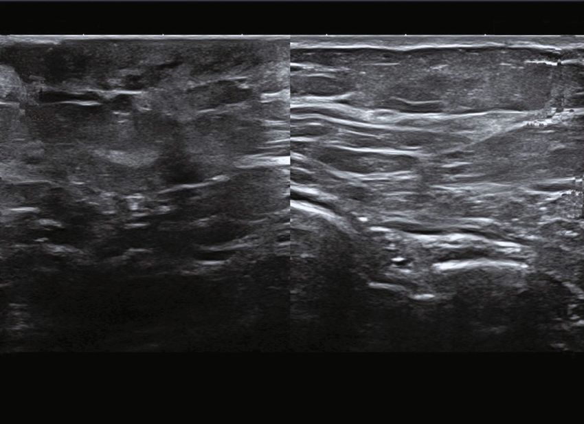
2                                                                                                             Case Reports in Radiology

                                    (a) LE, CC view, right breast            (b) LE, CC view, left breast

                                      (c) LE, MLO view, right breast        (d) LE, MLO view, left breast

Figure 1: (a–d) Bilateral LE images in a craniocaudal (CC) and medial oblique (MLO) views. Posttreatment changes at the right inner breast
are best appreciated on the CC view (arrow, (a)). A separate tissue marker is demonstrated in the right upper outer quadrant is related to a
previous benign biopsy.

3. Clinical Findings                                                    mide, and Herceptin), followed by 16 fractions of radiation
                                                                        therapy which were completed in September 2017. She pre-
The patient’s physical examination was conducted by a                   sented for her yearly routine mammographic surveillance in
breast surgeon. The patient presented with right breast                 September 2017, August 2018, and August 2019. Her surveil-
erythematous skin discoloration associated with multiple                lance appointment for August 2020 was postponed due to
dermal lesions extending medially with the total region of              COVID-19, and by November 2020, she presented with new
involvement spanning over 8 cm. No palpable lymph nodes                 symptoms and was diagnosed with angiosarcoma. Right breast
were noted on physical examination.                                     modified mastectomy was completed in March 2021.

4. Timeline                                                             5. Diagnostic Assessment
In early November 2016, the patient presented with a palpa-             Contrast-enhanced mammography was performed following
ble lump within the right breast. By the end of November                the administration of intravenous 87 ml Omnipaque 350 mg.
2016, she was diagnosed with right breast invasive mammary              Bilateral LE images (Figures 1(a)–1(d)) showed posttreat-
carcinoma, grade 2 (score 7/9), ER/PR-negative, and HER2-               ment changes at the right upper inner quadrant. Comparison
positive receptors. After her right breast lumpectomy in                to previous exams from one and three years prior showed a
January 2017, she was treated with chemotherapy and                     new mild increase in skin thickening and trabecular thicken-
HER2 inhibitor targeted therapy (Taxotere, cyclophospha-                ing in the right inner breast (Figures 2(a)–2(c)). Recombined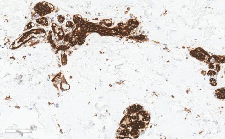
Case Reports in Radiology 3
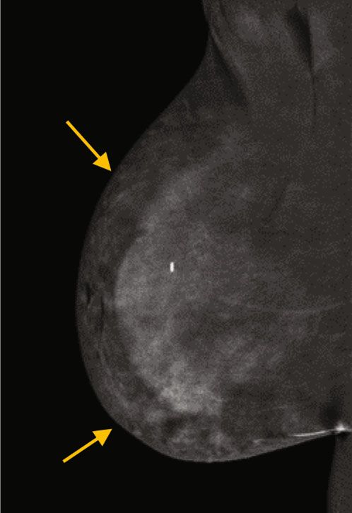
(a) Right CC view 2020 (b) Right CC view 2019
(c) Right CC view 2017
Figure 2: (a–c) Right LE image in a craniocaudal view (a) compared to the previous craniocaudal view from 2D mammography exams from
one year ((b), 2019), and 3 years ((c), 2017) before show new mild skin thickening (arrow, (a)) and trabecular thickening (arrowhead, (a)).
views (Figures 3(a)–3(d)) showed minimal bilateral back- uled for a right modified radical mastectomy, using a skin
ground parenchymal enhancement (BPE) with marked non- flap for skin closure due to the large defect. The postsurgical
mass enhancement noted on the right breast, subcutaneous pathology report described a 23:3 × 20 × 5:5 cm breast tissue
region. Targeted the US of the right breast demonstrated specimen with irregular skin lesions composed of multiple
marked skin thickening in the area of concern, associated confluent red papules extending over an area of 16 × 12:8
with increased vascularity (Figures 4(a)–4(c)). This demon- cm. Histologically, there was an infiltrative, cytologically
strates marked skin thickening with no corresponding intra- malignant, vasoformative neoplasm present within the
parenchymal breast lesion, similar to this case. Pathological dermis and superficial subcutis. The nipple and areola were
slides images are also provided (Figures 5(a)–5(d)). MRI involved. Mitotic activity was not observed. The lesional cells
and mammography images from a different patient are pro- stained diffusely positively for CD31 and ERG, while C-MYC
vided for teaching purposes (Figures 6(a)–6(c)). showed patchy positivity. All histologically confirmed resid-
Ultrasound-guided biopsy was performed targeting the ual radiation-induced angiosarcoma showed minimal to no
subcutaneous skin thickening, using a 14-gauge core biopsy histological response to preoperative neoadjuvant chemo-
needle. Pathology results confirmed an infiltrative, cytologi- therapy (paclitaxel). The surgical skin resection margins were
cally malignant, vasoformative neoplasm within the sampled at least 1.0 cm from the angiosarcoma.
tissues. The lesion cells stained positively for CD31, ERG, and
c-Myc. The c-Myc immunopositivity is characteristic of 6. Discussion
radiation-associated angiosarcoma.
Angiosarcoma of the breast is a rare entity which accounts
5.1. Therapeutic Intervention and Follow-Up. Since the for less than 1% of all breast malignancies and has a poor
patient had CEM, DCE-MRI was not necessary. Staging chest prognosis [2, 3, 6]. Primary and secondary forms of angiosar-
and abdominal CT study was obtained in January 2021 with coma are clinically distinct. Secondary angiosarcomas are
no evidence of distant metastasis. PET-CT was not war- usually induced either by radiation therapy or are detected
ranted. The patient received chemotherapy and was sched- in the setting of lymphedema. Angiosarcoma, in the setting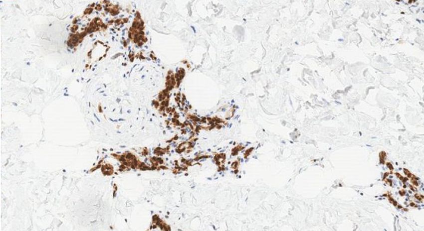
4                                                                                                       Case Reports in Radiology

                                  (a) CEDM, CC view, right breast     (b) CEDM, CC view, left breast

                                      (c) MLO view, right breast        (d) MLO view, left breast

Figure 3: (a–d) Bilateral recombined images in a craniocaudal (CC) and medial oblique (MLO) views. There is minimal background
parenchymal enhancement. Marked nonmass enhancement is noted predominantly within the inner aspect of the right breast, especially
in the subcutaneous peripheral areas (yellow arrows, (a, c)).

of lymphedema, is more likely to occur in breast cancer             Since these changes are expected in a patient with prior lump-
patients who underwent radical mastectomy [1, 6, 7].                ectomy and radiation treatment, one must be able to detect
Radiation-induced angiosarcoma generally occurs after               even the mildest interval differences. Angiosarcomas charac-
breast conservation surgery and radiation therapy and only          teristically result in high signal on T2 and low signal on T1
rarely occurs after mastectomy. The average time between            DCE-MRI-weighted images. Some studies report a rapid
radiation therapy and the development of angiosarcoma is            enhancement followed by rapid washout [8], while others
six years [1, 6]. However, several reports indicate that a diag-    report prolonged enhancement with no washout [2–6]. Given
nosis of angiosarcoma may occur as early as one to two years        the overlapping reported kinetic curves, a malignant entity
or as late as 41 years after treatment [6]. The mean age of pre-    should still be considered in the differential diagnosis [6] keep-
sentation is in the late 60s [3, 6]. It usually affects the dermis   ing in mind that noncancerous, postirradiation breast tissue
of the breast within the radiation field but may occasionally        changes are not expected to enhance on either CEM or
develop within the breast parenchyma [6]. On clinical pre-          DCE-MRI three years after treatment.
sentation, the breast can be firm on palpation and there                  In our department, contrast-enhanced mammography is
may be associated skin discoloration ranging from blue,             offered routinely to provide rapid assessment in different
red, and purple [1, 3, 5, 6]. Dimpling of the skin is sometimes     clinical settings, including symptomatic patients. In the pres-
present as an indication of tissue congestion.                      ent case, CEM demonstrated minimal BPE, as expected for
    Mammography and ultrasound of radiation-induced                 the patient’s age group. As with DCE-MRI, decreased BPE
angiosarcomas do not have characteristic pathognomonic              on CEM improves specificity (9). While CEM is not a
imaging features. Imaging findings may be as indolent as mild        dynamic scan, the kinetic curve for angiosarcoma is not spe-
skin thickening and trabecular changes, as seen in our case.        cific anyway, as discussed above. The main disadvantage of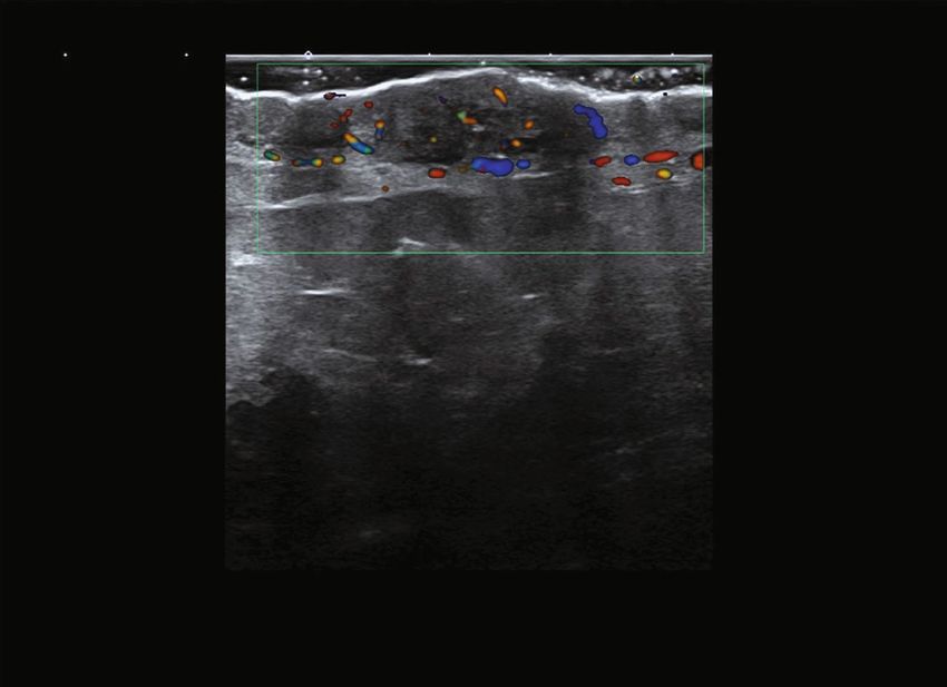
Case Reports in Radiology 5
0
T
Mlm
(0.8)
i24LX8
d19
30 fps
Qscan 1
G: 87
DR: 60
A: 3
P: 4
2
4 mm
3
4
RT 3:30 7 CM FN
Palpable
# 53
(a) Grayscale ultrasound of the right breast
Precision+ A pure+
0
T 3.4
Mlm
(0.9)
i24LX8
d19
8 fps
Qscan 1
G: 87
DR: 60
3.4
cm/s
CF 8.8
CG: 41
F: 5
2
4 mm
3
4
RT 3:30 7 CM FN
Palpable
#6
(b) Doppler ultrasound of the right breast
A pure+ Precision+ T Precision+ A pure+ T
0 0
Mlm Mlm
(0.7) (0.7)
i24LX8 i24LX8
d19 d19
31 fps 31 fps
Qscan Qscan
1 G: 87 G: 87 1
DR: 60 DR: 60
A: 3 A: 3
P: 4 P: 4
2 2
4 mm
3 3
4 4
LT 830 7CMFN
RT 3:30 7 CM FN
Palpable
# 172 # 99
(c) Grayscale ultrasound comparing the two breasts, demonstrating the area of concern within the right breast, and the corresponding contralateral side with
normal breast tissue and skin
Figure 4: (a–c) Selected ultrasound exam on the right breast shows marked skin thickening measuring up to 7.2 mm, in the area of concern
(a), associated with increased flow (b).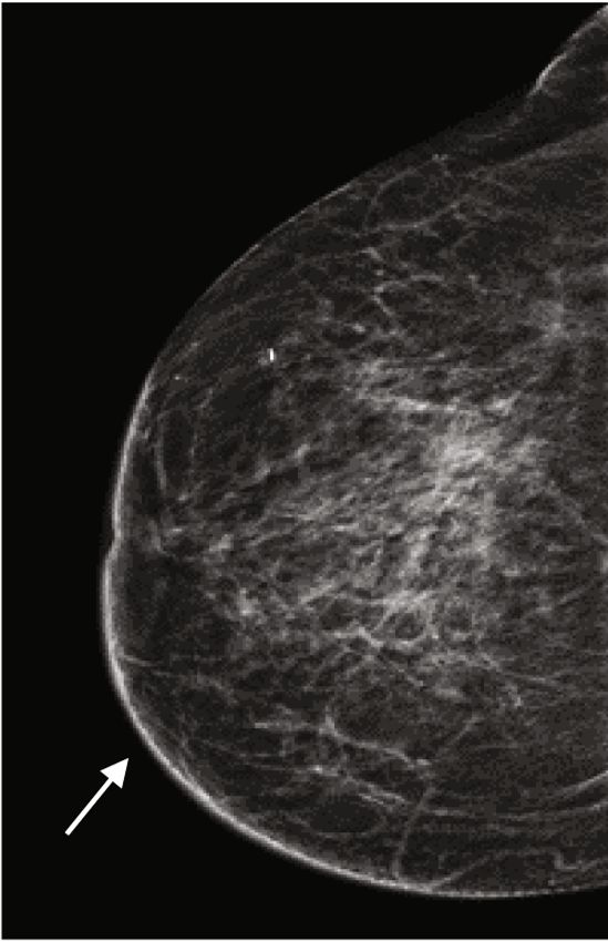
6 Case Reports in Radiology
(a) (b)
(c) (d)
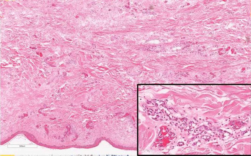
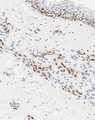
Figure 5: (a–d) Hematoxylin and eosin (H&E) stained section ((a), ×40 magnification) showing skin with underlying dermis containing an
infiltrative vasoformative neoplasm. The endothelial cells lining these vascular spaces are cytologically atypical (inset, H&E ×400
magnification). Immunohistochemistry staining (×200 magnification) showed diffuse positivity in the malignant cells for CD31 (b), patchy
staining for C-MYC (c), and nuclear immunoreactivity for ERG (d).
(a) T1 FS+C axial MRI (b) MIP reconstruction axial MRI (c) Right CC view
Figure 6: (a–c) This is a different case. An 80-year-old patient with remote history of right breast lumpectomy and radiation treatment due to
breast malignancy. Patient presented with similar clinical symptoms of skin changes and discoloration. Axial T1 with fat suppression (FS),
postcontrast injection (+C) MRI (Figure 6(a)), and MIP reconstruction (Figure 6(b)) demonstrating marked skin thickening and
enhancement. Corresponding right CC mammographic view (Figure 6(c)) demonstrating marked skin thickening with no discreet mass
within the breast.
CEM, compared to DCE-MRI, is providing only 2D views skin lesion with no evidence of intraparenchymal breast
which makes localizing and distinguishing subcutaneous tissue mass. Angiosarcoma, inflammatory process (benign
enhancement from BPE more difficult. In our case, decreased and malignant), and lymphoma were included in the differ-
BPE on the contralateral breast with marked asymmetric ential diagnoses.
enhancement in the peripheral subcutaneous regions The prognosis of angiosarcoma depends on the tumour
(Figures 3(a)–3(d)), along with new skin thickening, indi- size, histological grade, and presence of metastasis at the time
cated to us that this was an abnormal exam. The ultrasound of presentation. Early diagnosis may play a key role in
confirmed the feasibility of ultrasound-guided biopsy of a improving the morbidity and mortality of this condition.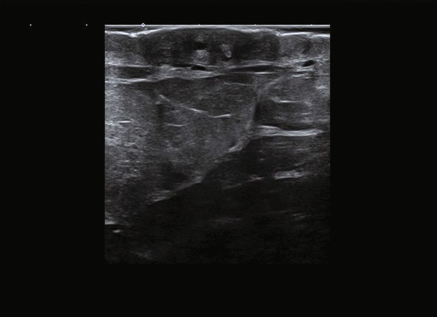
Case Reports in Radiology                                                                                                               7

Therefore, secondary breast angiosarcomas should be con-              [4] K. F. Ghaderi, J. Phillips, H. Perry, P. Lotfi, and T. S. Mehta,
sidered in the differential diagnosis when it is clinically                “Contrast-enhanced mammography: current applications and
appropriate. The combination of new skin thickening seen                  future directions,” Radiographics, vol. 39, no. 7, pp. 1907–
on LE images associated with enhancment seen on recom-                    1920, 2019.
bined views and patient medical history, made us consider             [5] M. S. Jochelson, K. Pinker, D. D. Dershaw et al., “Comparison of
radiation-induced breast angiosarcoma in the differential                  screening CEDM and MRI for women at increased risk for
diagnosis. The ability to perform second look ultrasound,                 breast cancer: a pilot study,” European Journal of Radiology,
ultrasound guided biopsy and CEM on the same day, made                    vol. 97, pp. 37–43, 2017.
it possible for us to reach the final diagnosis more rapidly.          [6] K. N. Glazebrook, M. J. Magut, and C. Reynolds, “Angiosar-
This points to the main advantages of CEM in comparison                   coma of the breast,” American Journal of Roentgenology,
to DCE-MRI.                                                               vol. 190, no. 2, pp. 533–538, 2008.
    Due to the infrequent incidence of breast angiosarcoma,           [7] A. Hui, M. Henderson, D. Speakman, and A. Skandarajah,
there are no randomized trials comparing wide local excision              “Angiosarcoma of the breast: a difficult surgical challenge,”
and breast-conserving approach with mastectomy as treat-                  Breast, vol. 21, no. 4, pp. 584–589, 2012.
ment options. Given the aggressiveness and poor prognosis             [8] R. F. Lim and R. Goei, “Angiosarcoma of the Breast,” Radio-
of this disease, total mastectomy with or without axillary                Graphics, vol. 27, Supplement 1, pp. S125–S130, 2007.
node dissection is the preferred surgical treatment. The
necessity for axillary node dissection is unclear since the
hematogenous spread is more common in angiosarcomas
than the lymphatic spread [8]. The reported median survival
period ranges from 14.5 to 37 months, with a 5-year survival
rate of 15% [6, 8].

7. Conclusion
This rare case of radiation-induced breast angiosarcoma in a
symptomatic patient could have been missed by routine
mammography alone, or the diagnosis could have been
delayed if breast MRI was required. This is an example where
a CEM study can replace DCE-MRI. The main imaging find-
ing that raised concern for breast angiosarcoma in this case
was the abnormal skin enhancement that was appreciated
on the CEM which led to further assessment with targeted
US and US-guided biopsy. This rare diagnostic case can be
added to the multitude of studies showing the advantages
of CEM in the diagnostic setting by allowing rapid diagnosis.

Consent
We reviewed the case with the ethical committee, and patient
informed consent was not required for the purpose of this
case study.

Conflicts of Interest
The author has no conflict of interest to declare.

References
[1] N. C. Hodgson, C. Bowen-Wells, F. Moffat, D. Franceschi, and
    E. Avisar, “Angiosarcomas of the breast: a review of 70 cases,”
    American Journal of Clinical Oncology, vol. 30, no. 6, pp. 570–
    573, 2007.
[2] Y. Kikawa, Y. Konishi, Y. Nakamoto et al., “Angiosarcoma of
    the breast - specific findings of MRI,” Breast Cancer, vol. 13,
    no. 4, pp. 369–373, 2006.
[3] R. J. Weinfurtner and S. Falcon, “Primary and secondary angio-
    sarcoma of the breast,” Contemporary Diagnostic Radiology,
    vol. 39, no. 18, pp. 1–5, 2016.
You can also read