Portal Vein Thrombosis in a Patient with Type 1 Diabetes Presenting as Acute Pyelonephritis
←
→
Page content transcription
If your browser does not render page correctly, please read the page content below
European Journal
                                                                             of Case Reports in
                                                                         Internal Medicine

                       Portal Vein Thrombosis in a Patient with Type 1 Diabetes
                                                  Presenting as Acute Pyelonephritis
                                   Zahra Abdulwahed Al Saeed1, Fatimah Mohammed Alabdrabalnabi1, AlAnoud AlAnazi1,2,
                                                Waleed Ibrahim Albaker1,2,3, Osma Abdullah Al-Sultan1,2,4
                                         1
                                             College of Medicine, Imam Abdulrahman Bin Faisal University, Dammam, Saudi Arabia
                          2
                              Department of Internal Medicine, College of Medicine, King Fahad University Hospital, Al Khobar, Saudi Arabia
        3
            Department of Endocrinology & Internal Medicine, College of Medicine, Imam Abdulrahman Bin Faisal University, Dammam, Saudi Arabia
               4
                   Department of Internal Medicine and Hematology, College of Medicine, King Fahad University Hospital, Al Khobar, Saudi Arabia

                                        Doi: 10.12890/2020_001391 - European Journal of Case Reports in Internal Medicine - © EFIM 2020

Received: 18/11/2019
Accepted: 31/12/2019
Published: 05/02/2020

How to cite this article: Al Saeed ZA, Alabdrabalnabi FM, AlAnazi A, Albaker WI, Al-Sultan OA. Portal vein thrombosis ina patient with type 1 diabetes
presenting as acute pyelonephritis. EJCRIM 2020;7: doi:10.12890/2020_001391.

Conflicts of Interests: The Authors declare that there are no competing interest
This article is licensed under a Commons Attribution Non-Commercial 4.0 License

ABSTRACT
Background: Few cases have been reported with respect to portal vein thrombosis in non-cirrhotic patients. Asymptomatic or non-specific
symptoms of portal vein thrombosis may lead to misdiagnosis or may delay the diagnosis until complications develop. We report a case of
portal vein thrombosis in a patient with type 1 diabetes presenting as acute pyelonephritis.
Case description: An 18-year-old female with type 1 diabetes on an insulin pump presented with epigastric abdominal pain for 3 days
associated with nausea and vomiting. She was a conscious, alert, young female who appeared to be in pain. Vital signs were stable with a
random blood sugar (RBS) level of 179 mg/dl. Abdominal examination revealed a soft and lax abdomen with tenderness in the epigastric area
and right renal angle, as well as no sign of rigidity or rebound tenderness. No signs of ascites, splenomegaly or hepatomegaly were noted.
Investigations showed a WBC count of 10.2, neutrophils at 65%, urine microsopy analysis revealed WBCs between 30–50 per high power
field, with culture showing >105 CFU/ml. All parameters of a thrombophilic screen were within normal values. Computed tomography
(CT) revealed reduced enhancement of the right kidney, likely indicating acute pyelonephritis, and left portal vein oedema with complete
occlusion. Local factors and prothrombotic disorders were ruled out. The patient was managed with ciprofloxacin, enoxaparin and warfarin.
Follow-up imaging revealed complete resolution of thrombosis.
Conclusions: Portal vein thrombosis is an uncommon condition in the absence of liver disease. Few case reports exhibit sepsis and portal
vein thrombosis. Sepsis can create a predisposed environment for hypercoagulability. To our knowledge, this is the first case report of
pyelonephritis with portal vein thrombosis.

LEARNING POINTS
• Until now, no cases have linked acute pyelonephritis to portal vein thrombosis.
• Suspect the presence of portal vein thrombosis in a diabetic patient presenting with unusual abdominal pain.
• Complete revascularization occurs with early treatment.

KEYWORDS
Portal vein thrombosis, acute pyelonephritis, type 1 diabetes

DOI: 10.12890/2020_001391                                                                       European Journal of Case Reports in Internal Medicine © EFIM 2020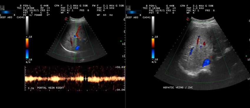
European Journal
of Case Reports in
Internal Medicine
INTRODUCTION
Portal vein thrombosis (PVT) refers to venous thrombosis that develops within the extrahepatic portal venous system which can extend to
the branches of the intrahepatic portal vein or up to the splenic vein and superior mesenteric vein[1]. PVT can occur either in association with
cirrhosis or malignancy of the liver or may present in the absence of liver disease. Few case studies report an association between sepsis and
PVT. We report the first case of PVT presenting with pyelonephritis in a type 1 diabetic patient.
CASE DESCRIPTION
An 18-year-old Saudi female known to have type I diabetes mellitus, who was on an insulin pump, presented to the emergency department
with epigastric abdominal pain of 3 days’ duration, which was non-radiating, colicky in nature and associated with attacks of nausea and
vomiting of food content. There was no haematemesis.
On examination: a conscious, alert, oriented young female appearing to be in pain, vital signs were stable with a temperature of 37.7°C and
a random blood sugar (RBS) level of 179 mg/dl. Abdominal examination revealed a soft and lax abdomen with tenderness in the epigastric
area and right renal angle. There were no signs of rigidity or rebound tenderness. Bowel sounds were present. There was no sign of ascites,
splenomegaly or hepatomegaly. The insulin pump was functioning and in place. Per rectal examination was normal.
The patient was initially evaluated by the general surgery team, with the impression of appendicitis. Abdominal ultrasound was normal; this
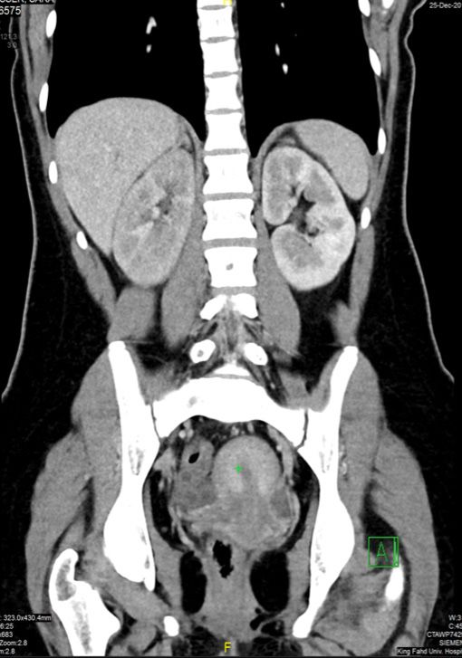
was followed by abdominal CT with contrast, which revealed reduced enhancement of the right kidney, likely indicating acute pyelonephritis.
In addition, the left portal vein was oedematous, with complete non-visualization of the supply to liver segment 2, in keeping with complete
left branch PVT; there was partial visualization of the right portal vein supplying liver segment 8 with surrounding oedema, in keeping with
partial non-occlusive right PVT (Figs. 1 and 2.)
Figure 1. CT scan of the abdomen showing intrahepatic periportal thickening and oedema Figure 2. Axial contrast-enhanced CT scan of the abdomen showing multiple areas
that is more severe at the left liver lobe ducts causing portal vein thrombosis of decreased enhancement (low attenuation) in the right kidney, and normal renal
arteries and veins. This is suggestive of acute pyelonephritisthrombosis
Laboratory investigations revealed a complete blood count showing haemoglobin of 10.7 g/dl, platelets of 255,000/mcl, a white blood cell
count of 10.2 × 10³ cells/mcl, neutrophils 65%. Renal and liver function results were within normal limits, as were amylase and lipase levels.
Haemoglobin A1c was 6.4%. The prothrombin time was 14.1 seconds (11.7–14.5), the INR was 1.48 and the activated partial thromboplastin
time was 30.9 seconds (24.1–34.7), the ESR was 48 mm/h (0–20) while CRP was 4.2 mg/dl (0.05–0.3).
DOI: 10.12890/2020_001391 European Journal of Case Reports in Internal Medicine © EFIM 2020European Journal
of Case Reports in
Internal Medicine
Urine analysis showed WBCs at 30–50 per high power field microscopy, pH=7, negative nitrite presence, while culture showed more than
10,000 colony forming units (CFU)/ml. A urine pregnancy test was negative.
The serological testing, including work-up for underlying thrombophilia, is shown in Table 1.
Gastrojejunoscopy and colonoscopy findings were normal. Biopsy revealed chronic non-specific inflammation, preservation of villus
architecture and no evidence of intraepithelial lymphocytosis or malignancy.
The patient was managed with ciprofloxacin and anticoagulation therapy (enoxaparin) followed by warfarin. She was then discharged on 8
mg of warfarin with a therapeutic INR. The long-term plan was to continue anticoagulation treatment for 6–12 months and follow up in the
outpatient clinic with gastroenterology and haematology assessment; the follow-up imaging showed complete resolution of thrombosis
(Fig. 3).
Figure 3. Follow-up Doppler and colour flow mapping with no sonographic evidence of thrombosis. The portal vein at the porta hepatis and its main right and left branches show
normal colour filling
Test Value Reference Range Interpretation
Anti-gliadin 0.9 CUEuropean Journal
of Case Reports in
Internal Medicine
DISCUSSION
Venous thromboembolism is a condition with varying aetiologies, often involving genetic predisposition, acquired or environmental factors[2].
In PVT, there are 3 major aetiologies: malignant thrombosis, chronic liver diseases – particularly, cirrhosis with portal hypertension – and
non-cirrhotic and non-malignant PVT[3].
In a retrospective study carried out in 2006 that included 23,796 post-mortem autopsies, the majority of thromboses occurred secondary
to malignancy involving the hepatobiliary region[4]. PVT was present in 14.3% of patients with primary hepatic malignancy accompanied
by cirrhosis[4]. Almost one-third of patients with PVT had cirrhotic liver disease. Nevertheless, isolated systemic or local factors have been
reported to cause PVT, such as suppurative infections occurring in areas where the drainage is ultimately near the portal vein or 1 of its
tributaries. Appendicitis and diverticulitis are considered the most attributable causes of septic thrombophlebitis[5].
Pyelonephritis should be suspected when there is a fever ≥37.8°C with at least 3 out of the 5 following criteria present: (1) lower urinary
tract symptoms; (2) flank pain; (3) costophrenic angle tenderness; (4) peripheral blood with leucocytosis ≥20,000/mm3 or neutrophils ≥65%;
(5) urinary analysis with WBCs ≥10 per high power field [6]. Our patient had 4 out of 5 of these criteria. CT is the initial study suggested by
the American College of Radiology in the assessment of pyelonephritis that has complex presentation as in diabetic or immunocompromised
patients [7]. Most of the studies comparing CT and ultrasonography (US) or Doppler US have shown that CT is superior to US in detecting
parenchymal abnormalities. Moreover, CT has higher sensitivity than Doppler US (81.0% and 33.3%, respectively)[8].
Pyelonephritis with renal vein thrombosis (RVT) has been reported in fewer than 10 cases in the literature, to the best of our knowledge [9–15].
Until now, no cases have linked acute pyelonephritis to PVT; instead, previous cases consider acute pyelonephritis as a cause of local
inflammation and sepsis and then, theoretically, it can cause thromboembolism like other factors.
It appears that RVT caused by acute pyelonephritis occurs in the right renal vein 60% of the time. Moreover, 80% of cases were related
to E. coli and Klebsiella pneumoniae[9–15]. These agents are Gram-negative endotoxin-releasing bacteria that stimulate thrombosis. The
mechanisms by which hypercoagulability is promoted include the prompting of a change in the surface of the endothelium and decreasing
the amount of anticoagulants. Tissue factor gene expression is also increased and cell production in the endothelium of the fibrinolytic
inhibitor plasminogen activator inhibitor-1 is enhanced [9].
In our patient, it is likely that local inflammation resulted in sepsis secondary to pyelonephritis.
The clinical presentation differs based on several factors, including: acuity, the degree of occlusion and the presence of malignant or benign
PVT. PVT commonly presents with abdominal pain, diarrhoea, rectal bleeding, vomiting, lactic acidosis, splenomegaly, anorexia and fever,
and sepsis can be variably present [16].
US is usually the investigation modality of choice for PVT. It shows solid, hyperechoic material in a distended portal vein or its tributaries,
the presence of collateral vessels or a cavernoma[17]. Colour Doppler imaging can show the absence of flow in part or all of the vasal lumen,
with sensitivity and specificity ranging from 66% to 100%[17,18]. The CT scan usually shows PVT as a hypodense filling defect in the portal vein
lumen, with partial or complete occlusion on contrast-enhanced scans[19]. Moreover, CT is useful for the identification of the possible cause
of the thrombosis or potential complications [20].
The next step after a diagnosis of PVT has been made is to start an extensive investigation of the cause of the thrombosis, which may include
local abdominal factors and prothrombotic disorders.
Our patient was extensively assessed for the presence of cirrhosis and local abdominal causes of thrombosis, which were negative. A
thrombophilia screen including testing for inherited genetic mutation for a hypercoagulable state was all normal, and no detectable
mutations were found. No apparent cause for the thrombosis was recognized except the presence of sepsis.
The aim of the treatment is to reverse or prevent advancement of thrombosis in the portal venous system and to treat complications
for established PVT. Early initiation of anticoagulation therapy within 30 days of symptoms manifesting is recommended, as there is no
spontaneous recanalization reported except in acute pancreatitis [18]. Turnes and colleagues found that early anticoagulation therapy could
achieve recanalization in 12 out of 27 patients (44%) without cirrhosis and malignancy compared to 0 out of 11 patients who were not
given anticoagulation treatment[21]. Recanalization decreased from 69% when anticoagulation therapy was instituted within the first week
to 25% when instituted in the second week[18, 21]. Early anticoagulation therapy had been initiated in our patient with successful complete
revascularization and resolution of thrombosis in subsequent follow-up imaging.
CONCLUSIONS
PVT is an uncommon condition in the absence of liver disease. Few cases report an association between sepsis and PVT. Sepsis can create a
predisposed environment for hypercoagulability. Moreover, it may trigger the coagulation cascade for thrombus formation. The management
of PVT should be individualized and should weigh the benefits and risks for every patient.
DOI: 10.12890/2020_001391 European Journal of Case Reports in Internal Medicine © EFIM 2020European Journal
of Case Reports in
Internal Medicine
REFERENCES
1. Senzolo M, Riggio O, Primignani M. Vascular disorders of the liver: recommendations from the Italian Association for the Study of the Liver (AISF) ad hoc committee. Dig Liver
Dis 2011;43(7):503–514.
2. Rosendaal FR. Venous thrombosis: a multicausal disease. Lancet 1999;353(9159):1167–1173.
3. Denninger MH, Chait Y, Casadevall N, Hillaire S, Guillin MC, Bezeaud A, et al. Cause of portal or hepatic venous thrombosis in adults: the role of multiple concurrent factors.
Hepatology 2000;31(3):587–591.
4. Ogren M, Bergqvist D, Bjorck M, Acosta S, Eriksson H, Sternby NH. Portal vein thrombosis: prevalence, patient characteristics and lifetime risk: a population study based on
23,796 consecutive autopsies. World J Gastroenterol 2006;12(13):2115–2119.
5. Tang R, Tian X, Xie X, Yang Y. Intestinal infarction caused by thrombophlebitis of the portomesenteric veins as a complication of acute gangrenous appendicitis after
appendectomy: a case report. Medicine (Baltimore) 2015;94(24):e1033.
6. Kim Y, Seo MR, Kim SJ, Kim J, Wie SH, Cho YK, et al. Usefulness of blood cultures and radiologic imaging studies in the management of patients with community-acquired acute
pyelonephritis. Infect Chemother 2017;49(1):22–30.
7. Nikolaidis P, Dogra VS, Goldfarb S, Gore JL, Harvin HJ, Heilbrun ME, et al. ACR Appropriateness Criteria® Acute Pyelonephritis. J Am Coll Radiol 2018;15(11S):S232–S239.
8. Yoo JM, Koh JS, Han CH, Lee SL, Ha US, Kang SH, et al. Diagnosing acute pyelonephritis with CT, Tc-DMSA SPECT, and Doppler ultrasound: a comparative study. Korean J Urol
2010;51(4):260–265.
9. Yildiz H, Van Nieuwenhove S, Doyen M, Yombi JC. Acute pyelonephritis and renal vein thrombosis: a case report and review of the literature. J Infect Chemother
2016;22(11):759–761.
10. Bassilios N, Tassart M, Restoux A, Bigot JM, Rondeau E, Sraer JD. Inferior vena cava thrombosis due to acute pyelonephritis. Nephrol Dial Transplant 2004;19(4):981–983.
11. Kumar S, Singh SK, Mavuduru RS, Acharya NC, Agarwal MM, Jha VK, et al. Acute pyelonephritis with renal vein and inferior vena cava thrombosis in a case of
hyperhomocysteinemia. Int Urol Nephrol 2009;41(1):185–188.
12. Eijsten A, Leisinger HJ, Jucker A. Unilateral pyonephrosis with septic thrombosis of the renal vein and vena cava. Urol Int 1986;41(1):77–79.
13. Mamzer-Bruneel MF, Anglicheau D, Correas JM, Skhiri H, Jacobs F, Chretien Y, et al. [Renal venous thrombosis: a forgotten complication of acute pyelonephritis.] Presse Med
1997;26(28):1334–1336.
14. Novelli L, Raynaud A, Pellerin O, Carreres T, Sapoval M. Percutaneous manual aspiration embolectomy of renal vein thrombosis due to acute pyelonephritis. Cardiovasc Intervent
Radiol 2007;30(5):1075–1078.
15. Harris LA, Van Every MJ, Fundell LJ. Acute bilateral renal vein thrombosis secondary to sepsis from pyelonephritis. Int Braz J Urol 2012;38:132–134.
16. Sogaard KK, Astrup LB, Vilstrup H, Gronbaek H. Portal vein thrombosis; risk factors, clinical presentation and treatment. BMC Gastroenterol 2007;7:34.
17. Ponziani FR, Zocco MA, Campanale C, Rinninella E, Tortora A, Di Maurizio L, et al. Portal vein thrombosis: insight into physiopathology, diagnosis, and treatment. World J
Gastroenterol 2010;16(2):143–155.
18. Chawla Y, Duseja A, Dhiman RK. Review article: the modern management of portal vein thrombosis. Aliment Pharmacol Ther 2009;30(9):881–894.
19. Lee HK, Park SJ, Yi BH, Yeon EK, Kim JH, Hong HS. Portal vein thrombosis: CT features. Abdom Imaging 2008;33(1):72–79.
20. Kocher G, Himmelmann A. Portal vein thrombosis (PVT): a study of 20 non-cirrhotic cases. Swiss Med Wkly 2005;135(25–26):372–376.
21. Turnes J, Garcia-Pagan JC, Gonzalez M, Aracil C, Calleja JL, Ripoll C, et al. Portal hypertension-related complications after acute portal vein thrombosis: impact of early
anticoagulation. Clin Gastroenterol Hepatol 2008;6(12):1412–1417.
DOI: 10.12890/2020_001391 European Journal of Case Reports in Internal Medicine © EFIM 2020You can also read