Pilimiction, A Rare Manifestation of Ovarian Teratoma: A Case Report
←
→
Page content transcription
If your browser does not render page correctly, please read the page content below
Pilimiction, A Rare Manifestation of Ovarian
Teratoma: A Case Report
Feysel Hassen Issack ( feyselh@gmail.com )
SPHMMC: St Paul's Hospital Millennium Medical College https://orcid.org/0000-0002-9066-6919
Seid Mohammed Hassen
SPHMMC: St Paul's Hospital Millennium Medical College
Seid Kedir Hassen
SPHMMC: St Paul's Hospital Millennium Medical College
Kaleab Habtemichael Gebreselassie
SPHMMC: St Paul's Hospital Millennium Medical College
Ferid Ousman Mummed
SPHMMC: St Paul's Hospital Millennium Medical College
Ibsa Kedir Hassen
SPHMMC: St Paul's Hospital Millennium Medical College
Case report
Keywords: Pilimiction, Adnexal mass, Bladder teratoma, Trichiuria, Case report
Posted Date: September 20th, 2021
DOI: https://doi.org/10.21203/rs.3.rs-915549/v1
License: This work is licensed under a Creative Commons Attribution 4.0 International License.
Read Full License
Page 1/10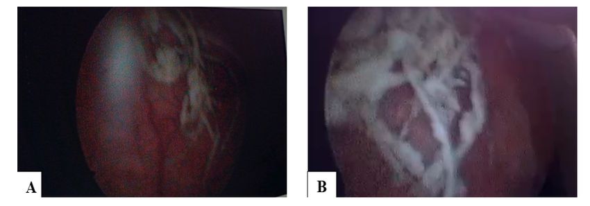
Abstract
Introduction: Adnexal teratoma involving the urinary bladder is a very rare condition. Presentation is
variable ranging from irritative LUTS (lower urinary tract symptoms) to pilimiction or trichiuria (passage
of hair in the urine).

Case presentation: We report a case of 42-year-old woman who presented with pilimiction and lower
abdominal pain. Contrast enhanced computed tomography scan (CECT) and Cystoscopy were used for
the diagnosis. Tumor markers were negative. Right side salpingo-oophorectomy and partial bladder wall
excision were performed. Histopathology of the specimen showed features consistent with mature
teratoma. The Patient reported improvement of symptoms in the subsequent follow up visits.

Conclusion: Pilimiction is a pathognomonic sign of bladder teratomas. Therefore, it is wise to think of
this pathology in patients who report passage of hair through the urine (trichiuria or pilimiction), as in our
case.

Cystoscopy and cross-sectional imaging aided in the initial diagnosis. However, definitive diagnosis was
provided by histopathology.

Introduction
Teratomas are tumors consisting of three germ layers seen commonly during childhood. Mature
teratomas are benign and demonstrate well differentiated tissues such as sebaceous glands, hair and
teeth. Adnexal teratoma involving the urinary bladder is a very rare condition. The clinical presentation
may vary from irritative Lower Urinary Tract Symptoms (LUTS) or urinary retention to pilimiction (passage
of hair in the urine). We hereby present a case of mature teratoma of the ovary involving the urinary
bladder, which primarily manifested with pilimiction. The condition is rare and there are only few similar
reports in the literature.

Case Presentation
42 years old Para I female patient, was referred to a tertiary hospital after she presented with passage of
hair in the urine and lower abdominal pain for a year. Otherwise, there was no compliant of urinary
frequency, urgency or pain on urination. She did not report fever, foul smelling vaginal discharge or pain
during coitus. She has not had any known chronic medical illness.

On physical examination the patient had stable vital signs. There was no remarkable positive physical
finding other than an old Pfannenstiel surgical scar. Pelvic exam was negative for any visible genital
lesions, cervical motion tenderness or palpable adnexal mass.

She underwent a joint evaluation and workup by urology and gynecologic oncology units. On laboratory
investigation, CBC (complete blood count) was normal and hematocrit was 40.3%. Urine analysis was

                                                   Page 2/10
remarkable for microscopic hematuria in the range of 3–5 RBC/HPF (Normal ≤ 2 RBCs/HPF). But the
other parameters of urine analysis were in the normal range. The serum Creatinine, blood urea nitrogen
(BUN), serum electrolytes and liver enzymes were also normal. Serologic tests for HIV, hepatitis B and C
as well as treponema were negative.
Abdominopelvic ultrasound examination showed a bizarre shaped tubular echogenic shadow in the
lumen of the bladder that looks like a foreign body. Further imaging with Contrast enhanced CT scan of
abdomen and pelvis showed a complex heterogeneous mass lesion in the right adnexa measuring
5cm*3.7 cm that has multiple calcifications and fat attenuations inside it. The lesion invaded the right
superolateral surface of the bladder with a defect in the bladder mucosa. It was concluded as right
adnexal mass lesion with possible bladder invasion (Fig. 1: CECT of the abdomen and pelvis. A: Coronal
reformatted image B: Axial image).
Cystoscopy was also done and revealed a whitish papillary mass with hair like component over right
posterior wall of the bladder near the dome (Fig. 2: Cystoscopy. A: Far view B: Near view).
Further workup with tumor markers using serum beta HCG, serum LDH, CA-125, and CEA didn’t reveal any
clue toward a specific diagnosis.
A joint decision was made to surgically explore the patient. The pelvis was entered through the previous
Pfannenstiel incision. The finding was a 4cm*6cm right ovarian mass with solid and cystic areas
containing hair, bone and teeth. Right side salpingo-oopherectomy and partial cystectomy bladder repair
was done. The excised mass was sent for histopathological analysis.
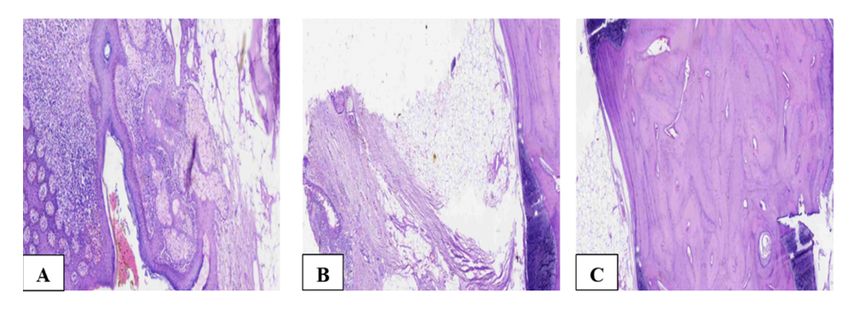
Excisional biopsy report showed stratified squamous epithelium, adnexal structures, fatty and bony
tissue fragments confirming the diagnosis of mature ovarian teratoma (Fig. 3: A, B, &C - scanned pictures
of the histopathology specimen).
On subsequent follow up visits, the patient reported improvement of symptoms and physical examination
was unremarkable.
Discussion
Mature teratomas are the commonest benign tumors of the ovary. They account for 20–50 % of all
ovarian tumors and are more prevalent in premenopausal females. In clinical practice, they are
characterized by a unilateral involvement, which is often on the right side, although up to 10% cases can
be bilateral [3, 4]. Pathologically, ovarian teratomas are germ cell tumors. The word was first used by
Virchow in 1863 and was derived from the Greek ‘teras’, meaning ‘monster’ [1, 2, 5].
Mature ovarian teratomas are indolent and asymptomatic tumors. Their diagnosis is often incidental
either during routine pelvic examination or abdomino-pelvic imaging performed for other indications.
However, some patients may present with symptoms that are often secondary to tumor related
complications. These include acute abdomen, abdominal lump, LUTS or sepsis. Very rare clinical features
Page 3/10such as passage of hair in the urine (pilimiction), gross hematuria, passage of hair through the anal
orifice, small bowel obstruction and fistula into the rectum are also reported in the literature [5, 6, 7].
Torsion is the commonest complication of mature ovarian teratoma occurring in 16% of the cases. Other
uncommon complications include tumor rupture (1–4%), malignant transformation (1–2%), infection
(1%), invasion into adjacent viscera (< 1%) and very rarely, autoimmune hemolytic anemia and
paraneoplastic syndrome [5–8]. Invasion and rupture of the tumor might involve adjacent pelvic and
abdominal structures most commonly the urinary bladder. There are also case reports of involvement of
rectum, vagina, small intestine, sigmoid colon, anterior abdominal wall, and peritoneal cavity [9].
The anatomic proximity of urinary bladder to the ovaries makes it vulnerable to direct involvement by
tumors of ovarian origin. The clinical presentation of this rare occurrence depends on the extent of
bladder involvement and biologic nature of the tumor. According to our literature review, superficial
involvement of the bladder wall often presents with irritative LUTS such as frequency and urgency. On the
other hand, deeper invasion into the bladder lumen by the teratoma manifest itself with urinary tract
infection (UTI), hematuria and LUTS. Many of these features are non-specific and can be easily
overlooked. Pilimiction, however, is a rare but pathognomonic feature of full thickness bladder wall
invasion by ovarian teratoma. At times, the hair in the bladder lumen might create an obstructive ball at
the bladder outlet and manifest as acute urinary retention [1, 4, 5, 9].
Pilimiction was first reported in 1700 by Wallace. Its presence is a specific and diagnostic indicator of
ovarian teratoma and fistula formation. Localized ovarian teratomas do not pose a diagnostic difficulty
on their own. However, involvement of the urinary bladder is often diagnosed late unless patients present
with pilimiction like the case in our patient.
In most of similar cases reported so far, the definitive diagnosis was made through the use of cystoscopy,
computed tomography (CT) scan, or laparotomy [1, 5, 10]. There are also few reports on laparoscopic
diagnosis and management of mature teratoma with bladder involvement [10].
Previous reports attributed the pathogenesis of bladder involvement to malignant transformation of the
teratoma at some point in time leading to aggressive invasion of adjacent pelvic organs [17]. However,
with detailed research of the cases and pathology specimens, it was shown that benign teratomas can
also cause fistula formation with nearby structures. Intermittent leakage of tumor contents can lead to
chronic inflammatory process and adhesion formation resulting in fistulation. This is particularly
common during tumor necrosis, torsion, and infection. Chronic pressure of the tumor on adjacent organs
is also suspected as a possible mechanism for fistula formation [9, 10].
The urologist has a vital role in the management of such conditions. A joint involvement of a
gynecologist and urologist is recommended. Surgical resection of the lesion and ipsilateral fallopian tube
together with partial bladder resection is recommended. A malignant transformation should be ruled out
with histopathological examination of the surgical specimen [1].
Page 4/10Conclusion
Though very rare as a presenting symptom, pilimiction is pathognomonic sign of primary or secondary
bladder teratomas. Therefore, it is imperative to consider teratoma involving the urinary bladder in any
patient who reports passage of hairlike particles through the urine as in the case of our patient.
In addition to the clinical history, cystoscopy and cross-sectional imaging aid in the diagnosis of bladder
teratoma. Definitive diagnosis is provided by histopathology of the surgical specimen.
We performed open surgical excision of the primary tumor in the right ovary and part of the involved
bladder wall.
Abbreviations
BUN
Blood urea nitrogen
CA-125
Carbohydrate antigen-125
CBC
Complete blood count
CEA
Carcino-embryonic antigen
CECT
Contrast enhanced computed tomography
CT
Computed tomography
HCG
Human chorionic gonadotrophin
HPF
High power field
LDH
Lactate dehydrogenase
LUTS
Lower urinary tract symptoms
RBC
Red blood cells
UTI
Urinary tract infection
Declarations
Page 5/10Acknowledgements
Not applicable
Availability of data and materials
All the generated data are included in this article.
Authors' contributions
SMH and FHI diagnosed the case and conceived the idea. SMH and SKH operated the patient. SKH and
IKH were involved in post-operative follow up of the patient. FHI, FOM, KHG, and IKH compiled patient
clinical data. FOM and KHG organized the literature review. FHI, KHG, and FOM prepared draft of the
manuscript. SMH critically revised and edited the manuscript. FHI was engaged in the correspondence
and submission of the article. All the authors read and approved the final manuscript before submission.
Funding
The authors received no funding for writing of this article.
Ethics approval and consent to participate
No institutional review board approval was required.
Consent for publication
Written informed consent was obtained from the patient for publication of the clinical data and will be
made available to the editor upon request.
Competing interests
The authors declare that they have no competing interests.
References
1. Godara R, Karwasra RK, Garg P, Sharma NP. A diagnostic symptom of ovarian dermoid cyst. Internet
J Gynecol Obstet. 2006;6(1):62–9.
2. Nagamani T, Vani I, Nimmana SP, Kumar YM. Ovarian cystovesical fistula causing pilimiction: an
unusual complication of ovarian cyst. Ann Women Child Health. 2015 Dec 18;1(1):C1-4.
3. Hamza M, Yasmeen T, Nadeem IA, Fatima N, Fatima S, Huzaifa M. Ovarian dermoid cyst presenting
with unusual complaint of hair coming out of the anal orifice-A case report. JPMA. 2020 Jan
14;2019.
4. Kizaki Y, Nagai T, Ohara K, Gomi Y, Akahori T, Ono Y, Matsunaga S, Takai Y, Saito M, Baba K, Seki H.
Ovarian mature cystic teratoma with fistula formation into the rectum: a case report. SpringerPlus.
Page 6/102016 Dec;5(1):1–6.
5. Tandon A, Gulleria K, Gupta S, Goel S, Bhargava SK, Vaid NB. Mature ovarian dermoid cyst invading
the urinary bladder. Ultrasound in Obstetrics and Gynecology: The Official Journal of the
International Society of Ultrasound in Obstetrics and Gynecology. 2010 Jun;35(6):751–3.
6. Sardesai S, Raghoji V, Dabade R, Shaikh H. Benign Cytic Teratoma of Ovary Perforating into the
Urinary Bladder: A Rare Case. The Journal of Obstetrics Gynecology of India. 2012;1(62):54–5.
7. Bhasin SK, Malik SM, Sharma G, Gupta SK. Ovarian dermoid presenting as acute intestinal
obstruction: a rare case report and review of literature. International Surgery Journal. 2016 Dec
13;2(2):283–5.
8. Guo H, Yin K, Wang Y, Tong X, Yang H, Xia M, Shuang W. Mature cystic ovarian teratoma invading the
bladder: A rare case report. Translational Surgery. 2018 Jul 1;3(3):62.
9. Naqvi KZ, Abdullah A, Jabeen M, Iqbal F, Edhi M. Ovarian dermoid causing pilimiction. J Coll
Physicians Surg Pak. 2015 Jan 1;25(1):71 – 2.
10. Vaishnav A, Sarkar D, Pal DK. Bladder teratoma with pilimiction in a male adolescent. Urology
Annals. 2020 Jul;12(3):286.
Tables
Page 7/10Table 1
Important dates in this case
Date Events and Reports and findings
activities
November Referred to a
04, 2020 tertiary
Hospital
November Initial surgical History: Presented with passage of hair in the urine and lower
04, 2020 OPD visit and abdominal pain of 01year duration.
evaluation Physical examination: stable vital signs. An old Pfannenstiel surgical
scar was seen.
November Initial Complete blood count was normal with Hematocrit of 40.3%, White
04, 2020 laboratory cell count of 8.6 *103.
investigations Urinalysis showed urine PH of 6, 3–5 RBC/HPF (normal,Date Events and Reports and findings
activities
March 31, Operated The pelvis entered though the previous Pfannenstiel incision. There
2021 was a 4cm*6cm right ovarian mass with solid and cystic areas
containing hair, bone and teeth. It has dense adherence to dome of the
bladder. Upon gentle dissection off the bladder wall, it was seen that it
has a direct communication with the bladder lumen. Right side
salpingo-oophorectomy and partial cystectomy was done.
The excised mass was sent for histo-pathological analysis
March 31, Post- On post op period patient had stable vital signs. The Foley catheter
2021 to operative was kept for 5 days. Subsequently, the urine started clearing. She was
April 06, course discharged after 07 days of hospital stay.
2021
April 15, Excisional The excised biopsy specimen showed a stratified squamous
2021 biopsy report epithelium, adnexal structures, and fatty and bony tissue fragments
with a conclusion as Mature Teratoma.
April 21, Follow up The patient reported improvement of symptoms and physical
2021 visit to examinations were unremarkable
urology and
gynecology
oncology
clinic
Figures
Figure 1
CECT of the abdomen and pelvis. A: Coronal reformatted image B: Axial image
Page 9/10Figure 2
Cystoscopy. A: Far view B: Near view
Figure 3
A, B, &C - scanned pictures of the histopathology specimen
Supplementary Files
This is a list of supplementary files associated with this preprint. Click to download.
CAREchecklistEnglishFilled.pdf
Page 10/10You can also read