Primary Thyroid Lymphoma in a Patient with Hashimoto Disease
←
→
Page content transcription
If your browser does not render page correctly, please read the page content below
International Journal of
                 Case Reports & Short Reviews
Case Report

Primary Thyroid Lymphoma in a Patient with
Hashimoto Disease -
Portmann-Baracco A1* and Rodriguez-Hurtado D2
1
    Internal Medicine, Cayetano Heredia University, Lima, Peru
2
    Cayetano Heredia University, Arzobispo Loayza Hospital, Lima, Peru

*Address for Correspondence: Portmann-Baracco A, Internal Medicine, Cayetano Heredia
University, Lima, Peru, Tel: +51-98-94-05-840; ORCiD: https://orcid.org/0000-0003-3489-3751;
E-mail:

Submitted: 21 May 2019; Approved: 25 June 2019; Published: 27 June 2019

Cite this article: Portmann-Baracco A, Rodriguez-Hurtado D. Primary Thyroid Lymphoma in a Patient
with Hashimoto Disease. Int J Case Rep Short Rev. 2019;5(2): 037-040.

Copyright: © 2019 Portmann-Baracco A, et al. This is an open access article distributed under
the Creative Commons Attribution License, which permits unrestricted use, distribution, and
reproduction in any medium, provided the original work is properly cited.

                                          ISSN: 2638-9355
International Journal of Case Reports & Short Reviews ISSN: 2638-9355
ABSTRACT
Introduction: Primary Thyroid Lymphoma (PTL) is a rare disease with an incidence of 2 cases per million habitants per year and
is associated with autoimmune thyroiditis. The diagnosis represents a challenge in the clinical practice because of its low incidence and
nonspeci ic clinical presentation.
Case Presentation: A 65 year old female previously healthy was admitted complaining about a non-tender neck mass that started
developing 2 years ago associated with dysphagia, odynophagia, cough and dyspnea. On examination, a large non-tender mass measuring
approximately 6x5cm was detected on the left side of the neck. Laboratory exams showed a sub clinic hypothyroidism and pernicious anemia.
Ultrasound and CT showed a heterogeneous mass with irregular borders which compressed adjacent structures. FNAC showed nonspeci ic
thyroiditis (Bethesda II). Pathology with immunohistochemistry of incisional biopsy revealed Diffuse Large B Cell Lymphoma (DLBCL). The
patient received R-CHOP chemotherapy and had a favorable evolution.
Discussion: Primary thyroid lymphoma is a disease rarely found in the clinical practice, is important to have a high index of suspicion
to assure its diagnosis. It usually presents as a fast-growing neck mass producing compression symptoms and is highly associated with
autoimmune thyroiditis. Ultrasound is the irst diagnosis work-up but con irmation is obtained by tissue biopsy. Although FNAC has a limited
effect on PTL, recent advances of immunophenotyping have increased its capacity to almost 80-100%. The most common type is B-cell derived
non-Hodgkin’s lymphoma, mainly including DLBCL, MALT lymphoma or a mixed type. PTL is sensitive to chemotherapy; the treatment
regimen and prognosis depend of the age of the patient, the subtype of lymphoma and the stage of disease.
Keywords: Thyroid lymphoma; Hashimoto thyroiditis; Fine needle aspiration biopsy
ABBREVIATIONS Peripheral lymph node examination was normal. The rest of the exam
was not contributory.
PTL: Primary Thyroid Lymphoma; DLBCL: Diffuse Large B-Cell
Lymphoma; FNAC: Fine Needle Aspiration Cytology; MALT: The thyroid function test showed a sub-clinic hypothyroidism
Mucosa-Associated Lymphoid Tissue (TSH 7.12 mIU/L, FT4: 1.15ng/dl; T3: 80.07ng/dl). The anti-
thyroid peroxidase and anti-thyroglobulin antibodies were positive,
INTRODUCTION indicating Hashimoto’s thyroiditis. The complete blood count
showed a macrocytic anemia, normal platelets and leucocytes. Serum
Primary Thyroid Lymphoma (PTL) is a rare disease with an
vitamin B-12 was decreased (81.09 ng/dl), anti-gastric parietal cell
incidence of 2 cases per million habitants per year. It represents
antibody and anti-intrinsic factor antibody were positive, indicating
less than 5% of all thyroid neoplasms and approximately 2% of all
pernicious anemia.
extra-nodal malignancies [1]. It occurs 3-4 times more frequently in
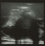
women between 50-80 years old [2]. About 60-90% of all patients with The first ultrasound of the thyroid gland done outside the hospital
this lymphoma present with autoimmune thyroiditis (Hashimoto showed a large round-shaped mass on the left lobe of the thyroid
disease) before or at the time of the diagnosis [3]. gland with irregular borders and heterogeneous echogenicity with
hypoechoic nodules. The mass markedly displaced the trachea to the
Most tumors are B-cell derived non-Hodgkin’s lymphoma,
right side. The second ultrasound (Figure 1), done in the hospital,
mainly including the Diffuse Large B-Cell Lymphoma (DLBCL), the
showed a hypoechoic formation on the left lobule of the thyroid gland
Mucosa-Associated Lymphoid Tissue (MALT) or a mixed type [4].
measuring 43x44mm. Nasolaryngoscopy showed left vocal cord
The diagnosis of this disease represents a challenge in the clinical
paralysis, laryngeal displacement to the right side, normal motility of
practice because of its low incidence and the nonspecific clinical
the right vocal cord and low right piriform sinus volume.
presentation commonly found in other thyroid disorders.
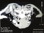
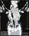
CT scanning of the neck and chest with contrast (Figure 2)
CASE PRESENTATION revealed an isodense mass with heterogeneous enhancement and
A 65 year old female previously healthy was admitted to the irregular borders in the left lobule of the thyroid gland. The lesion
Arzobispo Loayza Hospital (HAL) complaining about a non- measured 5.7 x 7.8 x 11.1 cm and it reached the thyroid cartilage
tender neck mass that started developing 2 years ago and had been
increasing in size. A year previous to the hospital admission it
was associated with dry cough and moderate dyspnea which then
progressed to dyspnea at rest. 7-8 months later the patient presented
odynophagia and dysphagia to solids and then to liquids, hoarseness
and cough with hemoptysis. A month previous to the admission
the patient went to another medical institution where a FNAC and
a thyroid ultrasound were realized and an unknown treatment
was given. Because of the exacerbation of all symptoms in spite of
the treatment given, she decided to go to the emergency room. The
patient was vitally stable and afebrile (HR: 61x’, RR: 20x’, BP: 110/70),
she needed supplementary oxygen with a nasal cannula to maintain
oxygen saturation over 97%. There was a large non-tender mass
on the left side of the neck measuring approximately 6x5cm with a Figure 1: Thyroid ultrasound showed a large round-shaped mass on the left
hard consistency, irregular shape, and low mobility to deglutition. lobe of the thyroid gland.
SCIRES Literature - Volume 5 Issue 2 - www.scireslit.com Page - 038International Journal of Case Reports & Short Reviews ISSN: 2638-9355
in its cranial portion and the middle and posterior mediastinum in per million habitants per year and it represents less than 5% of all
its caudal portion at T4 vertebral level. The lesion compressed and thyroid neoplasms [1].
infiltrated the left brachiocephalic vein in its middle portion and
It usually presents as a fast-growing neck mass and produces
the internal jugular vein in its medial portion at C6-C7 level. At T2
compression symptoms like dysphagia, hoarseness and dyspnea.
level CT showed narrowing of the trachea (diameter of 1.3mm) and
B symptoms such as fever, night sweats and weight loss are rarely
esophageal wall collapse. No lymph nodes were seen.
present, approximately in 3-10% of all patients. 30% of patients
The first FNAC of the thyroid gland done outside the hospital have increased levels of TSH and half of those have sub-clinical
showed round cells without cytoplasm with a lymphoid appearance, hypothyroidism [5,6].
no follicular thyroid cells were found. Those characteristics were
There is an important association between PTL and autoimmune
compatible with a reactive lymphoid tissue. A month later the second
thyroiditis. Approximately 0.6% of all patients with diagnosis
FNAC was done inside the hospital and it showed cytology compatible
of Hashimoto disease develop thyroid lymphoma. However the
with nonspecific thyroiditis (Bethesda II) with abundant mature non-
prevalence of autoimmune thyroiditis in patients with PTL is 80-
atypical lymphocytes and some homogeneous epithelial cells.
90%. For this reason, in spite of the low rate of progression to thyroid
A week later the patient underwent an incisional biopsy lymphoma, physicians should have a high index of suspicion of this
of the cervical tumor which revealed a round cell tumor. disease in those patients with Hashimoto disease, usually between the
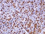
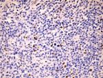
Immunohistochemistry revealed CD20 (+), CD3 (-), Ki67 70%, BCL- sixth and seventh decade of life, who developed a fast-growing neck
2 (-), BCL-6 (-), CD10 (+), MUM-1 (+). Pathologist final diagnosis mass [5]. Recently, the discovery of a clonal relationship between
was Germinal Center B-Cell like (GCB) DLBCL (Figure 3). Hashimoto disease and PTL is helpful to understand the pathogenesis
of the tumor. Similar clonal IgVH bands only present in a minority
of patients with Hashimoto disease have been found in PTL which
supports the hypothesis of clonal evolution of thyroid lymphoma
from Hashimoto thyroiditis. Three nucleotide replacements in IgVH
genes were found in Hashimoto thyroiditis sequence, each one
leading to an amino acid change [7].
Ultrasonography is the initial diagnostic modality used in the
workup of thyroid enlargement. Most patients with PTL present a
heterogeneous hypoechoic parenchyma with intervening echogenic
septa-like structures while others present less frequently features
such as markedly hypoechoic masses or a mixed pattern [8]. Once
Figure 2: CT scanning of the neck and chest with contrast: axial and sagittal the diagnosis has been confirmed, the next step is to make a staging
view. of the disease, which can be done with a full-body CT scan. Also
PET-SCAN has been implemented for the diagnosis and therapeutic
following [9].
Diagnosis confirmation is obtained by tissue biopsy and FNAC
is the initial diagnostic modality used to evaluate thyroid gland [10].
However, a study by Isik A, et al. found that the decision to make a
FNAC depends of the opinion of the physician rather than the up-to-
date guidelines about approaching thyroid nodules and perioperative
thyroid surgery [11]. FNAC has a limited effect on PTL because
cytological differentiation of thyroid lymphoma from lymphocytic
thyroiditis and anaplastic carcinoma is difficult [12]. For this reason it
usually requires a core needle or open biopsy to confirm the diagnosis
and the subtype. Most tumors are B-cell derived non-Hodgkin’s
lymphoma, mainly including the Diffuse Large B-Cell Lymphoma
(DLBCL), the Mucosa-Associated Lymphoid Tissue (MALT) or a
mixed type [4]. FNAC is used in the diagnosis of Hashimoto disease
which results in single or multiple nodules. Microscopic evaluation of
F these nodules show typical cytomorphologic features such as diffuse
mature and mixed lymphoid population, epithelioid histiocytes,
Figure 3: Pathology and Immunohistochemistry of thyroid gland tissue metaplasic Hürtle cells and macrophages [13].
showing Germinal Center B-Cell like (GCB) DLBCL A. HE round cell tumor B.
CD 20 (+) C. CD 10 (+) D. BCL 6 (-) E. Mum1 (+) F. Ki67 70%. Sharma A, et al. [5] reported that 26.2% of the FNAC of patients
with PTL were previously interpreted as benign, especially in the
MALT subtype. Core biopsy showed a better sensibility (93%) than
DISCUSSION the FNAC (71%). The implementation of ancillary studies such as
Primary thyroid lymphoma is a disease rarely found in the flow cytometry to the FNAC makes this technique more accurate to
clinical practice, is important to have a high index of suspicion to differentiate reactive tissues from malignant tissues. While suspicion
assure its diagnosis. The annual incidence of this disease is of 2 cases of lymphoma can be based on morphology, flow patterns are essential
SCIRES Literature - Volume 5 Issue 2 - www.scireslit.com Page - 039International Journal of Case Reports & Short Reviews ISSN: 2638-9355
for diagnosis and sub classification. Swart et al. found a sensitivity of ACKNOWLEDGEMENT
96.9% and a specificity of 86.7% by adding flow cytometry to FNAC
[14]. Recent advances of immunophenotyping have increased the We extend our thanks to Dr. Cesar Chian for providing the
pathology and immunohistochemistry of thyroid gland tissue.
capacity of FNAC to detect PTL to almost 80-100% depending of the
quality of the sample taken [3]. Although this technique is the first REFERENCES
histologic test to diagnose PTL, sometimes an open incision biopsy
1. Pedersen RK, Pedersen NT. Primary non-Hodgkin’s lymphoma of the thyroid
will still be necessary [15].
gland: a population based study. Histopathology. 1996; 28: 25-32. https://bit.
The treatment of primary thyroid lymphoma is still controversial ly/2Fw4p9k
because of lack of studies with a high quality of evidence. PTL is 2. Graff Baker A, Roman SA, Thomas DC, Udelsman R, Sosa JA. Prognosis of
sensitive to both chemotherapy and radiotherapy and the regimen primary thyroid lymphoma: Demographic, clinical, and pathologic predictors
depends of the lymphoma subtype. Because of its aggressive clinical of survival in 1,408 cases. Surgery. 2009; 146: 1105-1115. https://bit.
ly/2YhEdac
course DLBCL regimen is multimodal including a combination of
monoclonal antibodies (Rituximab) and chemotherapy (CHOP 3. Stephanie Aleskow Stein, Leonard Wartofsky. Primary thyroid lymphoma: a
regimen). On the other side, a single-modality treatment such as clinical review. The Journal of Clinical Endocrinology & Metabolism. 2013; 98:
3131-3138. https://bit.ly/2LdqNIp
surgery alone, radiotherapy alone or a combination of both may
be used to treat MALT lymphoma [10]. Pyke et al. [16] showed no 4. Efstathios T. Pavlidis, Theodoros E. Pavlidis. A review of primary thyroid
difference in remission or in survival rates between the combination lymphoma: molecular factors, diagnosis and management. Journal of
Investigative Surgery. 2017; 32: 137-142. https://bit.ly/2X1voVc
of debulking surgery with external beam radiotherapy and surgical
biopsy with radiotherapy. Therefore, a recent analysis of the National 5. Sharma A, Jasim S, Reading CC, Ristow KM, Villasboas Bisneto
Cancer Database showed that surgery therapy such as lobectomy JC, Habermann TM, et al. Clinical presentation and diagnostic challenges of
thyroid lymphoma: a cohort study. Thyroid. 2016; 26: 1061-1067. https://bit.
and total or subtotal thyroidectomy had a significant improvement
ly/2Yb9snh
in survival rates (HR 0.58) as well as beam radiation (HR 0.67),
multi-agent (HR 0.4) and single agent chemotherapy (HR 0.43). 6. Watanabe N, Noh JY, Narimatsu H, Takeuchi K, Yamaguchi T, Kameyama K,
et al. Clinicopathological features of 171 cases of primary thyroid lymphoma:
Immunotherapy benefit was not found statistically significant [17].
a long-term study involving 24553 patients with Hashimoto’s disease. Br J
The prognosis of PTL depends of the age of the patient, the Haematol. 2011; 153: 236-243. https://bit.ly/2xizusB
subtype of lymphoma and the stage of disease. DLBCL subtype has 7. Moshynska OV, Saxena A. Clonal relationship between hashimoto thyroiditis
a lower survival rate than that of MALT subtype. Patients have a and thyroid lymphoma.J Clin Pathol. 2008; 61: 438-444. https://bit.ly/2N9Zy49
life expectancy of 9 years after the diagnosis and mortality is usually 8. Nam M, Shin JH, Han BK, Ko EY, Ko ES, Hahn SY, et al. Thyroid lymphoma:
associated with the lymphoma (44%), non-oncologic causes such correlation of radiologic and pathologic features. J Ultrasound Med. 2012; 31:
as cardiovascular or cerebrovascular disease (35%) and additional 589 -594. https://bit.ly/2ZL9QZM
non-thyroid malignancies (13%). The treatment of PTL such as 9. Stein SA, Wartofsky L. Primary thyroid lymphoma: a clinical review. J Clin
chemotherapy, radiotherapy and surgery improves the prognosis of Endocrinol Metab. 2013; 98: 3131-3138. https://bit.ly/2LbrOAR
this disease. Five years after diagnosis, disease- specific survival was
10. Walsh, Lowery, Evoy, McDermott EW, Prichard RS. Thyroid lymphoma:
86% for stage I, 81% for stage II, and 64% for stage III/IV [2]. For recent advances in diagnosis and optimal management strategies.
this reason, early diagnosis of this uncommon disease is important to Oncologist. 2013; 18: 994-1003. https://bit.ly/2xebCGM
have a better prognosis.
11. Isik A, Firat D, Yilmaz I, Peker K, Idiz O, Yilmaz B, et al. A survey of current
RESULT approaches to thyroid nodules and thyroid operation. International Journal of
Surgery. 2018.
During the hospitalization the patient developed oppressive
12. Sarinah B, Hisham AN. Primary lymphoma of the thyroid: diagnostic and
chest pain radiating up into her neck. ECG showed no alteration, therapeutic considerations. Asian J Surg. 2010; 33: 20-24. https://bit.
echocardiography showed type I diastolic dysfunction, mild ly/2J9n9wJ
enlargement of the left auricle and mild regurgitation of the mitral
13. Paksoy N,Yazal K. Cervical lymphadenopathy associated with hashimoto’s
valve and tricuspid valve. thyroiditis: an analysis of 22 cases by fine needle aspiration cytology. Acta
Cytol. 2009; 53: 491-496. https://bit.ly/2Ydp6ye
The patient was treated by the Oncology department with
R-CHOP chemotherapy. The tumor was reduced and the symptoms 14. Swart GJ, Wright C, Brundyn K, Mansvelt E, du Plessis M, ten Oever D,
such as dysphagia, odynophagia and dyspnea disappeared. et al. Fine needle aspiration biopsy and flow cytometry in the diagnosis of
lymphoma. Transfus Apher Sci. 2007; 37: 71-79. https://bit.ly/2KCOkTO
CONCLUSION
15. Mack LA, Pasieka JL. An Evidence-based approach to the treatment of
A case of a patient who came to the emergency room with a thyroid lymphoma. World J Surg. 2007; 31: 978-986. https://bit.ly/2X6Z1o4
cervical mass that started developing and increasing 2 years ago 16. Pyke CM, Grant CS, HabermannTM, Kurtin PJ, van Heerden JA, Bergstralh
associated with compression symptoms is presented. The patient EJ, et al. Non-Hodgkin’s lymphoma of the thyroid: Is more than biopsy
is diagnosed with subclinic hypothyroidism and autoimmune necessary? World J Surg. 1992; 16: 604-609. https://bit.ly/2xeuKo8
thyroiditis and then an incisional biopsy was taken which revealed 17. Vardell Noble V, Ermann DA, Griffin EK, Silberstein PT. Primary thyroid
a Germinal Center B-Cell like (GCB) DLBCL. The patient received lymphoma: An analysis of the national cancer database. Cureus. 2019; 11:
R-CHOP chemotherapy and had a favorable evolution. 4088. https://bit.ly/2Yb6Zch
SCIRES Literature - Volume 5 Issue 2 - www.scireslit.com Page - 040You can also read


