Fetal Acalvaria Malformation: Antenatal Diagnosis in 4 Cases and Review of the Literature - Scholars Middle East Publishers
←
→
Page content transcription
If your browser does not render page correctly, please read the page content below
Scholars International Journal of Obstetrics and Gynecology
                                                                                                   Abbreviated Key Title: Sch Int J Obstet Gynec
                                                                                            ISSN 2616-8235 (Print) |ISSN 2617-3492 (Online)
                                                                                  Scholars Middle East Publishers, Dubai, United Arab Emirates
                                                                                                 Journal homepage: https://saudijournals.com

                                                                                                                              Case Report

Fetal Acalvaria Malformation: Antenatal Diagnosis in 4 Cases and
Review of the Literature
Imane Attar1*, Hekmat Chaara1, Hind Adadi1, Sofia Jayi1, Fatima-Zahra Fdili Alaoui1, Moulay Abdelilah Melhouf1
1
 Department of Gynecology and Obstetrics Ii, Chu Hassan Ii Fes, Morocco

DOI: 10.36348/sijog.2021.v04i04.005                               | Received: 06.03.2021 | Accepted: 03.04.2021 | Published: 15.04.2021
*Corresponding author: Imane Attar

Abstract
Acalvaria Is a rare congenital disease considered as a post-neurulation defect: It consists of the absence of Calvary bones,
dura mater and associated muscles in the presence of a normal skull base and facial bones normal. Currently, there is no
identified cause of Acalvaria. The main putative pathogenesis is the problematic migration of the membranous
neurocranium from the normal positioning of the immature ectoderm. Although the malformation has been fatal to date
with only a few survivors, the prenatal diagnosis of Acalvaria is of rather remarkable importance as it allows clinicians to
plan appropriate and timely management. So that this fetus can benefit from surgical advances.
Keywords: Acalvaria; antenatal ultrasound sign; differential diagnosis; prognosis.
Copyright © 2021 The Author(s): This is an open-access article distributed under the terms of the Creative Commons Attribution 4.0 International
License (CC BY-NC 4.0) which permits unrestricted use, distribution, and reproduction in any medium for non-commercial use provided the original
author and source are credited.

                                                                             ultrasound in the prenatal diagnosis of this rare entity
INTRODUCTION                                                                 while raising the issue of differential diagnosis which
         Acalvaria is an extremely rare malformation                         presents itself as the main challenge for the obstetrician,
characterized by the absence of the bones of the skull,                      as well as 'an overview on the prognosis will be
dura mater and scalp muscles. The base of the skull and                      reported.
facial features are fully formed and generally appear
normal [1]. It occurs at a frequency of 1 in 100,000
births [2]. Its causality is poorly understood and no                        CLINICAL OBSERVATIONS
concrete aetiology has been described [3].                                   Case N ° 1
                                                                                      This is a new born female, from a non-
         This is an anomaly accessible to antenatal                          consanguineous marriage, without ATCD (in particular
diagnosis from the 12th week of gestation in knowledge                       no diabetes, no exposure to a teratogenic material), the
of the different differential diagnoses, of which                            pregnancy was not followed without any notion of
anencephaly remains the most important [4]. This                             premedication by acid folic, with a single ultrasound
malformation can be associated with several                                  performed at 35 WA where the diagnosis of an
abnormalities, which makes a detailed morphological                          anencephaly was made and then referred to our
examination mandatory in order to rule out any                               structure for specialist advice. Focused morphological
associated malformation which may further aggravate                          exploration of the neural tube objectified a well-formed
the postnatal prognosis [1].                                                 brain with cerebral convolutions, an interhemispheric
                                                                             fissure, lateral ventricles covered with a thick
         Since then, Acalvaria has been widely                               membrane that corresponds to the skin while the bone
regarded as a fatal anomaly [5]. But with advances in                        matrix was absent (Figure-1A). The facial structures
foetal medicine, prenatal identification of this anomaly                     appeared normal. The two orbital cavities were placed
has enabled clinicians to plan appropriate management                        symmetrically of equal size and shape (Figure-1B). The
resulting in a few surviving cases [1, 2, 6, 7].                             brain showed a normal vascular pattern with normal
                                                                             function of Willis circle on the Doppler scan. No spinal
        Through four clinical observations and a                             defect was detected. The study of long bones seemed
review of the literature, we will prove the relevance of                     necessary to rule out osteogenesis imperfecta which

Citation: Imane Attar et al (2021). Fetal Acalvaria Malformation: Antenatal Diagnosis in 4 Cases and Review of the Literature.             103
Sch Int J Obstet Gynec, 4(4): 103-107.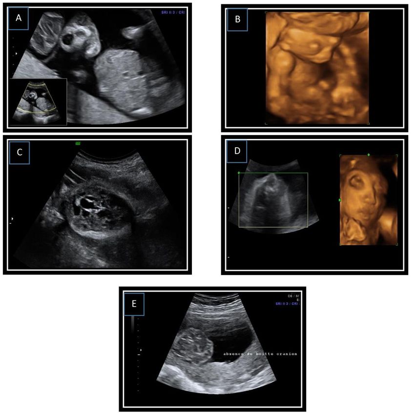
Imane Attar et al; Sch Int J Obstet Gynec, Apr. 2021; 4(4): 103-107
was normal; and the length of the femur was consistent folic acid from conception, referred in our training by
with the gestation period. her attending physician for suspected neural tube
closure abnormalities and whose morphological
After informing the couples that it is a fatal ultrasound performed (25 week of amenorrhoea)
malformation and given our religious context the objectified a complete protrusion of the early fetal brain
Therapeutic Abortion was not discussed so a above the remaining skull bones suggesting an
conservative attitude was adopted, with regular Acalvaria malformation (Figure 1 C, D), while the rest
monitoring until the end to ensure fetal well-being, but of the morphology was unremarkable. Regular
essentially to detect the appearance of cerebral lysis monitoring was recommended until term with
which is only observed in Anencephaly, that will push delivering through a cesarean section (for a surgical
us to question our initial diagnosis. basin) giving birth to a newborn baby girl with
Acalvaria who died 1 hour after.
At 39 weeks the patient gave birth vaguely to a
eutrophic mature newborn baby girl with normal Case N ° 3
hemodynamic constants, the morphological Mrs EL YF aged 29, not consanguineous
examination objectified a brain covered with a thick marriage, followed for type I diabetes, first childbirth
layer of skin, displaced towards the back due to the (folic acid administered from the 1st trimester), referred
absence of supporting cranial bones including frontal, in our training for follow-up, whose morphological
temporal, occipital and parietal bones, The facial ultrasound in the 2nd trimester (26SA) objectified : the
structure and the rest of the body were completely presence of a bone defect involving the entire skull with
normal. The evolution 2 hours later was towards death. a complete protrusion of the fetal brain suggesting an
isolated Acalvaria (Figure-1E). During monitoring, the
Case N ° 2 patient presented an antepartum fetal death and
Mrs HN, 36 years old, non-consanguineous delivered vaginally giving birth to a baby girl with
marriage, first childbirth put on supplementation with Acalvaria.
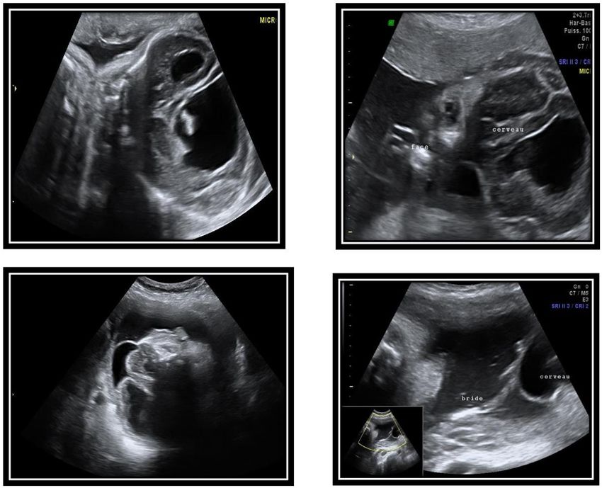
Fig-1: A-B) 2D and 3D ultrasound images illustrating agenesis of the cranial limb with preserved cerebral parenchyma in
relation to Acalvaria in the first case .C-D) 2D and 3D ultrasound images illustrating agenesis of the cranial box with cerebral
parenchyma preserved in report with an Acalvaria in the second case. E) 2D ultrasound image illustrating agenesis of the skull
with preserved cerebral parenchyma in relation to an Acalvaria in third case
© 2021 |Published by Scholars Middle East Publishers, Dubai, United Arab Emirates 104Imane Attar et al; Sch Int J Obstet Gynec, Apr. 2021; 4(4): 103-107
Case N ° 4 cerebral parenchyma and a ventriculomegaly linked to
Mrs AN, 36 years old, non-consanguineous the uterine wall by amniotic bands suggesting a partial
marriage, with a history of two diabetic brothers on Acalvaria secondary to an amniotic band (Figure-2), the
Oral Antidiabetics, first childbirth, referred in our patient reached full term gave birth vaginally to a new
training for management of a pregnancy associated with born baby girl whose Examination of the nervous
gestational diabetes discovered at 25 weeks by oral system revealed partial agenesis of the occipital skull
glucose tolerance test at 25 weeks + 04 days. Whose with adhesion between the cranial defect, the amnion
morphological study this time has objectified a and a layer skin covering brain tissue.
posterior segmental bone defect with protrusion of the
Fig-2: Ultrasound images showing partial agenesis of the cranial vault calling for Acalvaria, with adhesion between the cranial
defect and the amnion covering the brain tissue
DISCUSSION epidemiological investigation shows female
Lemire in 1988 divided neural tube defects predilection, as is the case in our series [2, 3].
into open (neurulation defects) and closed (post-
neurulation defects). Open lesions, thought to occur This is a form of congenital malformation that
before neural tube closure in the embryonic stages, are result from poor migration of the membranous
much more common in foetal and pediatric neurocranium (which gives rise to muscle and bone)
neuropathology and include anencephaly and with a normally placed embryonic ectoderm (from
myelomeningocele. Post-neurulation neural tube defects which the skin and scalp are formed). As a result, the
are less common, including Acalvaria. The Acalvaria is brain is only covered with a layer of intact skin without
characterized by the absence of flat bones of the skull, a calvarium [9]. In cases associated with the amniotic
dura mater and associated muscles with the presence of band, the early rupture of the amnion is the triggering
normal cranial contents and facial bones. Although event. The amnion can twist like a cord trapping the
some cases may show abnormal development of brain head of the foetus [10]. Our fourth case illustrates this
tissue [8]. association.
Acalvaria occurs in less than 1 in 100,000 So far, no etiology has been described. It has
births, only 23 possible cases have been reported in the not been shown that taking folic acid prevents
English literature. Usually it is reported as a fatal Acalvaria, which distinguishes this pathology from
congenital malformation with only rare cases (three in neural tube defects, confirmed by our study seen that 3
number) describing prolonged survival. The of our patients were on folic acid. Nevertheless, four of
© 2021 |Published by Scholars Middle East Publishers, Dubai, United Arab Emirates 105Imane Attar et al; Sch Int J Obstet Gynec, Apr. 2021; 4(4): 103-107
our patients were diabetic, this pathology which is often Other pathologies can be confused with Acalvaria such
implicated in the abnormalities of the neural tube but no as [2]:
direct link with Acalvaria has been proven. There were Encephalocele: the cranial vault is still detected
no chromosomal abnormalities associated with and part of the brain exteriorized.
Acalvaria, but other congenital anomalies can be Severe osteogenesis imperfecta or Familial
associated with it such as holoprosencephaly, hypophosphatemia can lead to inadequate
hydrocephalus as observed for our 4th case, visualization or ossification of the bones of the
micropolygyria, hypertelorism and cleft lip or cleft calvarium, leading to a misdiagnosis of Acalvaria
palate, for this the morphological analysis of the foetus An additional condition that can be confused at
must be thorough before predicting the prognosis [1,3]. autopsy with Acalvaria is aplasia cutis congenita
Regarding our cases, no associated malformations were (ACC). It is estimated that 20% of cases of ACC
detected. may have an underlying bony and dura defect thus
causing possible diagnostic confusion.
Detection of Acalvaria should be possible
from the 12th week of gestation by trans-vaginal The management of Acalvaria is not codified.
ultrasound. Early diagnosis is important for better The initial treatment is mainly conservative and aims at
management of the pregnancy. In our study, the mean supportive care as well as the management of
age of diagnosis was 28 WA and no case was suspected associated anomalies, no recommendation is declared in
in the first trimester due to the delay in consultation [1]. terms of surgical management. The treatment consists
of an increase in soft tissues associated with a skin graft
On ultrasound, the Acalvaria appears as an which will facilitate the protection of the dura by a
absence of posterior shading to the structures of the layer of soft tissue while maintaining a separate layer of
head due to the absence of the bony skull and dura skin for future interventions, subsequently proceeded by
mater and muscles with the presence of cranial contents a bone graft when the child reaches school age. There
covered by an echogenic layer that corresponds to the are only two cases published in the literature that have
skin that protects the brain from degeneration with a benefited from conservative treatment associated with
normal facial floor, the whole achieves an aspect of extensive surgical reconstruction [11, 12]
"Mickey Mousse". Although some cases may show
abnormal development of brain tissue, it is usually Usually, babies with this defect do not survive
developed in the majority of cases. Anechoic areas after birth. However, there have been cases of survival.
representing necrosis or hemorrhage are also possible. The first surviving case was reported in 2004 in Japan.
This ultrasound description was the rule for making the He was treated by surgical closure of the scalp defect,
diagnosis in our 4 patients. in addition, there was associated hydrocephalus, so
shunt surgery was also performed. At follow-up, the
A new means of antenatal diagnosis was child had severe developmental delay with mental
suggested for Acalvaria is the dosage of alpha- retardation, last time reported alive at the age of 11 [3].
fetoprotein (AFP) which should be normally low in
these cases and whose rise is probably caused by the Unfortunately, in our series all our new-borns
destruction of the tissue of the central nervous system died after giving birth, which confirms the fatality of
by the amniotic fluid, something observed in the this anomaly.
anencephaly, but this means of diagnosis remains a
subject of debate since it can be distorted by the CONCLUSION
occurrence of AFP leakage through the thin layer of Acalvaria is a rare congenital disease reported
ectoderm covering the brain. to be fatal in most newborns. It is an anomaly that
remains accessible to antenatal diagnosis in knowledge
The differential diagnosis of Acalvaria remains of the different differential diagnosis that presents the
a real challenge for the obstetrician. Acrania is the main challenge for the obstetrician, her prognosis
closest differential diagnosis and the two have been remains poor but there remains a malformation that
used interchangeably in the medical literature on deserves special attention so that it can benefit from
several occasions. In Acalvaria, the brain being normal progress. surgical nowadays.
covered by the scalp and can potentially be treated with
a chance of being compatible with life while the Conflicts of interest: The authors declare no conflict of
Acrania is lethal with brain tissue totally exposed to the interest.
outside. An incorrect initial diagnosis can be caught up
with surveillance if the brain maintains its shape Contributions from the authors
throughout pregnancy [2] All the authors contributed to the conduct of
this work. All authors also declare that they have read
and approved the final version of the manuscript.
© 2021 |Published by Scholars Middle East Publishers, Dubai, United Arab Emirates 106Imane Attar et al; Sch Int J Obstet Gynec, Apr. 2021; 4(4): 103-107
REFERENCE skin. Journal of the American College of
1. Harris, C. P., Townsend, J. J., & Carey, J. C. Surgeons, 217(3), 533-555.
(1993). Acalvaria: a unique congenital 8. Asai, M., Kitamura, H., Yanagibashi, T., Asukai,
anomaly. American journal of medical K., & Katagiri, N. (1998). Case of acrania
genetics, 46(6), 694-699. associated with congenital
2. Harris, C. P., Townsend, J. J., & Carey, J. C. medulloblastoma. European Journal of Obstetrics
(1993). Acalvaria: a unique congenital & Gynecology and Reproductive Biology, 81(1),
anomaly. American journal of medical 115-117.
genetics, 46(6), 694-699. 9. Chandran, S., Lim, M. K., & Yu, V. Y. H. (2000).
3. Kurata, H., Tamaki, N., Sawa, H., Oi, S., Fetal acalvaria with amniotic band
Katayama, K., Mochizuki, M., ... & Nakamura, H. syndrome. Archives of Disease in Childhood-Fetal
(1996). Acrania: report of the first surviving and Neonatal Edition, 82(1), F11-F13.
case. Pediatric neurosurgery, 24(1), 52-54. 10. Sepulveda, W., De La Maza, F., & Meagher, S.
4. E c (2020). An Unusual First‐ Trimester Ultrasound
Sonographic diagnosis of fetal acrania. Journal of Presentation of the Acrania‐ Anencephaly
clinical ultrasound, 19(6), 363-366. S q nc : Th “T r sh T rban” S n Journal of
5. Bang, R. L., Ghoneim, I. E., Gang, R. K., & Al Ultrasound in Medicine, 39(4), 829-832.
Najjadah, I. (2003). Treatment dilemma: 11. Hawasli, A. H., Beaumont, T. L., Vogel, T. W.,
conservative versus surgery in cutis aplasia Woo, A. S., & Leonard, J. R. (2014). Acalvaria:
congenita. European journal of pediatric case report. Journal of Neurosurgery:
surgery, 13(02), 125-129. Pediatrics, 14(2), 200-202.
6. Gupta, V., & Kumar, S. (2012). Acalvaria: A rare 12. Bang, R. L., Ghoneim, I. E., Gang, R. K., & Al
congenital malformation. Journal of pediatric Najjadah, I. (2003). Treatment dilemma:
neurosciences, 7(3), 185. conservative versus surgery in cutis aplasia
7. Kamel, R. A., Ong, J. F., Eriksson, E., Junker, J. congenita. European journal of pediatric
P., & Caterson, E. J. (2013). Tissue engineering of surgery, 13(02), 125-129.
© 2021 |Published by Scholars Middle East Publishers, Dubai, United Arab Emirates 107You can also read