Flexion-extension standing radiographs underestimate instability in patients with single-level lumbar spondylolisthesis: comparing flexion-supine ...
←
→
Page content transcription
If your browser does not render page correctly, please read the page content below
Original Article

Flexion-extension standing radiographs underestimate instability
in patients with single-level lumbar spondylolisthesis: comparing
flexion-supine imaging may be more appropriate
Nathan J. Lee, Justin Mathew, Jun S. Kim, Joseph M. Lombardi, Andrew C. Vivas, Jay Reidler,
Scott L. Zuckerman, Paul J. Park, Eric Leung, Meghan Cerpa, Mark Weidenbaum, Lawrence G. Lenke,
Ronald A. Lehman, Zeeshan M. Sardar

Department of Orthopaedics, Columbia University Medical Center, The Spine Hospital at New York-Presbyterian, New York, NY, USA
Contributions: (I) Conception and design: NJ Lee, ZM Sardar; (II) Administrative support: M Weidenbaum, LG Lenke, RA Lehman, ZM Sardar; (III)
Provision of study materials or patients: E Leung, M Cerpa, LG Lenke, RA Lehman, ZM Sardar; (IV) Collection and assembly of data: NJ Lee, J
Mathew, E Leung, M Cerpa; (V) Data analysis and interpretation: NJ Lee, J Mathew, E Leung, M Cerpa; (VI) Manuscript writing: All authors; (VII)
Final approval of manuscript: All authors.
Correspondence to: Nathan J. Lee, MD. Columbia University Medical Center, 161 Fort Washington Avenue, New York, NY 10032, USA.
Email: njl2116@cumc.columbia.edu.

                 Background: Generally, most spine surgeons agree that increased segmental motion viewed on flexion-
                 extension radiographs is a reliable predictor of instability; however, these views can be limited in several ways
                 and may underestimate the instability at a given lumbar segment.
                 Methods: Consecutively collected adult (≥18 years old) patients with symptomatic single-level lumbar
                 spondylolisthesis were reviewed from a two-surgeon database from 2015 to 2019. Routine standing lumbar
                 X-rays (neutral, flexion, extension) and supine lumbar MRI (sagittal T2-weighted imaging sequence) were
                 performed. Patients were excluded if they had prior lumbar surgery, missing radiographic data, or if the time
                 between X-rays and MRI was >6 months.
                 Results: All 39 patients with symptomatic, single-level lumbar spondylolisthesis were identified. The mean
                 age was 57.3±16.7 years and 66% were female. There was good intra- and inter-rater reliability agreement
                 between measured values on the presence of instability. The slip percentage (SP) difference was significantly
                 highest in the flexion-supine (FS) (5.7 mm, 12.3%) and neutral standing-supine (NS) (4.3 mm, 8.7%) groups,
                 both of which were significantly higher compared with the flexion-extension (FE) group (1.8 mm, 4.5%,
                 P8% was observed more frequently in FS (79.5%) and NS (52.6%)
                 groups compared with FE group (16.7%, P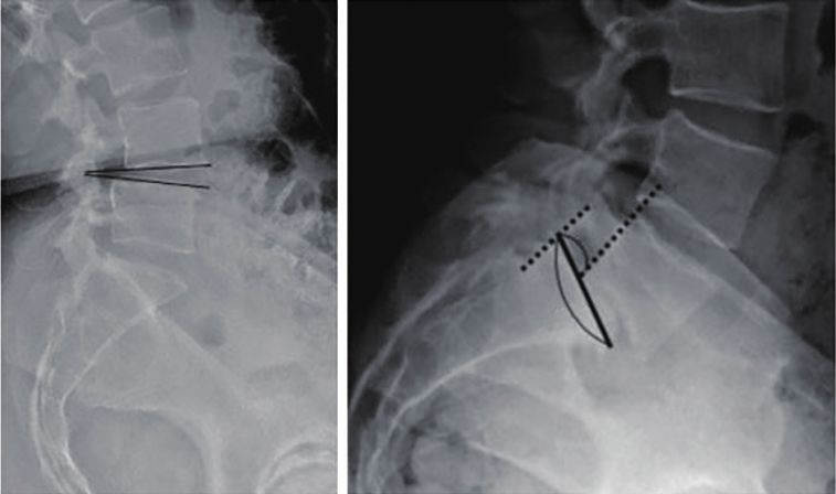
Journal of Spine Surgery, Vol 7, No 1 March 2021                                                                                     49

Introduction                                                     Table 1 The differences in slip, slip percentage, and segmental angle
                                                                 for each view
Lumbar spondylolisthesis is a common cause of low
                                                                                             Mean slip difference
back pain and radicular leg pain, which often warrants                                       between (SD)         Mean segmental
operative intervention (1). However, there continues to          Patient position                                 angle difference
                                                                                             Distance,
be considerable debate among spine surgeons regarding                                                    SP (%) (SD), angle
                                                                                                mm
the optimal surgical management of lumbar stenosis in
                                                                 Flexion and neutral          1.8 (2.0) 4.4 (4.0)        4.1 (4.3)
the presence of spondylolisthesis, namely decompression
                                                                 standing, FN
alone versus decompression and fusion (2-4). The
                                                                 Neutral and extension, EN 0.9 (1.9) 1.8 (4.2)           3.6 (4.0)
decision regarding whether or not to fuse most often
depends on the surgeon’s assessment of lumbar segmental          Flexion and extension, FE 1.8 (1.9) 4.5 (3.8)           5.1 (4.6)
stability. In the appropriate patient, fusion has been shown     Neutral and supine (NS)      4.3 (3.0) 8.7 (6.2)        3.9 (4.1)
to successfully halt the progression of spondylolisthesis,
                                                                 Flexion and supine (FS)      5.7 (2.8) 12.3 (6.4)       4.7 (4.4)
reduce pain, and improve patient-reported outcomes
                                                                 P value
50 Lee et al. Utility of flexion-supine views for spondylolisthesis
Table 2 Ventral instability (slip % difference cut-off >8%) breakdown
for each view
Patient positioning No Yes
FN 82.1% 17.9%
A
EN 91.0% 9.0%
B
FE 83.3% 16.7%
NS 47.4% 52.6%
FS 20.5% 79.5%
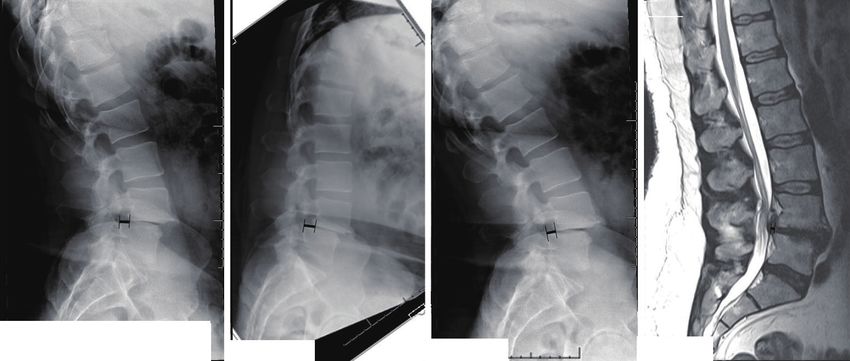
Figure 1 Slip percentage (A/B) on left and segmental slip angle on
P valueJournal of Spine Surgery, Vol 7, No 1 March 2021 51 for NS (P
52                                                                                            Lee et al. Utility of flexion-supine views for spondylolisthesis

                                                     L
                                                                                                         Extention
                                                                                                              L

                                                                              L
                                                                                   n
                                                                                io
                                                                              ex
                                                                              Fl
                    Slip Difference, mm (Slip Percentage, %)
                    10.4 (25.2%)                               12.6 (31.2%)            9.8 (23.9%)                   2.7 (7.0%)

Figure 2 Example of patient with standing-neutral, flexion, extension, supine/MRI views (from left to right). slip differences FN (2.2 mm),
FE (2.8 mm), NS (7.7 mm), FS (9.9 mm). Slip percentage differences FN (6.0%), FE (7.3%), NS (18.2%), FS (24.2%).

technicians felt this positioning was logistically demanding                             isthmic disease), specific disc levels (L4/5 versus L5/S1),
and more difficult to explain to patients than the positioning                           and number of disc levels involved,
required for standardized views.
   In comparison with these other stress views, the
                                                                                         Conclusions
standing-supine and flexion-supine views provide a safe,
reproducible method that can be readily integrated into                                  Standing flexion-extension views have long been the
the routine clinical work-up for these patients. MRI scans                               standard technique used to assess instability in lumbar
were used to assess supine parameters since they are already                             spondylolisthesis. This study demonstrates that
available for almost every patient that undergoes surgery for                            assessment of this instability is more accurately identified
spinal stenosis and spondylolisthesis. Imaging in the supine                             by comparing standing lateral radiographs with supine
position requires minimal effort from the patient, relaxes                               sagittal MRI images. An example of this is illustrated
muscles and soft tissues which may otherwise block true                                  in Figure 2. Ventral instability based on SP >8% was
lumbar instability from the patient, and excludes potential                              observed more frequently in FS (79.5%) and NS (52.6%)
examiner and patient bias (31).                                                          groups compared with FE group (16.7%, P
Journal of Spine Surgery, Vol 7, No 1 March 2021 53
Footnote original work is properly cited (including links to both the
formal publication through the relevant DOI and the license).
Reporting Checklist: The authors have completed the
See: https://creativecommons.org/licenses/by-nc-nd/4.0/.
STROBE reporting checklist. Available at http://dx.doi.
org/10.21037/jss-20-631
References
Data Sharing Statement: Available at http://dx.doi.
1. Kalichman L, Kim DH, Li L, et al. Spondylolysis and
org/10.21037/jss-20-631
spondylolisthesis: prevalence and association with low
back pain in the adult community-based population. Spine
Conflicts of Interest: All authors have completed the ICMJE
2009;34:199-205.
uniform disclosure form (available at http://dx.doi.
2. Weinstein JN, Lurie JD, Tosteson TD, et al. Surgical
org/10.21037/jss-20-631). LGL serves as an unpaid
compared with nonoperative treatment for lumbar
editorial board member of Journal of Spine Surgery from Oct
degenerative spondylolisthesis. four-year results in
2019 to Oct 2021. SZ serves as an unpaid editorial board
the Spine Patient Outcomes Research Trial (SPORT)
member of Journal of Spine Surgery from Oct 2019 to Sep
randomized and observational cohorts. J Bone Joint Surg
2021. LGL reports personal fees from Medtronic, grants
Am 2009;91:1295-304.
and personal fees from DePuy-Synthes Spine, personal fees
3. Simmonds AM, Rampersaud YR, Dvorak MF, et
from K2M, non-financial support from Broadwater, non-
al. Defining the inherent stability of degenerative
financial support from Seattle Science Foundation, grants
spondylolisthesis: a systematic review. J Neurosurg Spine
and non-financial support from Scoliosis Research Society,
2015;23:178-89.
non-financial support from Stryker Spine, non-financial
4. Herkowitz HN, Kurz LT. Degenerative lumbar
support from The Spinal Research Foundation, grants from
spondylolisthesis with spinal stenosis. A prospective
EOS, grants from Setting Scoliosis Straight Foundation,
study comparing decompression with decompression and
personal fees from Fox Rothschild, LLC, personal fees
intertransverse process arthrodesis. J Bone Joint Surg Am
from Quality Medical Publishing, other from Evans Family
1991;73:802-8.
Donation, other from Fox Family Foundation, grants and
5. Noorian S, Sorensen K, Cho W. A systematic review of
non-financial support from AOSpine, outside the submitted
clinical outcomes in surgical treatment of adult isthmic
work. RAL reports consultant/royalty fees from Medtronic,
spondylolisthesis. Spine J 2018;18:1441-54.
royalty fees from Stryker, research grants from Department
6. Ghogawala Z, Dziura J, Butler WE, et al. Laminectomy
of Defense, outside the submitted work. ZMS reports
plus Fusion versus Laminectomy Alone for Lumbar
Stryker Spine (past), outside the submitted work. The
Spondylolisthesis. N Engl J Med 2016;374():1424-34.
authors have no other conflicts of interest to declare.
7. Chan AK, Bisson EF, Bydon M, et al. Laminectomy alone
versus fusion for grade 1 lumbar spondylolisthesis in 426
Ethical Statement: The authors are accountable for all
patients from the prospective Quality Outcomes Database.
aspects of the work in ensuring that questions related
J Neurosurg Spine 2018;30:234-41.
to the accuracy or integrity of any part of the work are
8. Forsth P, Olafsson G, Carlsson T, et al. A Randomized,
appropriately investigated and resolved. This study was
Controlled Trial of Fusion Surgery for Lumbar Spinal
deemed exempt from the institution’s IRB since only
Stenosis. N Engl J Med 2016;374():1413-23.
deidentified radiographic data was assessed. The study was
9. Ong KL, Auerbach JD, Lau E, et al. Perioperative
conducted in accordance with the Declaration of Helsinki (as
outcomes, complications, and costs associated with lumbar
revised in 2013).
spinal fusion in older patients with spinal stenosis and
spondylolisthesis. Neurosurg Focus 2014;36:E5.
Open Access Statement: This is an Open Access article
10. Chang W, Yuwen P, Zhu Y, et al. Effectiveness of
distributed in accordance with the Creative Commons
decompression alone versus decompression plus fusion
Attribution-NonCommercial-NoDerivs 4.0 International
for lumbar spinal stenosis: a systematic review and meta-
License (CC BY-NC-ND 4.0), which permits the non-
analysis. Arch Orthop Trauma Surg 2017;137:637-50.
commercial replication and distribution of the article with
11. Zhong ZM, Deviren V, Tay B, et al. Adjacent segment
the strict proviso that no changes or edits are made and the
disease after instrumented fusion for adult lumbar
© Journal of Spine Surgery. All rights reserved. J Spine Surg 2021;7(1):48-54 | http://dx.doi.org/10.21037/jss-20-63154 Lee et al. Utility of flexion-supine views for spondylolisthesis
spondylolisthesis: Incidence and risk factors. Clin Neurol do we need extension radiographs in routine exams? Eur
Neurosurg 2017;156:29-34. Spine J 2014;23:96-101.
12. Dupuis PR, Yong-Hing K, Cassidy JD, et al. Radiologic 22. Iguchi T, Ozaki T, Chin T, et al. Intimate relationship
diagnosis of degenerative lumbar spinal instability. Spine between instability and degenerative signs at L4/5 segment
1985;10:262-76. examined by flexion-extension radiography. Eur Spine J
13. Iguchi T, Kanemura A, Kasahara K, et al. Lumbar 2011;20:1349-54.
instability and clinical symptoms: which is the more 23. Morgan FP, King T. Primary instability of lumbar
critical factor for symptoms: sagittal translation or segment vertebrae as a common cause of low back pain. J Bone
angulation? J Spinal Disord Tech 2004;17:284-90. Joint Surg Br 1957;39-b:6-22.
14. Cho BY, Murovic JA, Park J. Imaging correlation of the 24. Quinnell RC, Stockdale HR. Flexion and extension
degree of degenerative L4-5 spondylolisthesis with the radiography of the lumbar spine: a comparison with
corresponding amount of facet fluid. J Neurosurg Spine lumbar discography. Clin Radiol 1983;34:405-11.
2009;11:614-9. 25. Pearcy M, Portek I, Shepherd J. The effect of low-back
15. Caterini R, Mancini F, Bisicchia S, et al. The correlation pain on lumbar spinal movements measured by three-
between exaggerated fluid in lumbar facet joints and dimensional X-ray analysis. Spine 1985;10:150-3.
degenerative spondylolisthesis: prospective study of 52 26. Chaput C, Padon D, Rush J, et al. The significance of
patients. J Orthop Traumatol 2011;12:87-91. increased fluid signal on magnetic resonance imaging
16. Pitkanen MT, Manninen HI, Lindgren KA, et al. in lumbar facets in relationship to degenerative
Segmental lumbar spine instability at flexion-extension spondylolisthesis. Spine 2007;32():1883-7.
radiography can be predicted by conventional radiography. 27. Wood KB, Popp CA, Transfeldt EE, et al. Radiographic
Clin Radiol 2002;57:632-9. evaluation of instability in spondylolisthesis. Spine
17. Leone A, Guglielmi G, Cassar-Pullicino VN, et al. 1994;19():1697-703.
Lumbar intervertebral instability: a review. Radiology 28. Luk KD, Chow DH, Holmes A. Vertical instability in
2007;245:62-77. spondylolisthesis: a traction radiographic assessment
18. Thome C, Zevgaridis D, Leheta O, et al. Outcome after technique and the principle of management. Spine
less-invasive decompression of lumbar spinal stenosis: 2003;28:819-27.
a randomized comparison of unilateral laminotomy, 29. Dennis Hey HW, Choong DAW, Lin AZ, et al. Patient
bilateral laminotomy, and laminectomy. J Neurosurg Spine and radiographer assessment of slump sitting flexion
2005;3:129-41. compared to conventional standing forward bending
19. Epstein NE. Decompression in the surgical management flexion. J Spine Surg 2018;4:750-6.
of degenerative spondylolisthesis: advantages of a 30. Hey HW, Lau ET, Lim JL, et al. Slump sitting X-ray
conservative approach in 290 patients. J Spinal Disord of the lumbar spine is superior to the conventional
1998;11:116-22; discussion 123. flexion view in assessing lumbar spine instability. Spine J
20. Blumenthal C, Curran J, Benzel EC, et al. Radiographic 2017;17:360-8.
predictors of delayed instability following decompression 31. Cabraja M, Mohamed E, Koeppen D, et al. The analysis
without fusion for degenerative grade I lumbar of segmental mobility with different lumbar radiographs in
spondylolisthesis. J Neurosurg Spine 2013;18:340-6. symptomatic patients with a spondylolisthesis. Eur Spine J
21. Pieper CC, Groetz SF, Nadal J, et al. Radiographic 2012;21:256-61.
evaluation of ventral instability in lumbar spondylolisthesis:
Cite this article as: Lee NJ, Mathew J, Kim JS, Lombardi JM,
Vivas AC, Reidler J, Zuckerman SL, Park PJ, Leung E, Cerpa M,
Weidenbaum M, Lenke LG, Lehman RA, Sardar ZM. Flexion-
extension standing radiographs underestimate instability in
patients with single-level lumbar spondylolisthesis: comparing
flexion-supine imaging may be more appropriate. J Spine Surg
2021;7(1):48-54. doi: 10.21037/jss-20-631
© Journal of Spine Surgery. All rights reserved. J Spine Surg 2021;7(1):48-54 | http://dx.doi.org/10.21037/jss-20-631You can also read