We are IntechOpen, the world's leading publisher of Open Access books Built by scientists, for scientists
←
→
Page content transcription
If your browser does not render page correctly, please read the page content below
We are IntechOpen,
  the world’s leading publisher of
         Open Access books
  Built by scientists, for scientists

 4,300
Open access books available
                                        116,000
                                       International authors and editors
                                                                                    130M Downloads

                                           Our authors are among the

      154
  Countries delivered to
                                         TOP 1%
                                             most cited scientists
                                                                                   12.2%
                                                                            Contributors from top 500 universities

                            Selection of our books indexed in the Book Citation Index
                                   in Web of Science™ Core Collection (BKCI)

               Interested in publishing with us?
          Contact book.department@intechopen.com
                           Numbers displayed above are based on latest data collected.
                                For more information visit www.intechopen.com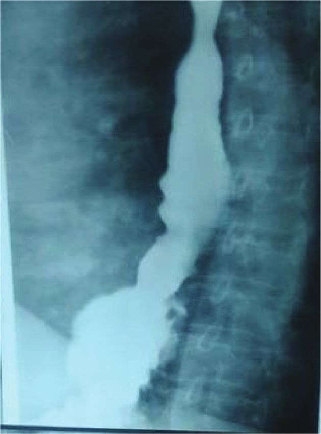
Chapter

Introductory Chapter:
Gastroesophageal Reflux Disease
Ali I. Yahya

1. Introduction

    Gastroesophageal reflux disease (GERD) occurs frequently in developed
countries. The number of cases, in fact, is increasing in the Middle East
countries. In western countries, its occurrence ranges from 10 to 20% of the
population who may present with typical or atypical symptoms or with com-
plications. Although GERD was described by Asher Winkelstein, an American
gastroenterologist, in 1935, it had appeared among patients earlier than that
time. Nowadays, cases of GERD are common among obese individuals, patients
with gallbladder disease, and those individuals under stress. It has also become
a common clinical problem that commonly affects young adults, both male and
female, of 40 years old.

2. History of GERD

   1855—Bowditch Rokitansky reported that esophagitis was due to gastroesopha-
geal reflux. Allison and Barrett found the association between hiatus hernia and
gastroesophageal reflux.
   1828—Charles Millard in Paris noticed the first case of esophagitis in child.
   1879—Heinrich Quincke reported that ulceration in the esophagus was due to
gastroesophageal reflux.
   1906—Tilston described the typical symptoms of esophagitis.
   1920—Joseph Sheehan described the endoscopic findings of esophagitis.
   1921—Porter Vinson noted the association between stricture and esophagitis.
   1934—Hampel introduced the term peptic esophagitis.
   1956—Rudolf Nissen performed a successful fundoplication for patient, who
suffered from GERD, with hiatus hernia. Patient was cured from the complaint.

3. Anatomy and Physiology

    At the lower end of the esophagus is a sphincter which is formed by a change
in the muscles of the esophagus. This sphincter controls the flow of esophageal
contents to the stomach. Different factors related to the anatomy and physiology
of the sphincter prevent the reflux of gastric contents into the esophagus. Among
these factors include the following:

    1. High pressure zone: Pressure at the lower esophageal sphincter area is high
       than stomach pressure (gastric pressure is +4 to +6, at the lower esophageal
       sphincter is +24 mmgh, and in the thoracic esophagus is −6). Because of the high

1
Gastroesophageal Reflux Disease - Theory and Research

      pressure at the sphincter, reflux is prevented. There are specific factors which
      will increase the tone of the sphincter, as well as factors like taking fatty meals,
      chocolate, smoking, and oral contraceptives that will reduce the tone of the
      sphincter.

    2. The length of the lower esophageal sphincter is 3 cm which is divided into
       abdominal part and thoracic part. If the abdominal part is less than 2 cm,
       patient will get reflux. Other factors like change of mucosa, the muscular
       coat of the stomach which will have oblique muscles in addition to the other
       two types of circular and longitudinal, crural effect and angulation of the
       esophagus to the stomach which is called Angle of His are not important in the
       prevention of the gastroesophageal reflux.

    3. Other factors that increase the effect of acid on the esophagus. Among these
       factors include the delayed gastric emptying. The increasing amount of the
       food in the stomach will lead to absorption of the sphincter and will increase
       the reflux. Reduced mucus and reduced saliva will lead to reduced bicarbonate
       which will reduce the effect of the acid refluxate.

4. Clinical presentation of gastroesophageal reflux disease

   GERD appears with typical symptoms or rarely by atypical symptoms, which
resemble cardiac symptoms and have been called cardiac symptoms.

4.1 Typical symptoms

    Typical symptoms which appear among 70% of patients include the following:

    1. Retrosternal pain (i.e., heart burn): It is the most common symptom which will
       be more manifested when patient is lying down or after meal and is seen among
       80% of patients.

    2. Regurgitation: It is a symptom observed when gastric or esophageal content comes
       in the mouth effortlessly. Regurgitation of gastric content will reach tracheobron-
       chial tree and will induce hoarseness of voice which is usually experienced in the
       morning. This hoarseness could be due to reflux of gastric content into the larynx
       or due to vagal irritation and will induce reflex spasm of the vocal cords. This
       symptom is seen among 50% of patients [1–6].

    3. Dysphagia: This is observed among 20% of patients with gastroesophageal
       reflux disease.

    Some rare presenting complaints, with rate of occurrence among patients, are as
follows:

    1. Abdominal pain: 30%

    2. Belching: 30%

    3. Coughing: 20%

    4. Wheezing: 10%

2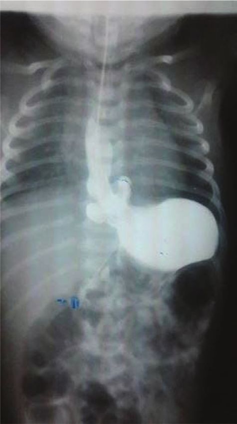
Introductory Chapter: Gastroesophageal Reflux Disease
DOI: http://dx.doi.org/10.5772/intechopen.84879
4.2 Atypical symptoms
Atypical symptoms are those where the patient will present with symptoms
not related to gastrointestinal system: coughing, wheezing, recurrent pharyn-
gitis, laryngitis, and chest pain. Its acute onset may resemble acute myocardial
infarction.
Patient may present with complicated gastroesophageal reflux disease—
some symptoms include the following: stricture, Barrett esophagus, lung damage in
the form of pneumonia, and lung fibrosis if condition goes chronic.
5. Investigations
1. Upper gastrointestinal endoscopy: Upper gastrointestinal endoscopy is very
important to exclude other serious disease which may mimic reflux, like tumors [7].
Upper gastrointestinal endoscopy can confirm hiatus hernia (Figure 1). Esophagitis
will be experienced by 50% of patients with GERD. It can also diagnose Barrett
esophagus and esophageal peptic stricture (Figure 2).
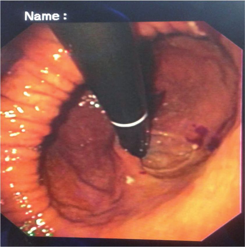
2. Contrast barium study: It is applied to detect hiatal hernia and esophageal
stricture [8]. Barium contrast study can show hiatus hernia which can be small
or large sac (Figures 3–5).
3. PH monitoring: 24 pH monitoring is very important in atypical presentation
of gastroesophageal reflux disease [9, 10]. It can confirm or exclude the disease,
with a diagnostic rate of 70–90%. It is not indicated in patients with esophagitis.
Figure 1.
The use of upper gastrointestinal endoscopy showing hiatus hernia in patient with GERD.
3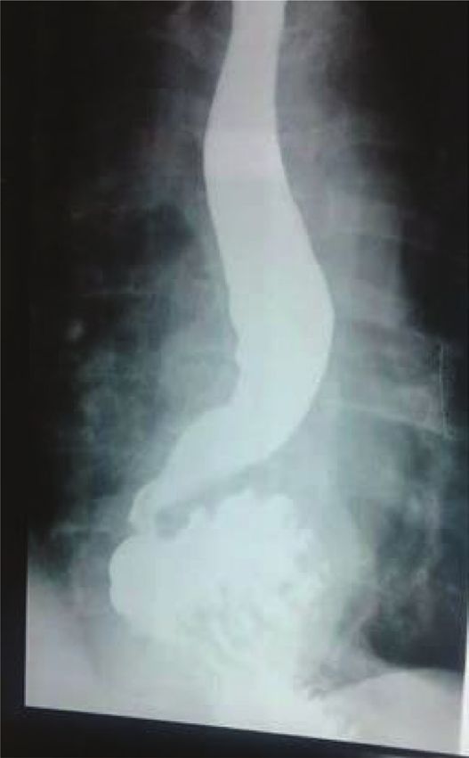
Gastroesophageal Reflux Disease - Theory and Research

    4. Gastroesophageal scintigraphy: Where drink like orange juice or milk is
       labeled with technetium and is used to study reflux, this test is rarely used
       for diagnosis of GERD. It is used in small children where we can study the
       reflexate to the lung, and the test is easy in small babies in comparison to
       other invasive tests. Gastroesophageal scintigraphy is used for patient who
       presents with atypical reflux symptoms like recurrent upper respiratory
       symptoms.

Figure 2.
The use of upper gastrointestinal endoscopy showing peptic stricture.

Figure 3.
Barium metal showing hiatus hernia.

4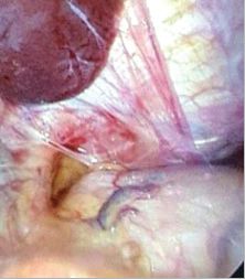
Introductory Chapter: Gastroesophageal Reflux Disease
DOI: http://dx.doi.org/10.5772/intechopen.84879

Figure 4.
Barium metal showing child hiatus hernia.

Figure 5.
Hiatus hernia with esophageal spasm.

    5. Multichannel impedance pH monitoring: It is a gold standard technique for
       diagnosis of GERD; it is more superior and more sensitive in diagnosing GERD
       than usual pH monitoring.

5
Gastroesophageal Reflux Disease - Theory and Research

    6. Manometry: It is a very important investigation to exclude motility disorders
       like achalasia and is indicated in patient who presents with atypical symptoms
       of gastroesophageal reflux disease. High-resolution manometry is more sensi-
       tive and superior than ordinary manometry in diagnosing esophageal motility
       disorders.

6. Treatment

    Reasons for treating GERD:

    1. Heart burn is a troubling symptom and affects patient life.

    2. Complications of GERD may cause esophagitis which will result to bleeding.
       Predisposition to Barrett esophagus that may turn to malignancy is 40–60 times
       seen in patient with reflux esophagitis-induced Barrett [11–13].

6.1 Nonsurgical treatment of gastroesophageal reflux disease

    1. Change of lifestyle

        a. Avoid having late meals, heavy meals, spicy or fatty meals, drinking alcohol,
           and smoking.

        b. Reduce weight; avoid tight clothes around the waist.

        c. Avoid drugs which reduce the tone of sphincter.

    2. Medical

   Medical treatment where drugs are used to neutralize the effect of the reflux on
esophageal mucosa:

    a. Antacids: Drugs that will neutralize the acid effect include the following—cal-
       cium, aluminum, and magnesium compounds. These are best taken after meals.
       Their effect is brief; and once they get emptied from the stomach, the symptoms
       may come back. These need to be given on an hour base to neutralize the acid
       effect.

    b. Histamine antagonists: There are receptors on the acid-producing cells which
       are stimulated by histamine to produce acid. These receptors are blocked by
       histamine-blocking drugs which act on H2 receptors. These drugs are best
       taken before meals. These include cimetidine which can be given 400–800 mg
       daily, ranitidine given 150 mg twice daily, and famotidine given 20–40 mg
       twice daily.

    c. Proton-pump inhibitors: These include omeprazole, esomeprazole, lansoprazole,
       pantoprazole, and rabeprazole. Their dosages range from 20 to 40 mg daily. PPI
       will cure the esophagitis up to 90%; 80% will recur within 1 year if treatment is
       stopped [14–17].

6
Introductory Chapter: Gastroesophageal Reflux Disease
DOI: http://dx.doi.org/10.5772/intechopen.84879
7. Surgery
Indication of surgery:
1. Failure of medical treatment.
2. Development of complications of the drugs.
3. Association with large hiatal hernia.
4. Atypical presentation with positive 24 h pH records.
5. Patients do not like to take drug for long life to control the symptoms.
7.1 Surgery for GERD
It can be done by lengthening the lower esophageal sphincter to create valve-like action
to prevent refluxing of gastric contents in the esophagus. Procedure is done by wrapping
the stomach around the lower esophagus [18–20], either full wrap of 360° (which is
named after Nissen) or partial wrap of 270°, either done posteriorly or anteriorly.
Fundoplication was previously performed by open surgery. Nowadays, most
operations are done laparoscopically (Figures 6 and 7), with excellent outcome on
short-term and long-term follow-ups.
Patient will stop taking the drugs. All patients should be seen by gastroenterolo-
gist, ENT specialist, and surgeons before surgery, especially for those patients who
come with atypical symptoms of GERD.
For many years, open surgery has been used for hiatus hernia but was rarely
applied for GERD without hernia. Many operations can be done, either abdominal
approach or thoracic approach. The common operation is the Nissen fundoplication
which has been used since 1950, with a success rate of 80–90%. Its complication
rate ranges from 10 to 15% and includes difficulty in swallowing and flatulence
which may go for some time than ease off.
7.2 Endoluminal surgery (NOTES)
It is also called incisionless surgery. Endoscopic treatment of GERD is still under
investigation:
1. Natural orifices transendoscopic surgery [21–25]
2. Endoscopic augmentation of lower esophageal sphincter, either by radio
frequency application or injection of ethylene vinyl alcohol in the region of the
lower esophageal sphincter [26–30].
7.3 Esophageal magnet ring
It is a new technique where there is no need to make wrapping around the
lower esophagus by the stomach. This is a magnet ring fixed around the lower
esophagus [31, 32]. It is not widely used and still under trial where magnet ring
is fixed laparoscopically around the esophageal sphincter. It moves out once food
comes in, and it comes back when the food enters the stomach. It has benefit
7Gastroesophageal Reflux Disease - Theory and Research over Nissen fundoplication. The patient can belch, vomit, and have no gas bloat syndrome, and it is reversible. The technique, however, is still under long-term trials. Figure 6. Laparoscopic view of big hiatus hernia in patient presented with GERD. Figure 7. Laparoscopic view of hiatus hernia in patient came with GERD symptoms. 8
Introductory Chapter: Gastroesophageal Reflux Disease
DOI: http://dx.doi.org/10.5772/intechopen.84879

Author details

Ali I. Yahya
Zliten University Hospital, Alasmaria University, Zliten, Libya

*Address all correspondence to: aliyahyaz60@hotmail.com

© 2019 The Author(s). Licensee IntechOpen. This chapter is distributed under the terms
of the Creative Commons Attribution License (http://creativecommons.org/licenses/
by/3.0), which permits unrestricted use, distribution, and reproduction in any medium,
provided the original work is properly cited.

9
Gastroesophageal Reflux Disease - Theory and Research

References

[1] Gao Y et al. A study of esophageal           pH monitoring with multichannel
function and reflux characteristics              impedance, esophageal manometry,
of gastroesophageal reflux disease               radiology and scintigraphy in
in patients presenting with chronic              gastroesophageal reflux disease? The
cough. Zhonghua Nei Ke Za Zhi.                   Turkish Journal of Gastroenterology.
2011;50(11):931-934                              2017;28(Suppl. 1):S16-S21

[2] Gyawali CP, Fass R. Management               [10] Gokturk S et al. Gastroesophageal
of gastroesophageal reflux disease.              reflux in asymptomatic patients
Gastroenterology. 2018;154(2):302-318            with diabetes: An impedance study
                                                 diabetes, obesity and gastroesophageal
[3] Broers C, Tack J, Pauwels A. Review          reflux. Experimental and Clinical
article: Gastro-oesophageal reflux               Endocrinology & Diabetes.
disease in asthma and chronic                    2018;2018:22
obstructive pulmonary disease.
Alimentary Pharmacology &                        [11] Wetscher GJ, Gadenstaetter M,
Therapeutics. 2018;47(2):176-191                 Klingler PJ, Weiss H, Obrist P, Wykypiel
                                                 H, et al. Efficacy of medical therapy
[4] Bor S et al. Prevalence of                   and antireflux surgery to prevent
gastroesophageal reflux disease in               Barrett's metaplasia in patients with
patients with asthma and chronic                 gastroesophageal reflux disease. Annals
obstructive pulmonary disease. Journal           of Surgery. 2001;234(5):627-632
of Gastroenterology and Hepatology.
2010;25(2):309-313                               [12] Corey KE, Schmitz SM, Shaheen NJ.
                                                 Does a surgical antireflux procedure
[5] Iliaz S et al. Does gastroesophageal         decrease the incidence of esophageal
reflux increase chronic obstructive              adenocarcinoma in Barrett's
pulmonary disease exacerbations?                 esophagus? A meta-analysis. The
Respiratory Medicine. 2016;115:20-25             American Journal of Gastroenterology.
                                                 2003;98(11):2390-2394
[6] Chen Y, Xiong L, Zeng J, Wei YG, Tan Y.
Gastroesophageal reflux disease is               [13] Lord RV. Does antireflux surgery
associated with high risk of obstructive         prevent progression of Barrett's esophagus?
sleep apnea syndrome. Zhonghua Nei               Minerva Chirurgica. 2011;66(1):1-6
Ke Za Zhi. 2018;57(11):824-829
                                                 [14] Saifullah AM, Ahmed F, Shil BC,
[7] Gyawali CP, Kahrilas PJ, Savarino E,         Banik RK, Saha SK, Chowdhury M,
Zerbib F, Mion F, Smout A, et al.                et al. Comparative study of Alginate and
Modern diagnosis of GERD: the Lyon               Omeprazole in symptomatic treatment
Consensus. Gut. 2018;67(7):1351-1362             of non-erosive gastroesophageal reflux
                                                 disease. Mymensingh Medical Journal.
[8] Khatami A et al. A comparison between        2018;27(4):771-775
gastroesophageal ultrasonography vs.
barium swallow in determining the                [15] Higuera-de-la-Tijera F. Efficacy
pattern of gastroesophageal reflux               of omeprazole/sodium bicarbonate
in a pediatric population. Medical               treatment in gastroesophageal reflux
Ultrasonography. 2015;17(1):22-27                disease: A systematic review. Medwave.
                                                 2018;18(2):e7179
[9] Vardar R, Keskin M. Indications
of 24-h esophageal pH monitoring,                [16] Freston JW. Therapeutic choices
capsule pH monitoring, combined                  in reflux disease: Defining the criteria

10
Introductory Chapter: Gastroesophageal Reflux Disease
DOI: http://dx.doi.org/10.5772/intechopen.84879
for selecting a proton pump inhibitor. fundoplication with Esophyx (Tif 2.0)
The American Journal of Medicine. and factors affecting outcomes in GERD
2004;117(Suppl. 5A):14s-22s patients followed for up to 6 years: A
prospective single-center study. Surgical
[17] Azzam RS. Are the persistent Endoscopy. 2015;29(9):2770-2780
symptoms to proton pump
inhibitor therapy due to refractory [24] Huang X, Chen S, Zhao H, Zeng X,
gastroesophageal reflux disease or to other Lian J, Tseng Y, et al. Efficacy of
disorder? Arquivos de Gastroenterologia. transoral incisionless fundoplication
Nov 2018;55(Suppl 1):85-91 (TIF) for the treatment of GERD:
A systematic review with meta-
[18] Papasavas PK, Keenan RJ, Yeaney analysis. Surgical Endoscopy.
WW, Caushaj PF, Gagne DJ, 2017;31(3):1032-1044
Landreneau RJ. Effectiveness of
laparoscopic fundoplication in relieving [25] Richter JE, Kumar A, Lipka s,
the symptoms of gastroesophageal Miladinovic B, Velanovich V. Efficacy
reflux disease (GERD) and eliminating of laparoscopic Nissen fundoplication
antireflux medical therapy. Surgical vs transoral incisionless fundoplication
Endoscopy. 2003;17(8):1200-1205 or proton pump inhibitors in patients
with gastroesophageal reflux disease:
[19] Wetscher GJ, Glaser K, Gadenstaetter A systematic review and network
M, Profanter C, Hinder RA. The effect meta-analysis. Gastroenterology.
of medical therapy and antireflux 2018;154(5):1298.e7-1308.e7
surgery on dysphagia in patients with
gastroesophageal reflux disease without [26] De Moura EGH et al.
esophageal stricture. American Journal Endoscopic polymer injection and
of Surgery. 1999;177(3):189-192 endoluminal plication in treatment
of gastroesophageal reflux disease:
[20] Shaw JM, Bornman PC, Callanan MD, Evaluation of long-term results.
Beckingham IJ, Metz DC. Long-term Endoscopy International Open.
outcome of laparoscopic Nissen and 2018;6(5):E630-E636
laparoscopic Toupet fundoplication
for gastroesophageal reflux disease: A [27] Kinoshita Y et al. Gastroesophageal
prospective, randomized trial. Surgical reflux after endoscopic injection
Endoscopy. 2010;24(4):924-932 sclerotherapy. The American Journal of
Gastroenterology. 1992;87(3):282-286
[21] Mayor MA, Fernando HC.
Endoluminal approaches to [28] Deviere J et al. Endoscopic
gastroesophageal reflux disease. implantation of a biopolymer in
Thoracic Surgery Clinics. the lower esophageal sphincter for
2018;28(4):527-532 gastroesophageal reflux: A pilot
study. Gastrointestinal Endoscopy.
[22] Cadiere GB, Buset M, Muls V, Rajan A, 2002;55(3):335-341
Rosch T, Eckardt AJ, et al. Antireflux
transoral incisionless fundoplication [29] Deviere J et al. Nonresorbable
using Esophy X: 12-month results copolymer implantation for
of a prospective multicenter gastroesophageal reflux disease:
study. World Journal of Surgery. A randomized sham-controlled
2008;32(8):1676-1688 multicenter trial. Gastroenterology.
2005;128(3):532-540
[23] Testoni PA, Testoni S, Mazzoleni G,
Vailati C, Passaretti S. Long-term [30] Yeh RW, Triadafilopoulos G.
efficacy of transoral incisionless Endoscopic antireflux therapy: The Stretta
11Gastroesophageal Reflux Disease - Theory and Research procedure. Thoracic Surgery Clinics. 2005;15(3):395-403 [31] Skubleny D, Switzer NJ, Dang J, Gill RS, Shi X, de Gara C, et al. LINX® magnetic esophageal sphincter augmentation versus Nissen fundoplication for gastroesophageal reflux disease: A systematic review and meta-analysis. Surgical Endoscopy. 2017;31(8):3078-3084 [32] Aiolfi A, Asti E, Bernardi D, Bonitta G, Rausa E, Siboni S, et al. Early results of magnetic sphincter augmentation versus fundoplication for gastroesophageal reflux disease: Systematic review and meta-analysis. International Journal of Surgery. 2018;52:82-88 12
You can also read