Clinical presentation and epidemiology of brain tumors firstly diagnosed in adults in the Emergency Department: a 10-year, single center ...
←
→
Page content transcription
If your browser does not render page correctly, please read the page content below
Original Article
                                                                                                                                       Page 1 of 5

Clinical presentation and epidemiology of brain tumors firstly
diagnosed in adults in the Emergency Department: a 10-year,
single center retrospective study
Ivan Comelli1, Giuseppe Lippi2, Valentina Campana1, Franco Servadei3, Gianfranco Cervellin1
1
    Emergency Department, University Hospital of Parma, Parma, Italy; 2Section of Clinical Biochemistry, University of Verona, Verona, Italy;
3
Department of Neurosurgery, Humanitas University and Research Institute, Milan, Italy
Contributions: (I) Conception and design: V Campana, G Cervellin; (II) Administrative support: None; (III) Provision of study materials or patients:
F Servadei, V Campana; (IV) Collection and assembly of data: V Campana, I Comelli; (V) Data analysis and interpretation: All authors; (VI)
Manuscript writing: All authors; (VII) Final approval of manuscript: All authors.
Correspondence to: Gianfranco Cervellin, MD. Emergency Department, Academic Hospital of Parma, Parma 43126, Italy.
Email: gianfranco.cervellin@gmail.com or gcervellin@ao.pr.it.

                  Background: Several patients with new onset brain tumors present to the Emergency Department (ED)
                  complaining for new symptoms. Although information exists on symptom prevalence in the entire population
                  of patients with brain tumors, little is known about the clinical presentation in ED. This retrospective study
                  was planned to investigate clinical presentation and epidemiology of brain tumors firstly diagnosed in a large
                  urban ED throughout a 10-year period.
                  Methods: All medical records of patients aged ≥18 years, discharged from our ED with a diagnosis of
                  brain tumor were retrieved from the electronic hospital database during a 10-year period (2006 to 2015).
                  The records were reassessed for selecting only brain tumors firstly diagnosed in the ED. The symptoms at
                  presentation were divided in six categories: (I) headache; (II) seizures; (III) focal signs; (IV) altered mental
                  status; (V) nausea/vomiting/dizziness; (VI) trauma. For all cases, the hospital record was retrieved, to obtain
                  histologic classification of tumors. Patients with inflammatory neoformations were excluded from the study.
                  Results: Overall, 205 patients with firstly diagnosed brain tumor were identified among 870,135 ED visits
                  (i.e.,
Page 2 of 5 Comelli et al. Brain tumors in the ED
diagnosed each year in the United States, and they are with a hospital discharge diagnosis of primary brain tumor,
associated with high rates of mortality (i.e., approximately were admitted in hospital through the ED. The presenting
3% of 5-year survival) (4). In the United States patients signs and symptoms included headache (56 patients), altered
with brain tumors have a monthly rate of Emergency mental status (51 patients), ataxia (41 patients), nausea
Department (ED) visit higher than that of the general or vomiting (37 patients), motor weakness (37 patients),
population (with a monthly mean cost of $48 vs. $3) (5). papilledema (28 patients), weakness (27 patients), cranial
Some regional registries (for example in Spain, Italy, and nerve palsies (26 patients), speech deficits (21 patients),
France) have also provided useful data (6-8), but clinical and visual deficits (20 patients), and sensory abnormalities (18
biological information are rarely investigated despite their patients). The average age was 43 years, with a range of
clear relevance. 3 days to 88 years. In this study, the majority of tumors were
It is generally acknowledged that the symptoms malignant astrocytomas, and the prevalent tumor location
caused by the presence of brain tumors can be physical was cortical (68 patients).
(e.g., focal signs, fatigue, headaches) or behavioral (e.g., In pediatric patients, brain tumors diagnosed in the
hallucinations, depression, anxiety, decreased attention and ED most commonly present with headache, symptoms
concentration, memory problems), mainly according to the related to hydrocephalus, nausea/vomiting, and gate
specific localization of the tumor (9). Nevertheless, precise disturbances (16).
information is almost lacking and, when available, it is also Therefore, due to the lack of information regarding the
quite contradictory. For example, in a study of 124 patients ED presentation of patients with brain tumors, the aim of
previously diagnosed with brain tumors, the most common this study was to investigate the clinical presentation and
symptoms that could be recorded were fatigue, sleep the epidemiology of brain tumors firstly diagnosed in a
disturbance, drowsiness, distress and dry mouth (10). Unlike large urban ED throughout a 10-year period.
these findings, six symptoms (i.e., fatigue, uncertainty about
the future, motor difficulties, drowsiness, communication
Methods
difficulties and headaches) were reported in >50% in a
sample of patients with glioma, with a considerably negative The University Hospital of Parma (Italy) is a 1,150-bed
impact on their quality of life (11). Weakness and headaches teaching general hospital, which serves a general population
were also the two main symptoms reported in a sample of of nearly 435,000 inhabitants, and is the only hospital in
92 patients with brain tumors by Davies et al. (12), whereas the geographical area of the city of Parma. The hospital is
depression has been identified as the single most important a level 2 trauma center, and also a referral center for stroke,
symptom predicting quality of life in a cohort of 73 patients neurosurgery, and acute myocardial infarction (AMI). In
with primary brain tumors by Pelletier et al. (13). More the local facility, children aged 14 years or younger are
recently, a study identified two clusters of symptoms in visited in the pediatric emergency room, and their medical
a group of newly diagnosed patients with brain tumors, information is recorded in a separate local database.
thus including a language cluster (i.e., difficulty reading or Adolescents aged 14–18 years are sometimes visited in the
writing and finding the right words) and a mood cluster ED, but on other occasions they directly access the Pediatric
(sadness, anxiety, depression) (14). Emergency Room, so that this part of the population has
A significant part of patients who were not previously been excluded from our study.
diagnosed with brain tumors present to the ED complaining All the records of patients aged ≥18 years, discharged
for new symptoms. Although, as aforementioned, from the local ED with a diagnosis of brain tumor have
information exists on the prevalence of signs/symptoms in hence been retrieved from the electronic hospital database,
the general population of patients with brain tumors, little during a 10-year period (i.e., between January 1st, 2006 to
is known about the clinical presentation in the ED. Only December 31st, 2015). All the records were independently
a single retrospective study, referred to a mixed pediatric reassessed by two of the authors (i.e., I Comelli and F
and adult population, has been published, describing the Servadei), and the classification was further supervised by
presenting signs and symptoms of patients with primary two other authors (i.e., G Cervellin and V Campana), with
brain tumors diagnosed in the ED to the best of our the aim of selecting only brain tumors which were not
knowledge (15). Briefly, a total of 101 patients, identified previously diagnosed outside the ED (i.e., only including
© Annals of Translational Medicine. All rights reserved. atm.amegroups.com Ann Transl Med 2017;5(13):269Annals of Translational Medicine, Vol 5, No 13 July 2017 Page 3 of 5
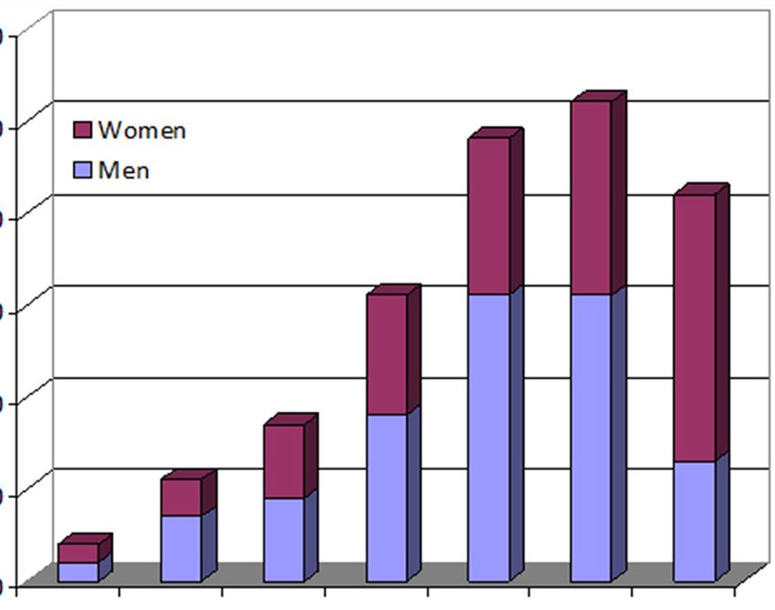
60
is >100%.
For all selected cases, the complete hospital record was
50 Women retrieved, to obtain additional information including the
Men
histologic classification of tumor. Due to the broad range
Number of patients
40
of histologic types (see section “Discussion”), we classified
30 all cases according to four main categories, for practical
purposes: (I) glial tumors (including astrocytomas,
20
glioblastomas, oligodendrogliomas etc.); (II) meningiomas;
10
(III) metastases (when neoplasm not formerly known); (IV)
others miscellaneous tumors (including medulloblastomas,
0 ependymal tumors, schwannomas, hemangiomas,
18–29 30–39 40–49 50–59 60–69 70–79 >80
years years years years years years years lymphomas etc.). Inflammatory neoformations (i.e.,
Age groups abscesses, mycetomas etc.) were obviously excluded from
Figure 1 Age of the study population.
the study. The differences between groups were evaluated
with one-way analysis of variance (ANOVA), using
Analyse-it (Analyse-it Software Ltd, Leeds, UK).
Table 1 Histological classification of brain tumors firstly diagnosed Due to the retrospective design of this study and
in the emergency department maintenance of anonymity of the patients, ethical
Tumor class Number (%) committee approval was unnecessary. Nevertheless, the
Glial tumors 95 (46.3)
study was approved by the local review Board and was
performed in accordance with the Declaration of Helsinki,
Meningiomas 45 (22.0)
under the terms of relevant local legislation.
Metastases 35 (17.1)
Others miscellaneous 30 (14.6)
Results
A total of 205 patients (111 men, 94 women) with a firstly
Table 2 Leading signs and symptoms of brain tumors firstly diagnosed brain tumor were identified among 870,135
diagnosed in the emergency department ED visits throughout the study period, thus representing
Main signs/symptoms Number (%) approximately 0.02% of the entire ED population. The
Focal signs 122 (59.5)
demographic characteristics of the patient population are
shown in Figure 1, whereas the histologically classified
Mental status alteration 51 (24.9)
groups are summarized in Table 1. Glial tumors were
Headache 30 (14.6) the most frequently diagnosed (i.e., 46.3%), followed by
Seizures 29 (14.1) meningiomas (21.9%), metastases (17.1%), and others
Trauma (occasional) 16 (7.8)
miscellaneous (14.7%). No significant differences were
observed between the mean age of patients with different
Nausea/vomiting/dizziness 9 (4.4)
histologically based groups (glioblastomas 65±16 years;
meningiomas 66±14.20 years; metastases 66±13 years;
other miscellaneous 66±19 years; P=0.972). The main
those which were firstly diagnosed in the ED). The signs and/or symptoms of presentation are reported in
symptoms at presentation were divided in six categories: Table 2, with focal signs accounting for over than 50% of
(I) headache; (II) seizures; (III) focal signs; (IV) altered all clinical pictures.
mental status; (V) nausea/vomiting/dizziness; (VI) trauma
(i.e., occasional diagnosis in course of CT execution for
Discussion
trauma). In several cases the clinical presentation included
more than one main symptom, e.g., headache plus altered The signs and symptoms found more frequently in our
mental status, so that the sum of the different rates population of primarily diagnosed brain tumor in the ED
© Annals of Translational Medicine. All rights reserved. atm.amegroups.com Ann Transl Med 2017;5(13):269Page 4 of 5 Comelli et al. Brain tumors in the ED
were focal signs, followed by mental status alteration, urban ED, so that the emergency physicians should always
seizures, and headache. Notably, a considerable part was be aware of this possibility.
serendipitously diagnosed (i.e., 7.8%, thus approaching 2
cases per year), when undergoing a CT scan for head injury.
Acknowledgements
This data substantially confirms that observed in a previous
study (17). Glial tumors were the most frequently diagnosed The authors acknowledge Drs. Marco Brambilla and Marco
in our sample, followed by meningiomas, metastases and Mignani of the University Hospital of Parma for the kind
others miscellaneous tumors. support in extracting data from electronic databases of the
The classification of brain tumors has been traditionally Institution.
characterized by a high degree of uncertainty, and has been
changed many times over the last two centuries. The first
Footnote
report about brain tumor classification was published by
Virchow in 1863 (18), whereas the vast majority of currently Conflicts of Interest: The authors have no conflicts of interest
used terms were introduced by Bailey and Cushing in to declare.
1926 (19). In 1949, Kernohan et al. simplified the
classification by reducing the number of brain tumor types Ethical Statement: The study was approved by the local
and introducing the concept of tumor grading (20). The review Board.
first World Health Organization (WHO) classification of
brain tumors was published in 1979 (21), and substantially
References
used the original terminology from Cushing and Bailey.
Revised WHO-based classifications have then been 1. Forman D, Bray F, Brewster DH, et al. editors. Cancer
published in 1993, 2000, 2007 and, finally, in 2016 (22). incidence in five continents, vol X. Lyon: IARC, 2014.
The current WHO classification is extremely complex and 2. Ostrom QT, Gittleman H, Fulop J, et al. CBTRUS
certainly practical for neurosurgeons and oncologists, but is statistical report: primary brain and central nervous system
not actually applicable in an emergency medicine context. tumors diagnosed in the United States in 2008-2012.
Considering the high number of histological subtypes, Neuro Oncol 2015;17:iv1-62.
nearly each subtype of brain tumor could indeed be 3. Crocetti E, Trama A, Stiller C, et al. Epidemiology of
considered as a rare one. Moreover, it has been previously glial and non-glial brain tumours in Europe. Eur J Cancer
demonstrated that there is a substantial inter-observer 2012;48:1532-42.
and inter-center discordance in the diagnosis of specific 4. American Cancer Society. Cancer facts and figures.
subtypes (23). This is the reason why we originally decided Available online: http://www.cancer.org. Last accessed
to simplify the classification of our patient population by April 15th 2017.
grouping similar tumor types in substantially homogeneous 5. Kutikova L, Bowman L, Chang S, et al. Utilization
groups. The mean age of our patients was found to be and cost of health care services associated with primary
higher than that previously reported (17), and this is malignant brain tumors in the United States. J Neurooncol
probably due to the exclusion of pediatric patients from our 2007;81:61-5.
study. All the age groups (i.e., >18 years) were represented 6. Baldi I, Gruber A, Alioum A, et al. Descriptive
in our population, with a higher prevalence of patients epidemiology of CNS tumors in France: results from the
aged more than 50 years, but with no significant differences Gironde registry for the period 2000-2007. Neuro Oncol
among the different histological subgroups. 2011;13:1370-8.
7. Ruiz-Tovar M, López-Abente G, Pollán M, et al. Brain
cancer incidence in the province of Zaragoza and Navarre
Conclusions
(Spain): effect of age, period and birth cohort. J Neurol Sci
Taken together, the results of our study show that first 1999;164:93-9.
diagnosis of brain tumors is not very frequent in the ED, 8. Caldarella A, Crocetti E, Paci E. Is the incidence of brain
but it cannot be considered a rare event for the wide and tumors really increasing? A population-based analysis from
heterogeneous population of patients admitted to a large a cancer registry. J Neurooncol 2011;104:589-94.
© Annals of Translational Medicine. All rights reserved. atm.amegroups.com Ann Transl Med 2017;5(13):269Annals of Translational Medicine, Vol 5, No 13 July 2017 Page 5 of 5
9. Shaw EG, Robbins ME. The management of radiation- 17. Darlix A, Zouaoui S, Rigau V, et al. Epidemiology for
induced brain injury. Cancer Treat Res 2006;128:7-22. primary brain tumors: a nationwide population-based
10. Armstrong TS, Gning I, Mendoza TR, et al. Clinical study. J Neurooncol 2017;131:525-46.
utility of the MDASI-BT in patients with brain metastases. 18. Virchow R. editor. Die Krankhaften Geschwulste. Berlin:
J Pain Symptom Manage 2009;37:331-40. Hirschwald, 1863.
11. Osoba D, Brada M, Prados MD, et al. Effect of disease 19. Bailey P, Cushing H. editors. A classification of tumors of
burden on health-related quality of life in patients with the glioma group on a histogenetic basis with a correlation
malignant gliomas. Neuro Oncol 2000;2:221-8. study of prognosis. Philadelphia: Lippincott, 1926.
12. Davies E, Clarke C. Early symptoms of brain tumours. J 20. Kernohan JW, Mabon RF, Svien HJ, et al. A simplified
Neurol Neurosurg Psychiatry 2004;75:1205-6. classification of the gliomas. Symposium on a new
13. Pelletier G, Verhoef MJ, Khatri N, et al. Quality of life simplified concept of gliomas. Mayo Clin Proc
in brain tumor patients: the relative contributions of 1949;35:71-5.
depression, fatigue, emotional distress, and existential 21. Zülch KJ, Avtsyn AP, Barnar RO, et al. editors.
issues. J Neurooncol 2002;57:41-9. Histological typing of tumours of the central nervous
14. Gleason JF, Case D, Rapp SR, et al. Symptom clusters in system. Geneva: Office of Publications, World Health
patients with newly-diagnosed brain tumors. J Support Organization, 1979.
Oncol 2007;5:427-33. 22. Louis DN, Ohgaki H, Wiestler OD, et al. editors. WHO
15. Snyder H, Robinson K, Shah D, et al. Signs and symptoms classification and grading of tumours of the central nervous
of patients with brain tumors presenting to the emergency system. Lyon: IARC Press, 2016.
department. J Emerg Med 1993;11:253-8. 23. van den Bent MJ. Interobserver variation of the
16. Lanphear J, Sarnaik S. Presenting Symptoms of Pediatric histopathological diagnosis in clinical trials on glioma: a
Brain Tumors Diagnosed in the Emergency Department. clinician’s perspective. Acta Neuropathol 2010;120:297-304.
Pediatr Emerg Care 2014;30:77-80.
Cite this article as: Comelli I, Lippi G, Campana V, Servadei
F, Cervellin G. Clinical presentation and epidemiology of
brain tumors firstly diagnosed in adults in the Emergency
Department: a 10-year, single center retrospective study. Ann
Transl Med 2017;5(13):269. doi: 10.21037/atm.2017.06.12
© Annals of Translational Medicine. All rights reserved. atm.amegroups.com Ann Transl Med 2017;5(13):269You can also read