Comparative Study of Metachromatic Staining Methods in Assessing the Exfoliative Cell Types During Oestrous Cycle in Sprague-Dawley Laboratory Rats
←
→
Page content transcription
If your browser does not render page correctly, please read the page content below
Int. J. Morphol.,
36(3):962-968, 2018.

           Comparative Study of Metachromatic Staining Methods
           in Assessing the Exfoliative Cell Types During Oestrous
                 Cycle in Sprague-Dawley Laboratory Rats

      Estudio Comparativo de los Métodos de Tinción Metacromática en la Evaluación de los Tipos
         de Células Exfoliativas Durante el Ciclo Estral en Ratas Sprague-Dawley de Laboratorio

                                               Stephanie Mohammed1 & Venkatesan Sundaram2

MOHAMMED, S. & SUNDARAM, V. Comparative study of metachromatic staining methods in assessing the exfoliative cell types
during oestrous cycle in Sprague-Dawley laboratory rats. Int. J. Morphol., 36(3):962-968, 2018.

        SUMMARY: This study was aimed at comparing the commonly used metachromatic stains viz., Papanicolaou stain, Wright-
Giemsa, Toluidine blue and Methylene blue in the assessment of cell types of the oestrous cycle in rats. Eight female Sprague-Dawley
rats aged 8-9 weeks were used for this assessment. Cotton Swabs were gently inserted in the animals vagina to obtain cells from which
they were then transferred to glass slides for staining and evaluation under microscopy. The different cell types were compared for their
morphological features and clarity of cellular detail under all four stains. The application, advantages and limitations of all stains were
then discussed. It was concluded that the selection of the most effective stain in the assessment of vaginal cytology depends on their
application to clinical or research which was based on the cellular detail of interest, time, cost and availability of each staining procedure.

           KEY WORDS: Papanicolaou; Wright-Giemsa; Methylene blue; Toluidine blue; Vaginal cytology.

INTRODUCTION

        In exfoliative vaginal cytology, epithelial cells                     quantification of each cell type, various staining procedures
undergo constant growth, shedding and replacement. The                        are readily employed. The proper choice of stain and
microscopic evaluation of these cells during exfoliation                      staining method is very crucial and selected based on the
provides information on the stages of the cycle, possible                     cellular detail of interest. The selection also depends on
infections, hormonal status and reproductive defects that                     the collection of sample, fixation, nuclear staining,
might be occurring within the female reproductive system.                     cytoplasmic staining and clearing (Ochei & Kolhatkar,
                                                                              2000). The most commonly used stains in the vaginal
        The short, regular but dynamic oestrous cycle of 4-                   exfoliative cytology are Papanicolaou stain, Wright-
5 days in rats has made them the ideal models for                             Giemsa, Toluidine blue and Methylene blue (Bancroft &
investigating the changes that occur in the reproductive                      Stevens, 1996).
system by vaginal exfoliative cytology (Cora et al. 2015).
These morphological changes due to the desquamation                                   This present study is aimed at comparing the
depict four major phases of the oestrus cycle which include                   commonly used metachromatic stains viz., Wright-Giemsa,
the Proestrus (P), Oestrous (O), Metestrous (M) and                           Toluidine blue, Methylene blue and Papanicolaou stain in
Diestrous (D) (Long & Evans, 1922). Throughout the                            the assessment of cell types of the oestrous cycle in rats.
oestrous cycle, the major cells observed are small and large
nucleated epithelial cells, anucleated, keratinized epithelial
cells and neutrophils. The presence of bacteria and                           MATERIAL AND METHOD
malignant cells are also identifiable during this process.

               To properly assess the morphology and                                   Eight female Sprague-Dawley rats aged 8-9 weeks

1
    Department of Preclinical Sciences, School of Medicine, Faculty of Medical Sciences, The University of The West Indies, Trinidad and Tobago.
2
    Department of Basic Veterinary Sciences, School of Veterinary Medicine, Faculty of Medical Sciences, The University of The West Indies, Trinidad.

962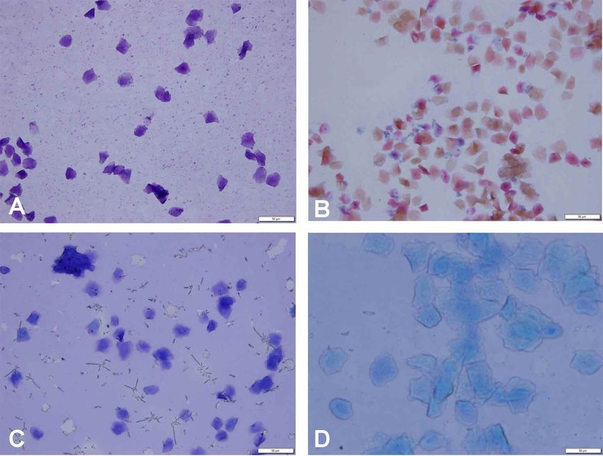
MOHAMMED, S. & SUNDARAM, V. Comparative study of metachromatic staining methods in assessing the exfoliative cell types during oestrous cycle in Sprague-Dawley laboratory rats.
                                                              Int. J. Morphol., 36(3):962-968, 2018.

were chosen for this study. The rats were exposed to a 12 h light/ 12 h dark
cycle with adequate food and water supply. All protocols involving the care
and use of live animals for experimentation and code of ethics were followed
and the work was approved by the institutional Ethical Committee.

        The vaginal swabs from all rats were taken at 9 am daily for 15 days
(three consecutive cycles and 120 vaginal swabs) to assess the oestrus cycle.
The cotton swab technique was used in which the sterilized vaginal swabs
were moist with saline, then inserted into the vagina, gently removing the
cells from the vaginal lumen (Fig. 1) and then transferring onto a clear glass
slide. The slides were then left to air-dry to get fixed for Wright-Giemsa,
Toluidine blue and Methylene blue staining whereas the slides were fixed
with 96 % ethanol for 30 min for Panipacolaou (Pap) stain. All the slides were
stained as per standard procedures (Suvarna et al., 2012; Cora et al.).

        The slides were then observed and analysed for the assessment of each
cell type and phase of the oestrous cycle and the digital images were obtained
with the aid of the Olympus BX51 system microscope and the Olympus DP71                                                     Fig. 1. Cotton swab used for taking vaginal
microscope digital camera.                                                                                                  cytology in Sprague-Dawley rat. The rat is
                                                                                                                            properly secured in an animal holder.

Fig. 2. Keratinized cells. Wrights-Giemsa stain (A), Pap Stain (B), Toluidine blue (C) and Methylene blue (D).

                                                                                                                                                                                963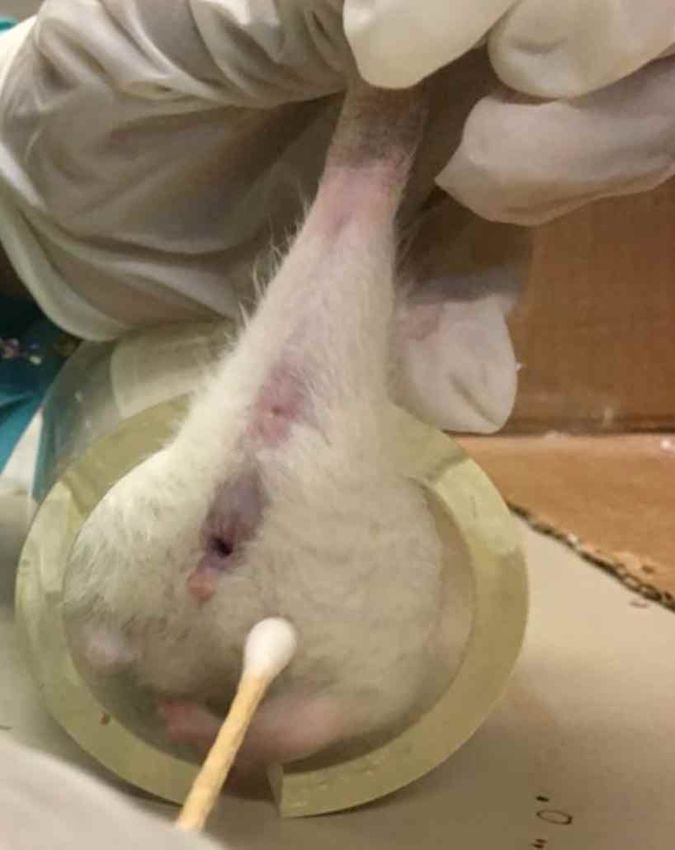
MOHAMMED, S. & SUNDARAM, V. Comparative study of metachromatic staining methods in assessing the exfoliative cell types during oestrous cycle in Sprague-Dawley laboratory rats.
                                                              Int. J. Morphol., 36(3):962-968, 2018.

RESULTS

Cell types of oestrous cycle: The following cell types were                                  cells with a high nuclear to cytoplasmic ratio and can be
identified based on the cytomorphology during the oestrous                                   seen clearly in clusters when stained with methylene blue.
cycle in the present study with the use of various                                           The intermediate cells varied in size and shape as some can
metachromatic stains.                                                                        be oval with prominent nucleus and others polygonal with
                                                                                             lesser nucleus to cytoplasmic ratio .These cells were clearly
1. Anucleated keratinized cells. The anucleated keratinised                                  shown with the Papanicolaou stain than other stains. The
cells were large, irregular shaped cornified cells with lack of                              superficial cells were the largest of all epithelial cells in the
visible nucleus (Fig. 2). All four stains gave proper clarity of                             present study and were polygonal and appeared as either
cornified cells. Papanicolaou stain however was able to show                                 flat or rolled up in shape. These cells were better visible
the various shades of orange, pink and blue associated with                                  with the Wright-Giemsa stain.
level of cornification. The presence of bacteria was also noticed
clearly with the use of toluidine blue in the present study.                                 3. Neutrophils. The neutrophils were small round cells with
                                                                                             mono and polymorph nuclear leucocytes. All four stains were
2. Epithelial cells. The epithelial cells consisted of three                                 able to show the neutrophils very clear whether in clusters
major cells viz, parabasal, intermediate and superficial (Fig.                               or singly (Fig. 4). The methylene blue gave better definition
3) in the present study. The parabasal cells were small round                                of neutrophils at the same magnification.

Fig. 3. Epithelial cells. Wright’s Giemsa stain (A), Papanicolaou (B), Toluidine blue (C) and Methylene blue (D). Superficial cells are
seen in (A), intermediate are clearly seen in (B) and parabasal cells in cluster (D).

964
MOHAMMED, S. & SUNDARAM, V. Comparative study of metachromatic staining methods in assessing the exfoliative cell types during oestrous cycle in Sprague-Dawley laboratory rats.
Int. J. Morphol., 36(3):962-968, 2018.
Fig. 4. Neutrophils for the Wrights Giemsa stain (A), Papanicolaou (B), Toluidine blue (C) and Methylene blue (D). The
neutrophils may be spaced out, single or clumped.
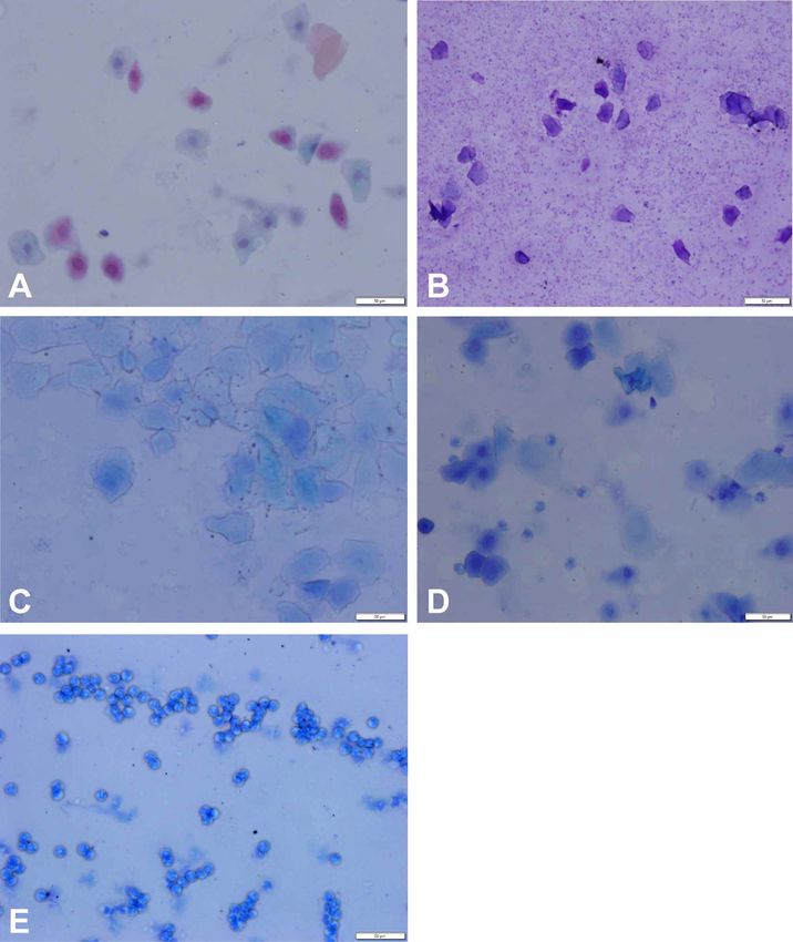
Description of Stages of Oestrus in Sprague-Dawley Metestrus. This stage showed a mixture of all major cell
rats with various stains. The following were the types, neutrophils, cornified and nucleated cells as shown
cytological detail of the normal oestrus cycle of female with toluidine blue stain (Fig. 5).
Sprague-Dawley rats observed in the present study.
Diestrus. This stage depicted a predominance of
Proestrus. This stage showed a predominance of small neutrophils as shown with methylene blue stain (Fig. 5).
round nucleated cells with uniform shape. The cells were
appeared either in clusters or individually as seen when
stained with Pap stain (Fig. 5). DISCUSSION
Oestrus. This stage was characterized by a progressive
shedding of cornified squamous epithelial cells that lack The anucleated cornified cells were prominent in all
visible nucleus, were irregular in shape and showed four stains performed in this study. Various shades of orange,
granular cytoplasm as shown with Wright-Giemsa and pink and blue associated with the level of cornification were
Methylene blue stain. The presence of bacteria was also observed from the Papanicolaou stain. The parabasal cells,
be seen clearly in this stage with methylene blue stain with a high nuclear to cytoplasmic ratio were clearly shown
(Fig. 5). in clusters with the use of methylene blue when compared
965MOHAMMED, S. & SUNDARAM, V. Comparative study of metachromatic staining methods in assessing the exfoliative cell types during oestrous cycle in Sprague-Dawley laboratory rats.
Int. J. Morphol., 36(3):962-968, 2018.
Fig. 5. Different stages of oestrus cycle in Sprague-
Dawley rats. A. Pap Stain for proestrus stage shows
many superficial nucleated cells. B. Wright’s Giemsa
stain for estrus stage gives many anucleated cells. C.
Methylene blue for late oestrus stage showing large
cornified cells with bacteria present in the cytoplasm
(arrow). D. Toluidine blue stain for metestrus showing
a mixture of all cells, cornified, large epithelial and
neutrophils (arrow). E. Methylene blue for diestrus
showing predominance of neutrophils.
with the toluidine blue. The intermediate cells were noticeably Wright’s-Giemsa stain. The neutrophils were visible regardless
visible with lesser nucleus to cytoplasmic ratio in the of the staining method. In the present study bacteria was clearly
Papanicolaou stain. The superficial cells were very clear in the evident in both the toluidine blue and methylene blue.
966MOHAMMED, S. & SUNDARAM, V. Comparative study of metachromatic staining methods in assessing the exfoliative cell types during oestrous cycle in Sprague-Dawley laboratory rats.
Int. J. Morphol., 36(3):962-968, 2018.
The Papanicolaou stain is considered a multi-chromatic components of cells and has been considered good in the
classical stain that provides colouring of differential acidophilic identification of bacteria and cell structure (Bancroft &
and basophilic cells with sharp nuclear staining. It stains the Stevens; Turgeon, 2005). The advantages of both the staining
cytoplasm of cells with various shades based on the level of methods are that they are very simple, less time consuming,
cornification. It requires fixation and the staining protocol takes do not require any special fixation and they show evidence
25-30 min. (Ochei & Kolhatkar; Cora et al.; Choudhary et al., of bacteria clearly as seen in this present study. Yet the poor
2012). Though the most recommended stain in vaginal quality of staining due to improper staining time and
cytology is the Pap stain based on the level of cellular details. deteriorated reagents are very common among these two
The wet fixation promotes the clarity of cellular details in this stains.
stain. The wet fixation sharpens the nuclear outlines and any
unusual chromatin patterns while air drying amplifies the cell In assessing the stages of oestrous cycle in rats, the
for more accurate evaluation. This happens because air drying toluidine blue and methylene blue will be very effective if
influences the structure of the nuclear chromatin which makes proper staining time and proper reagents were used when
it ideal for the comparison of normal and malignant cells. Due the time and cost is considered. Additionally, at present,
to this fact, it is widely used in traditional gynaecological unstained slides are also considered effective when examined
cytology and for screening of cervical cancer in humans. But, directly under the microscope and is considered reliable and
to assess the stages of oestrous cycle in rats with cytology did less consuming (Yener et al., 2007) but it needs little
not warrant the level of details depicted by the pap stain. In expertise.
addition, the cost and duration of other stains make them more
preferable than the Pap stain for the present study.
CONCLUSION
The Wright-Giemsa stain, a modified Romanowsky
stain is the combination of methylene blue with eosin red.
The generally accepted staining scheme produce purple All four stains have their own unique properties with
chromatin, blue leucocyte cytoplasm, purple neutrophils, many similarities and few varying limitations. The various
pink-red cells and purple granules. This occurs as a result of stains used in this study as seen can all be used in identifying
acid and base dying in poly cations and poly anions giving the various cell types and stages of the oestrus cycle.
rise to blue staining of ribosome rich cytoplasm (Horobin, However, the choice of staining depends mainly on the
2011). This stain is the most preferred stain for dried smears outcome desired as it will differ for clinical and research
as they allow better estimation of cell-nuclear ratio with purposes. The cellular detail of interest, the time, cost and
enhanced cytoplasmic details. This stain is used for non- availability will all play an important role in the choice of
gynaecological purposes such as staining of sputum and stain.
malignant cells (Conn, 1961; Lillie, 1969; Ochei & Kolhatkar)
and for vaginal infections and inflammatory conditions. The
advantage of this stain is that there is no special fixation MOHAMMED, S. & SUNDARAM, V. Estudio comparativo de
requirement and generally the time taken varies from 10-15 los métodos de tinción metacromática en la evaluación de los tipos
minutes depending on the strength of the stain. But this is a de células exfoliativas durante el ciclo estral en ratas Sprague-
major limitation as staining time can result in observable wrong Dawley de laboratorio. Int. J. Morphol., 36(3):962-968, 2018.
colours. The results are also dependent on the method of
RESUMEN: El presente estudio tuvo como objetivo com-
sample collection. In vaginal cytology, the major limitation parar las tinciones metacromáticas comúnmente utilizadas,
of this stain is the inability to highlight keratinization occurring Wright's-Giemsa, azul de toluidina, azul de metileno y tinción de
in the cytoplasm (Krafts & Pambuccian, 2011) which was Papanicolaou, en la evaluación de los tipos de células del ciclo
evident in the present study also. estral en ratas. El estudio se realizó en ocho ratas hembras Sprague-
Dawley, con edades entre 8 y 9 semanas, y se usaron hisopos
Toluidine blue which is considered a basic vaginales de algodón para preparar portaobjetos. Los diferentes
metachromatic dye with high affinity towards acidic tissues, tipos de células se compararon por sus características morfológicas
stains the nuclei dark blue and the polysaccharides violet y claridad en las cuatro tinciones. La aplicación, ventajas y limita-
ciones de todas las tinciones fueron discutidas. Se concluye que la
because of its strong binding capacity towards the nuclear
selección de la tinción más efectiva en la evaluación de la citología
material (DNA) in the nucleus and (RNA) in the cytoplasm vaginal depende de su uso, es decir, clínico o de investigación, el
(Sridharan & Shankar, 2012). This stain is very useful in detalle celular de interés, tiempo, costo y disponibilidad.
detecting vulvar cancer and lacerations in rape victims
(McCauley, 1987). A similar stain to toluidine blue is PALABRAS CLAVE: Papanicolaou; Wright-Giemsa;
methylene blue, a cationic stain that stains negatively charged Azul de metileno; Azul de toluidina; Citología vaginal.
967MOHAMMED, S. & SUNDARAM, V. Comparative study of metachromatic staining methods in assessing the exfoliative cell types during oestrous cycle in Sprague-Dawley laboratory rats.
Int. J. Morphol., 36(3):962-968, 2018.
REFERENCES Corresponding author:
Dr. Venkatesan Sundaram
Department of Basic Veterinary Sciences
Bancroft, G. D. & Stevens, A. Theory and Practice of Technique. 4th ed. Faculty of Medical Sciences
New York, Churchill Livingstone Inc., 1996. The University of The West Indies
Choudhary, P.; Sudhamani, S.; Pandit, A. & Kiri, V. Comparison of modified St. Augustine, Trinidad
ultrafast Papanicolaou stain with the standard rapid Papanicolaou stain TRINIDAD AND TOBAGO
in cytology of various organs. J. Cytol., 29(4):241-5, 2012.
Conn, H. J. Biological Stains: A Handbook on the Nature and Uses of the
Dyes Employed in the Biological Laboratory. 6th ed. Baltimore, Williams
Email: mailto:drvenkat1971@gmail.com
and Wilkins, 1961.
Cora, M. C; Kooistra, L. & Travlos, G. Vaginal cytology of the laboratory
rat and mouse: Review and criteria for the staging of the estrous cycle
using stained vaginal smears. Toxicol. Pathol., 43(6):776-93, 2015. Received: 24-01-2018
Horobin, R. W. How Romanowsky stains work and why they remain Accepted: 05-03-2018
valuable - including a proposed universal Romanowsky staining
mechanism and a rational troubleshooting scheme. Biotech. Histochem.,
86(1):36-51, 2011.
Krafts, K. P. & Pambuccian, S. E. Romanowsky staining in cytopathology:
history, advantages and limitations. Biotech. Histochem., 86(2):82-93,
2011.
Lillie, R. D. H. J. Conn's Biological Stains: A Handbook on The Nature
and Uses of the Dyes Employed in the Biological Laboratory. 8th ed.
Baltimore, Williams and Wilkins, 1969.
Long, J. A. & Evans, H. M. The Oestrous Cycle in the Rat and its Associated
Phenomena. Berkeley, University of California Press, 1922.
McCauley, J.; Guzinski, G.; Welch, R.; Gorman, R. & Osmers, F. Toluidine
blue in the corroboration of rape in the adult victim. Am. J. Emerg.
Med.,5(2):105-8, 1987.
Ochei, J. & Kolhatkar, A. Medical Laboratory Science: Theory and Practice.
New York, McGraw-Hill Education, 2000. pp.513-37.
Sridharan, G. & Shankar, A. A. Toluidine blue: A review of its chemistry
and clinical utility. J. Oral Maxillofac. Pathol., 16(2):251-5, 2012.
Suvarna, K. S.; Christopher, L. & Bancroft, J. Bancroft’s Theory and
Practice of Histological Techniques. 7th ed. New York, Churchill
Livingstone, 2012.
Turgeon, M.L. Clinical Hematology: Theory and Procedures. 4th ed.
Philadelphia, Lippincott Williams and Wilkins, 2005.
Yener, T.; Turkkani Tunc, A.; Aslan, H.; Aytan, H. & Cantug Caliskan, A.
Determination of oestrous cycle of the rats by direct examination: how
reliable? Anat. Histol. Embryol., 36(1):75-7, 2007.
968You can also read