A pilot study of mobile digital colposcopy in Japanese patients with cervical intraepithelial neoplasm
←
→
Page content transcription
If your browser does not render page correctly, please read the page content below
MOLECULAR AND CLINICAL ONCOLOGY 15: 207, 2021

                A pilot study of mobile digital colposcopy in Japanese
                   patients with cervical intraepithelial neoplasm
MASAKAZU SATO*, DAISUKE SHINTANI*, MIEKO HANAOKA, SHO SATO, MAIKO MIWA, AIKO OGASAWARA,
  AKIRA YABUNO, AKIRA KUROSAKI, HIROYUKI YOSHIDA, KEIICHI FUJIWARA and KOSEI HASEGAWA

                          Department of Gynecologic Oncology, Saitama Medical University International
                                        Medical Center, Hidaka, Saitama 350‑1298, Japan

                                         Received December 24, 2020; Accepted June 11, 2021

                                                     DOI: 10.3892/mco.2021.2370

Abstract. Digital colposcopy built around a smartphone is          definitions of the ‘same’, ‘almost the same’ and ‘different’,
becoming common, and this has advantages for telemedicine          respectively. The present results indicated that ≥75% cases
and data sharing by taking advantage of smartphone char‑           were equivalent in digital colposcopy and conventional
acteristics. However, digital colposcopy itself is not allowed     colposcopy. This suggests that digital colposcopy may not be
in clinical practice in Japan. The aim of the present study        inferior to conventional colposcopy.
was to investigate the feasibility of mobile digital colposcopy
incorporating a smartphone for management of cervical              Introduction
screening in Japanese patients. Patients who underwent
colposcopy at Saitama Medical University International             More than half a million women are diagnosed with cervical
Medical Center between July 2019 and February 2020                 cancer (1). Introduction of the Pap smear test has markedly
were enrolled in the present study. The inclusion criteria         decreased the mortality rate of cervical cancer, but cervical
were women aged 21‑65 years old referred for colposcopy            cancer is the fourth most common female malignancy
following the Japanese standard of care. Written informed          worldwide and is still one of the leading causes of death
consent was obtained from all patients. A total of 40 patients     worldwide (1‑5). Incidence and mortality vary widely with
(52 tests) were included in the study. Following the standard      geographic location, and ~90% of cervical cancers occur in
of care, acetic acid was applied to the cervix, which was          low‑income and middle‑income countries that lack organized
then visualized using a traditional colposcope, with biopsies      screening and HPV vaccination programs (1). To reduce
collected as necessary. The cervix was then visualized and         cervical cancer mortality, each management step ‑ vaccina‑
an imaged was captured using a mobile digital colposcope           tion, screening, colposcopy, and treatment ‑ is essential (1,4,5).
incorporating a smartphone (EVA System; Mobile ODT). All           Among these, colposcopy is frequently performed as a
images were collected before biopsy. Images were stored on a       detailed examination after an abnormal screening result (6,7).
secure cloud portal for subsequent evaluation by the provider      Colposcopy is a visual examination, in which acetic acid is
who performed the conventional colposcopy, and the diag‑           applied to the cervix and then the cervix is visualized using
noses were compared. The present study was approved by the         magnification through a colposcope. In Japan, colposcopy and
Institutional Review Board of Saitama Medical University           consecutive biopsy are performed for such patients as with
International Medical Center (Hidaka, Japan). The match            abnormal cytology or normal cytology and high‑risk HPV
rates for diagnoses were 75%. The match rates for the actual       infection.
(from conventional colposcopy) and assumed (from digital               Digital colposcopy built around a smartphone is
colposcopy) biopsy sites were 61, 16 and 23%, based on             becoming common, and this has advantages for telemedi‑
                                                                   cine and data sharing by taking advantage of smartphone
                                                                   characteristics (8‑12). Also, digital colposcopy is considered
                                                                   to be promising in that it can rapidly and relatively inexpen‑
                                                                   sively screen the patients (11). However, digital colposcopy
Correspondence      to: Dr Masakazu Sato, Department of            itself is not allowed in clinical practice in Japan. If digital
Gynecologic Oncology, Saitama Medical University International
                                                                   colposcopes can be shown to be comparable to traditional
Medical Center, 1397‑1 Yamane, Hidaka, Saitama 350‑1298, Japan
E‑mail: masakasatou‑tky@umin.ac.jp                                 colposcopes, then they can be useful to reduce the regional
                                                                   disparity through telemedicine and data sharing mentioned
*
    Contributed equally                                            above. Also, efficient data collection may be contributed
                                                                   for development of deep learning solutions (13). For this
Key words: cervical cancer, gynecologic imaging, human papilloma   reason, in this study we investigated the feasibility of using a
virus infection, cervical intraepithelial neoplasm                 mobile digital colposcope that incorporates a smartphone, in
                                                                   comparison with a traditional colposcope, in a population of
                                                                   Japanese patients.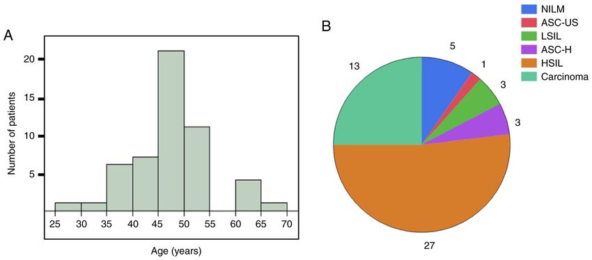
2 SATO et al: MOBILE DIGITAL COLPOSCOPY USE IN JAPAN
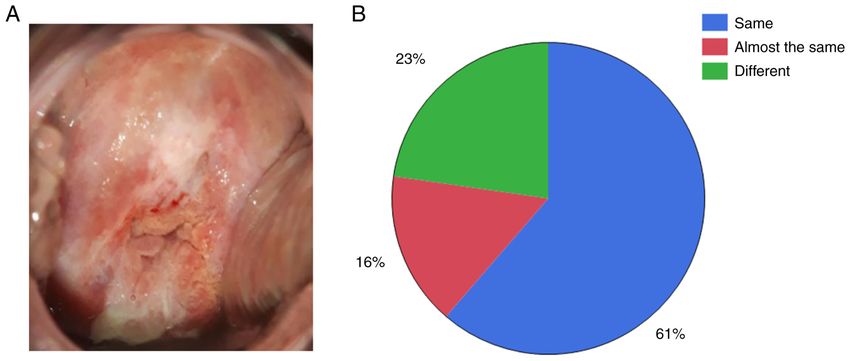
Figure 1. Patient background. (A) Age (years). (B) Cytology results in the same period as colposcopy. Saitama Medical University International Medical
Center (Hidaka, Japan) is a comprehensive cancer center, which is why most of the patients required a treatment intervention for HSIL or more. Cytology and
colposcopy are usually performed at the same time, which is why some results were NILM. NILM, negative for intraepithelial lesion or malignancy; ASC‑US,
atypical squamous cells of undetermined significance; LSIL, low grade squamous intraepithelial lesion; ASC‑H, atypical squamous cells cannot exclude HSIL;
HSIL, high grade squamous intraepithelial lesion.
Materials and methods that would have been determined using the digital images.
Diagnoses were classified as normal, cervical intraepithelial
Patient population. This study was approved by the Institutional lesion (CIN) 1, CIN2, CIN3, invasive cancer, adenocarcinoma
Review Board of Saitama Medical University International in situ (AIS), and unsatisfactory colposcopic findings (UCF).
Medical Center (approval no. 19‑026, 12/Jun/2019; Hidaka, Diagnoses that matched between conventional and digital
Japan). Patients who underwent colposcopy at Saitama colposcopy were defined as the ‘same’, and others were
Medical University International Medical Center between defined as ‘different’. Biopsy sites were compared by dividing
July 2019 and February 2020 were enrolled in the study. The the cervix into twelve clockface regions. If the actual (from
subjects were women aged 21‑65 years old who were referred conventional colposcopy) and assumed (from digital colpos‑
for colposcopy following the standard of care. The exclusion copy) biopsy sites matched, they were defined as the ‘same’; if
criteria were pregnancy, prior history of cervical cancer, prior the sites differed by one clockface region, they were defined as
history of a cervical excisional procedure, and hysterectomy. ‘almost the same’; and all others were ‘different’. The patho‑
Written informed consent was obtained from all subjects. A logical findings were confirmed by biopsy or conization, and
total of 40 patients (52 tests) were included in the study. Some the pathological diagnosis was confirmed by pathologists.
patients were tested two times through follow‑up. The clinical
background of the patients is shown in Fig. 1. The median Statistical analysis. JMP 15 (SAS Institute, Inc.) was used
age was 46 (range, 27‑65) years (Fig. 1A). Cytology results for statistical analysis. A paired t‑test was used to compare
are shown in Fig. 1B. Colposcopy was performed at initial the means of matched rates between pathological diagnosis
visit, just before conization (preoperative examination), or if and colposcopy diagnosis. The data are presented as the
abnormal cytology was detected. mean ± standard deviation. P
MOLECULAR AND CLINICAL ONCOLOGY 15: 207, 2021                                                                  3

Table I. Cases in which the results were ‘different’.

Age, years           Gravida, n           Para, n          Pathological diagnosis              Conventional colposcopy                 Digital colposcopy

30                         0                  0                   CIN1                                      CIN1                                CIN2
51                         2                  2               Adenocarcinoma                                CIN2                                CIN3
47                         2                  2                   CIN3                                      CIN2                                CIN3
48                         4                  2                   CIN3                                      CIN2                                CIN3
45                         4                  3                   CIN2                                      CIN2                                CIN3
47                         1                  1                   CIN3                                      CIN3                                CIN2
37                         0                  0                   CIN3                                      CIN3                                CIN2
50                         0                  0                   CIN2                                      CIN3                                CIN1
50                         0                  0                   CIN2                                      CIN3                                CIN2
47                         1                  1                   CIN3                                      CIN3                                UCF
47                         1                  1                   CIN3                                      CIN3                                CIN1
27                         1                  1                   CIN3                                      CIN3                                CIN1
46                         2                  2                    AIS                                      AIS                                 CIN2

CIN, cervical intraepithelial neoplasia; AIS, adenocarcinoma in situ; UCF, unsatisfactory colposcopic findings.

Figure 2. Digital colposcopy. (A) Representative image of digital colposcopy. (B) Match rate for actual (from conventional colposcopy) and assumed (from
digital colposcopy) biopsy sites. The match rates of the actual and assumed biopsy sites were 61% for ‘same’, 16% for ‘almost the same’ and 25% for ‘different’.

Figure 3. Comparison of diagnoses using conventional and digital colposcopy. (A) Diagnoses from conventional and digital colposcopy. (B) Match rate of
diagnoses. A comparison of diagnoses using conventional and mobile digital colposcopy gave match rates for diagnoses of ‘same’ (75%) and ‘different’ (25%).
CIN, cervical intraepithelial neoplasia; AIS, adenocarcinoma in situ; UCF, unsatisfactory colposcopic findings; CIS, carcinoma in situ; IC, invasive cancer.
4                                      SATO et al: MOBILE DIGITAL COLPOSCOPY USE IN JAPAN

55.7±6.9% (conventional colposcopy) and 46.1±6.9% (digital           deep learning application to colposcopy, using an image of
colposcopy). And there were no significant differences (t‑test,      only 150x150 pixels (13). This resolution can easily be achieved
P=0.33).                                                             by a mobile digital colposcope incorporating a smartphone.
                                                                     Therefore, we hope this form of digital colposcopy can be
Discussion                                                           applied to clinical practice and data collection in multiple
                                                                     centers.
Digital colposcopy built around a smartphone is becoming
common, and this has advantages for telemedicine and data            Acknowledgements
sharing by taking advantage of smartphone characteris‑
tics (10‑12). However, digital colposcopy itself is not approved     Not applicable.
in Japan. In this study, we investigated the feasibility of mobile
digital colposcopy using a smartphone for visualization, image       Funding
capture, and management in Japanese patients undergoing
cervical screening. To our knowledge, this is the first such         No funding was received.
study in a Japanese population.
    As shown in Figs. 2 and 3, the results from ≥75% of              Availability of data and materials
cases were equivalent between mobile digital colposcopy
and conventional colposcopy. There is a study in Japan that          The datasets used and/or analyzed during the current study are
reported the possibility of using smartphones to perform             available from the corresponding author on reasonable request.
colposcopy, and our results are consistent with the report (14).
The mobile digital colposcope used in this study has been            Authors' contributions
approved by the Food and Drug Administration (FDA) and is
in use in a number of Western countries (10‑12). These suggest       MS, DS, KF and KH designed the study. MS and DS analyzed
that mobile digital colposcopy was not inferior to conventional      the patient data and were major contributors in writing the
colposcopy even in Japanese population.                              manuscript. MS, DS, MH, SS and MM acquired the patient
    Our institution is a comprehensive cancer center, which is       data. AO, AY, AK and HY interpreted the data. MS, DS,
why most of the patients required a treatment intervention for       KF and KH confirm the authenticity of all the raw data. All
HSIL or more (Fig. 1). As such, there could be a selection bias      authors read and approved the final manuscript.
in patient enrollment. In addition, we seldom perform HPV
testing because it does not always contribute to the decision        Ethics approval and consent to participate
for the treatment.
    Another limitation is that the study included only a limited     The study was approved by the Institutional Review Board
number of cases, and further validation at multiple centers is       of Saitama Medical University International Medical Center
required.                                                            (approval no. 19‑026; 12/Jun/2019; Hidaka, Japan). Written
    Cases in which the results were ‘different’ in diagnosis         informed consent was obtained from all subjects.
were shown in Table I. Pathological CIN3 should not be
recognized as CIN2 by colposcopy, because treatment may be           Patient consent for publication
delayed. As mentioned in the Results, overall matched rates
between pathological diagnosis and colposcopy diagnosis were         Written informed consent for publication from each patient
55.7±6.9% (conventional colposcopy) and 46.1±6.9% (digital           was obtained.
colposcopy). And there were no significant differences (t‑test,
P=0.33). These data suggested that digital colposcopy was not        Competing interests
inferior to conventional colposcopy as a screening examination.
Still, our primary focus was not to compare the pathological         The authors declare that they have no competing interests.
diagnosis and the diagnosis from digital colposcopy, but rather
to compare the visual findings from conventional 3D colpos‑          References
copy and mobile digital colposcopy. The study protocol was that
in the examination using conventional colposcopy the clinician        1. Cohen PA, Jhingran A, Oaknin A and Denny L: Cervical cancer.
could adjust their view of the cervix and could also observe the         Lancet 393: 169‑182, 2019.
                                                                      2. Cook DA, Smith LW, Law J, Mei W, van Niekerk DJ, Ceballos K,
lesion through the period, whereas only one image was captured           Gondara L, Franco EL, Coldman AJ, Ogilvie GS, et al: Aptima
by mobile digital colposcopy. Capture of additional images may           HPV Assay versus Hybrid Capture(®) 2 HPV test for primary
have resulted in an even higher correlation rate.                        cervical cancer screening in the HPV FOCAL trial. J Clin
                                                                         Virol 87: 23‑29, 2017.
    Although the higher resolution of conventional colposcopy         3. Coste J, Cochand‑Priollet B, de Cremoux P, Le Galès C, Cartier I,
was helpful in observing the lesion in detail, we concluded that         Molinié V, Labbé S, Vacher‑Lavenu MC and Vielh P; French
digital colposcopy was not inferior to conventional colposcopy           Society of Clinical Cytology Study Group: Cross sectional study
                                                                         of conventional cervical smear, monolayer cytology, and human
as a screening test, and that this method provides sufficient            papillomavirus DNA testing for cervical cancer screening.
details for judging the biopsy site or diagnosis using acetic            BMJ 326: 733, 2003.
acid processing. We further speculate that digitally captured         4. Ginsburg O, Bray F, Coleman MP, Vanderpuye V, Eniu A,
                                                                         Kotha SR, Sarker M, Huong TT, Allemani C, Dvaladze A, et al:
images of the cervix can be used to train a machine learning             The global burden of women's cancers: A grand challenge in
algorithm. Indeed, we previously reported the possibility of             global health. Lancet 389: 847‑860, 2017.
MOLECULAR AND CLINICAL ONCOLOGY 15: 207, 2021 5
5. Peirson L, Fitzpatrick‑Lewis D, Ciliska D and Warren R: 12. Xue Z, Novetsky AP, Einstein MH, Marcus JZ, Befano B,
Screening for cervical cancer: A systematic review and Guo P, Demarco M, Wentzensen N, Long LR, Schiffman M
meta‑analysis. Syst Rev 2: 35, 2013. and Antani S: A demonstration of automated visual evalu‑
6. García‑Arteaga JD, Kybic J and Li W: Automatic colposcopy ation of cervical images taken with a smartphone camera. Int
video tissue classification using higher order entropy‑based J Cancer 147: 2416‑2423, 2020.
image registration. Comput Biol Med 41: 960‑970, 2011. 13. Sato M, Horie K, Hara A, Miyamoto Y, Kurihara K, Tomio K and
7. Khan MJ, Werner CL, Darragh TM, Guido RS, Mathews C, Yokota H: Application of deep learning to the classification of
Moscicki AB, Mitchell MM, Schiffman M, Wentzensen N, images from colposcopy. Oncol Lett 15: 3518‑3523, 2018.
Massad LS, et al: ASCCP Colposcopy Standards: Role of colpos‑ 14. Tanaka Y, Ueda Y, Okazawa A, Kakuda M, Matsuzaki S,
copy, benefits, potential harms, and terminology for colposcopic Kobayashi E, Yoshino K and Kimura T: ‘Smartscopy’ as an
practice. J Low Genit Tract Dis 21: 223‑229, 2017. alternative device for cervical cancer screening: A pilot study.
8. de Castro Hillmann E, Moreira Bacha O, Roy M, Paris G, BMJ Innov 3: 123‑126, 2017.
Berbiche D, Nizard V and Lopes Ramos JG: Cervical digital
photography: An alternative method to colposcopy. J Obstet
Gynaecol Can 41: 1099‑1107, 2019.
9. Louwers JA, Kocken M, ter Harmsel WA and Verheijen RH:
Digital colposcopy: Ready for use? An overview of literature.
BJOG 116: 220‑229, 2009. This work is licensed under a Creative Commons
10. Mink J and Peterson C: MobileODT: A case study of a novel Attribution-NonCommercial-NoDerivatives 4.0
approach to an mHealth‑based model of sustainable impact. International (CC BY-NC-ND 4.0) License.
MHealth 2: 12, 2016.
11. Thay S, Goldstein A, Goldstein LS, Govind V, Lim K and Seang C:
Prospective cohort study examining cervical cancer screening
methods in HIV‑positive and HIV‑negative Cambodian Women:
A comparison of human papilloma virus testing, visualization
with acetic acid and digital colposcopy. BMJ Open 9: e026887,
2019.You can also read