Concussion and Chronic Migraine in a Female Pediatric Patient with Chiari Malformation Type I and Syringohydromyelia
←
→
Page content transcription
If your browser does not render page correctly, please read the page content below
JSM Sports Medicine and Research
        Case Report                                                                                                                © Kaur D, et al. 2 2019

          Concussion and Chronic Migraine in a Female
        Pediatric Patient with Chiari Malformation Type I
                     and Syringohydromyelia
                                        Dilpreet Kaur, Jennifer W. McVige*, and Michael Lillis
                                                    Concussion Center, Dent Neurologic Institute, USA

            Abstract
                 Concussion in contact sports is a common occurrence, diagnosed at an increased frequency given new awareness regarding post-
            concussive injury. Both the decision to utilize neuroimaging in diagnosis and treatment, as well as the determination of return to play to
            contact sport, can be challenging and controversial. This case report discusses a 13-year-old female who sustained a concussion and
            experienced atypical neurologic symptoms. Chiari Malformation and syringohydromyelia were found incidentally and believed to have
            prolonged the recovery.

                 Keywords: Concussion; Headache; Chiari malformation; Contact sports

Abbreviations                                                                      Case Presentation
   CM: Chiari Malformation; CMI: Chiari Malformation Type I;                           A 13 year-old female athlete playing in a competitive soccer
PCP: Primary Care Physician; MRI: Magnetic Resonance Imaging                       tournament suffered a concussion with no loss of consciousness.
                                                                                   While playing defense, she was hit in the back of the head with
Introduction                                                                       a soccer ball by her own goalie. Directly after the event, she
    There has been an increased awareness of the dangers of                        experienced headaches, poor sleep, and nausea. She did not
                                                                                   experience imbalance or dizziness, attention and concentration
concussion in pediatric patients as a result of participation
                                                                                   deficits, or mood disturbance. The athlete continued to play
in contact sports [1–3]. Adolescent participation in soccer is
                                                                                   until she visited her primary care physician (PCP) 2 weeks after
known to be associated with increased risk of mild traumatic
                                                                                   the injury. The PCP recommended removal from play and a full
brain injury due to the high frequency of direct contact with the                  neurologic evaluation.
ball to the head [4]. Caring for patients with concussion can be
challenging given the various mechanisms of injury, their past                         One month after injury, she was seen at the Dent Neurologic
                                                                                   Institute’s concussion center in Amherst, New York. She
medical history, as well as the possibility of other, previously
                                                                                   presented with worsening symptoms including dull, daily
undiscovered, preexisting medical conditions which can extend
                                                                                   headaches at 5-6/10 on the pain scale, with intermittent spikes
recovery times. The literature in sports-related injuries shows 80-                in pain at 9/10. Her pain typically occurred behind and around
90% of concussion patients recover within 7-10 days [1,5,6]. The                   her eyes and was exacerbated with physical activity. However,
remaining 10-15% sub-population of more chronic concussion                         the patient experienced increased pain in the back of her neck
patients experience atypical symptoms and/or prolonged                             and occipital region when bending over, coughing, and physical
recovery. These patients are often treated in an outpatient                        exertion. The patient was diagnosed with concussion.
setting with a more complex evaluation and treatment plan
                                                                                       She had a preexisting history of mild, dull headaches, with
because of their chronicity and possible presence of underlying,                   photophobia and phonophobia, occurring on average 2 times
co-occurring health conditions [7].                                                a week. These headaches had no significant impact on her day-
                                                                                   to-day functioning or school attendance and did not require
  Submitted: 09 November 2019 | Accepted: 27 May 2019 | Published:
                                                                                   medication. There was a family history of migraines in her mom
  30 May 2019                                                                      and maternal aunts. It was suspected the patient had a past
                                                                                   history of migraines that were previously undiagnosed.
  *Corresponding author: Jennifer W McVige, Dent Neurologic Institute,
  3980, Sheridan Drive, Buffalo, NY, 14226, USA. Tel: (716) 250-2055. Fax:             Initially after the concussion, the athlete tried ibuprofen as
  (716) 250-6039; USA, Email:                                                      needed but did not experience relief. At the concussion center,
  Copyright: © 2019 Kaur D, et al. This is an open-access article                  treatment included amitriptyline, Relpax, Nasprosyn, and
  distributed under the terms of the Creative Commons Attribution                  magnesium oxide, as well as physical therapy and massage
  License, which permits unrestricted use, distribution, and reproduction in       therapy. The patient was non-responsive to treatment after a 3
  any medium, provided the original author and source are credited.                month period.
  Citation: Kaur D, McVige JW, Lillis M (2019) Concussion and Chronic Mi-
                                                                                      Subsequently, brain and cervical spine MRI scans on a 3
  graine in a Female Pediatric Patient with Chiari Malformation Type I and
  Syringohydromyelia. JSM Sports Med Res 3: 4.
                                                                                   Tesla magnet were ordered due to reports of focal numbness
                                                                                   and tingling down into her hands with increased pain when

 JSM Sports Med Res 3: 4                                                                                                                                 1/4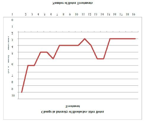
moving her neck. The brain MRI, shown in Figure 1, revealed low-
lying peg-shaped cerebellar tonsils extending 1.4 cm below the
foramen magnum, which is consistent with a diagnosis of Chiari
malformation type I (CMI) [8–10]. There was also straightening
of the normal lordotic curvature of the cervical spine indicative of
a paraspinal muscle spasm. The cervical MRI revealed a widening
of the central spinal canal from C6-C7 to C7-T1 consistent with
a diagnosis of syringohydromyelia. The syrinx measured 2.1
cm rostral caudal [Figure 2] with a maximum diameter of 0.28
cm [Figure 3]. Massimi et al., noted a high frequency temporal
association between CMI symptom onset and minor head injury
[11] which is consistent with the present case report.
    CMI can be managed using medical and surgical intervention.
McVige and Leonardo provide a step-wise flowchart for
clinical management of CMI and argue, “As headaches are the            Figure 1 T2 weighted sagittal image showing low lying peg-shaped
                                                                       cerebellar tonsils, measuring at 1.4 cm below the foramen magnum.
most common presenting symptom of CMI, they are typically
treated according to presenting phenotype. The decision for
surgical intervention is based on a combination of the clinical
presentation, neuroimaging and/or physiologic studies such as
somatosensory evoked potentials, swallow evaluations and sleep
studies [12].” The patient was referred to pediatric neurosurgery
to explore surgical options. After full evaluation and collaboration
with neurosurgery, a decision was made to aggressively treat her
headaches without surgery given that they were not typical of
Chiari-type headache [12,13]. The headache presentation was
atypical, as the patient consistently experienced pressure behind
her eyes especially with physical exertion, however, posterior
occipital and neck pain improved temporally with physical
therapy. Other notable atypical neurological symptoms included
numbness and tingling down her arms and occipital region pain.
The patient felt the postconcussive fogginess and concussion
headaches had resolved.
                                                                       Figure 2 T2 weighted sagittal image showing widening of the central
    At this point, her presentation seemed more consistent with
                                                                       spinal canal from C6-C7 to C7-T1. The syrinx measured 2.1 cm rostral
migraine, which was believed to be a preexisting condition. She        caudal.
eventually tried several treatments for migrainous headaches
including, Frova, Migranal, Propranolol, Butterbur, Topamax,
Cambia, and Sprix, as well as, occipital and supraorbital nerve
blocks, trigger point injections, and a series of sphenopalatine
blocks. The patient experienced minimal to no short-lived relief
from all previously referred migraine treatment options. The
patient was also physically examined for scoliosis as this is a
common comorbidity with CMI and syringohydromyelia [12], but
was not found to have this diagnosis. Her headaches were daily
and averaged 6-8/10 in the bifrontal/bitemporal with posterior
radiation. These headaches were described as “nagging” and
often negatively impacted her sleep. Due to her level of disability
and the fact that patient met criteria for chronic migraine with
failed pharmacotherapy, physical therapy and therapeutic
injection treatments, Botox injections were initiated [14].
    At the beginning of Botox injection treatment, she reported
daily headaches with pain averaging 6/10 and some days of
increased intensity “spikes” at 8/10. Over the past five years,
she has received a total 19 Botox injection treatments and at last
visit reported an average of 3 or fewer headaches days a month
with pain averaging 1/10 which she rates as a 90% improvement          Figure 3 T2 weighted transverse image showing the syrinx at the
                                                                       maximum diameter of 0.28 cm.
overall. Figures 4, 5 illustrate the improvement in her headache

 JSM Sports Med Res 3: 4                                                                                                             2/4
Discussion
The pathophysiology of CM is multifaceted and often cannot
be attributed to one theory or cause. While some research asserts
genetic predisposition other literature proposes a de novo
presentation. In fact, some patients with CM might have no family
history of CM or syringohydromyelia [8,10,12] as was the case for
this present patient. Furthermore, in the field of sports medicine,
there is generally an agreement regarding the dangers of CM.
Many researchers recommend that athletes with CM should
not return to contact sports [15,16]. In pediatric patients, most
doctors recommend that CM athletes, either with or without
syringohydromyelia, do not seek surgery before exhausting other
therapeutic options [12,17].
The present case shows a pediatric athlete who presented
with concussion and postconcussive headaches, as well as
an incidental finding of CMI with syringohydromyelia, and a
preexisting medical history of undiagnosed migraine headache
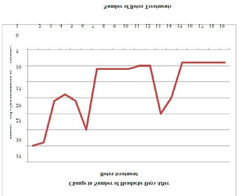
Figure 4 A longitudinal graph of the number of headache days on without aura. The patient’s migraine symptoms were well
average per month after the patient began Botox treatment. Note: managed until she sustained a concussion during soccer which
The spikes at treatments #6 and #13 are the result of extended delays exacerbated her headaches. Her diagnosis of CMI was unknown
between Botox treatments. until the headaches continued beyond an expected time period
and presented in a fashion atypical for migraines. When the
patient began to experience focal neurologic signs and atypical
headache, neuroimaging revealed the underlying cause of her
prolonged recovery. With the proper physical restrictions, due
to the CMI diagnosis, and treatment for chronic intractable
migraine headache, the patient was able to improve her headache
symptoms and quality of life.
This patient was not considered to be a good candidate
for neurosurgical intervention to treat CMI. Therefore, the
neurological treatment approach was targeted at decreasing
headache frequency and intensity by using a variety of
medications, injections, and physical therapies. After exhausting
several abortive and preventative therapeutic options, Botox
injections were chosen for treatment of her chronic migraines
[14,18]. While Botox treatment did not cure her pain completely,
it has significantly decreased her level of pain and headache
frequency, ultimately reducing the burden of her disabling
chronic migraines and pain.
In conclusion, this case suggests that advanced neuroimaging
Figure 5 A longitudinal graph of headache intensity on average per should be considered for individuals with focal atypical
month after the patient began Botox treatment. Pain scale represents
neurologic presentations, such as post- ocular pressure,
0 as no pain and 10 as severe pain. Note: The spikes at treatments #6
and #13 are the result of extended delays between Botox treatments. numbness and tingling in the upper extremities, and posterior
occipital pain, as well as prolonged recovery [12,19]. In addition,
alternative therapies should be entertained for post-concussive
frequency and intensity over the course of her Botox injection headache treatment if the patient does not respond to traditional
treatment. treatment. In this case, a patient with no family history of
Currently she participates in non-contact soccer training neurological deficits or complications had an underlying history
and other non-contact sports such as swimming, running, and of undiagnosed migraine coupled with congenital anomalies,
golf due to the CMI diagnosis. She regularly comes in for her CMI and syringohydromyelia, which exacerbated the patient’s
Botox injections and re-evaluations every 3 months to keep her headache presentation and required additional therapies. Given
headache frequency and intensity minimal. The athlete graduated the increasing popularity of participation in contact sports and
high school and is currently planning on attending college away related concussion injuries, patients with prolonged recovery
from home. and atypical presentation require an individually tailored
treatment plan.
JSM Sports Med Res 3: 4 3/4Acknowledgements 9. Chiari Malformation – Symptoms, Diagnosis and Treatments
[Internet]. 2018.
The authors thank the Dent Family Foundation for their
10. Chiari Malformation Type I | Johns Hopkins Medicine Health Library
continuous support. We also thank the patients of this study for
[Internet]. 2018.
their participation. Study funded through a grant by the Dent
Family Foundation. 11. Massimi L, Caldarelli M, Frassanito P, Di Rocco C. Natural history of
Chiari type I malformation in children. Neurol Sci. 2011; 32: 275–277.
References 12. McVige JW, Leonardo J. Imaging of Chiari Type I Malformation and
1. McCrory P, Meeuwisse WH, Aubry M, Cantu B, Dvořák J, Echemendia Syringohydromyelia. Neurol Clin. 2014; 32: 95–126.
RJ, et al. Consensus statement on concussion in sport: the 4th
13. Avellaneda Fernández A, Isla Guerrero A, Izquierdo Martínez
International Conference on Concussion in Sport held in Zurich,
M, Amado Vázquez ME, Barrón Fernández J, Chesa i Octavio E,
November 2012. Br J Sports Med. 2013; 47: 250–258.
et al. Malformations of the craniocervical junction (chiari type I
2. Kontos AP, Covassin T, Elbin RJ, Parker T. Depression and and syringomyelia: classification, diagnosis and treatment). BMC
Neurocognitive Performance After Concussion Among Male and Musculoskelet Disord. 2009; 10: 1.
Female High School and Collegiate Athletes. Arch Phys Med Rehabil.
14. Schaefer SM, Gottschalk CH, Jabbari B. Treatment of Chronic Migraine
2012; 93: 1751–1756.
with Focus on Botulinum Neurotoxins. Toxins. 2015; 7: 2615–2628.
3. Rivera RG, Roberson SP, Whelan M, Rohan A. Concussion Evaluation
15. Head Injury in Athletes | Neurosurgery | Oxford Academic [Internet].
and Management in Pediatrics. MCN Am J Matern Nurs. 2015; 40: 76.
2018.
4. Mueller FO, Kucera KL, Cox LM, Cantu RC. From the National Center
16. Miele VJ, Bailes JE, Martin NA. Participation in contact or collision
for Catastrophic Sport Injury Research At The University of North
sports in athletes with epilepsy, genetic risk factors, structural brain
Carolina at Chapel Hill. 2014; 66.
lesions, or history of craniotomy. Neurosurg Focus. 2006; 21: 9.
5. Graham R, Rivara FP, Ford MA, Spicer CM, Youth C on S-RC in, Board on
17. Schijman E, Steinbok P. International survey on the management of
Children Y, et al. Treatment and Management of Prolonged Symptoms
Chiari I malformation and syringomyelia. Childs Nerv Syst ChNS Off J
and Post-Concussion Syndrome [Internet]. National Academies Press
Int Soc Pediatr Neurosurg. 2004; 20: 341–348.
(US); 2014; 356.
18. Aurora S, Dodick D, Turkel C, DeGryse R, Silberstein S, Lipton R, et
6. McCrory P, Johnston K, Meeuwisse W, Aubry M, Cantu R, Dvorak J,
al. OnabotulinumtoxinA for treatment of chronic migraine: Results
et al. Summary and agreement statement of the 2nd International
from the double-blind, randomized, placebo-controlled phase of the
Conference on Concussion in Sport, Prague 2004. Br J Sports Med.
PREEMPT 1 trial. Cephalalgia. 2010; 30: 793–803.
2005; 39: 196–204.
19. Mechtler LL, Shastri KK, Crutchfield KE. Advanced Neuroimaging of
7. Hiploylee C, Dufort PA, Davis HS, Wennberg RA, Tartaglia MC,
Mild Traumatic Brain Injury. Neurol Clin. 2014; 32: 31–58.
Mikulis D, et al. Longitudinal Study of Postconcussion Syndrome: Not
Everyone Recovers. J Neurotrauma. 2017; 34: 1511–1523.
8. Hadley DM. The Chiari Malformations. J Neurol Neurosurg Psychiatry.
2002; 72: 38–40.
JSM Sports Med Res 3: 4 4/4You can also read