Primary Presentation of Ovarian Cancer with Bladder Outlet Obstruction/Chronic Urinary Retention in a 12-Year Old Female
←
→
Page content transcription
If your browser does not render page correctly, please read the page content below
Open Journal of Urology, 2021, 11, 233-239
                                                                                                 https://www.scirp.org/journal/oju
                                                                                                            ISSN Online: 2160-5629
                                                                                                              ISSN Print: 2160-5440

Primary Presentation of Ovarian Cancer with
Bladder Outlet Obstruction/Chronic Urinary
Retention in a 12-Year Old Female

Charles Azuwike Odoemene1*, Ijeoma Ezeome2, Okechukwu Charles Okafor3
1
 Alex Ekwueme Federal University Teaching Hospital, Abakaliki, Nigeria
2
 University of Nigeria Teaching Hospital, Enugu, Nigeria
3
 Department of Morbid Anatomy, University of Nigeria Teaching Hospital, Enugu, Nigeria

How to cite this paper: Odoemene, C.A.,         Abstract
Ezeome, I. and Okafor, O.C. (2021) Prima-
ry Presentation of Ovarian Cancer with          Urinary retention in women is rare and is more frequently described as case
Bladder Outlet Obstruction/Chronic Uri-         reports or small case series. The female/male ratio is 1:13 with about 3 cases
nary Retention in a 12-Year Old Female.         per 100,000 women every year We report a case of a 12-year old female stu-
Open Journal of Urology, 11, 233-239.
https://doi.org/10.4236/oju.2021.117021
                                                dent. She presented with progressive weight loss, worsening lower urinary
                                                tract symptoms with distended lower abdomen of 10 weeks duration. Physical
Received: April 29, 2021                        examination revealed a mobile tender firm pelvic mass, 18 centimeters (cm) ×
Accepted: July 6, 2021                          16 cm in size. Laboratory and imaging studies showed obstructive nephropa-
Published: July 9, 2021
                                                thy and uropathy respectively. She was worked up and had uneventful explo-
Copyright © 2021 by author(s) and               ratory laparotomy with right salpingo-oophorectomy, urinary bladder diver-
Scientific Research Publishing Inc.             ticulectomy and pelvic lymphadenectomy. Histopathology of the pelvic mass
This work is licensed under the Creative        showed ovarian dysgerminoma with lymph node metastasis. She responded
Commons Attribution International
                                                very well to chemotherapy and resumed her school activities. Bladder outlet
License (CC BY 4.0).
http://creativecommons.org/licenses/by/4.0/     obstruction is relatively rare in females and in the index patient, ovarian dys-
               Open Access                      germinoma is the cause leading to obstructive nephropathy and uropathy.

                                                Keywords
                                                Bladder Outlet Obstruction, Chronic Urinary Retention, Pelvic Mass,
                                                Ovarian Dysgerminoma, Chemotherapy

                                              1. Introduction
                                              Urinary retention implies a difficulty in passing urine or completely emptying
                                              the urinary bladder and it can be acute or chronic. Urinary retention in women
                                              is rare with about 3 cases per 100,000 women every year [1]. The female/male
                                              ratio is 1:13 [1] [2]. In short, urinary retention in females is more frequently de-

DOI: 10.4236/oju.2021.117021 Jul. 9, 2021                          233                                     Open Journal of Urology
C. A. Odoemene et al.
scribed as case reports or small case series with unusual causes [2]. Two com-
mon causes of chronic urinary retention in women are detrusor dysfunction and
obstruction [1]. Urinary symptoms such as hesitancy, straining, poor urinary stream
and feeling of incomplete emptying of the urinary bladder are clearly suggestive
of bladder outlet obstruction in women [3] [4] [5].
Anatomical non-iatrogenic causes of bladder outlet obstruction in females in-
clude pelvic organ prolapse, vaginal masses like cysts, urethral pathology like di-
verticulum, carcinoma, gynaecological lesions like large ovarian cysts, tumors, cer-
vical, uterine tumors and pregnancy due to uterine displacement [4].
We present the case of a 12-year old female student with ovarian cancer who
presented with lower urinary tract symptoms and chronic urinary retention with
urinary tract infection (UTI) and overflow incontinence to the urologist and her
successful management.
2. Case Report
Miss A, a 12-year old Nigerian student presented at the accident and emergency
department with about 3 months worsening symptoms of progressive weight loss,
daytime frequency, nocturia, overflow incontinence, feeling of incomplete emp-
tying of the bladder and lower abdominal pain. There was no haematuria.
She was cachectic, clinically pale, anicteric. The chest was unremarkable. There
was lower abdominal distension. A tender mobile mass 18 cm × 16 cm which
was firm in consistency was palpated. A working diagnosis of bladder outlet ob-
struction secondary to a pelvic mass was made. She was aseptically catheterized
with a size 12F (all silicone) two-way Foley catheter and 350 mililiters (mLs) of
turbid offensive urine drained.
Haematological investigations, serum electrolytes, urea and creatinine, etc. are
as shown in Table 1.
Abdominopelvic ultrasonography showed a solid pelvic mass posterior to and
compressing the urinary bladder with hypoechoic and highly echogenic areas
measuring 18.94 × 11.24 cm, there was also bilateral hydronephrosis with hy-
droureter.
Intravenous urography after normalization of serum electrolyte, urea and crea-
tinine showed bilateral hydronephrosis and hydroureter, bladder diverticulum in
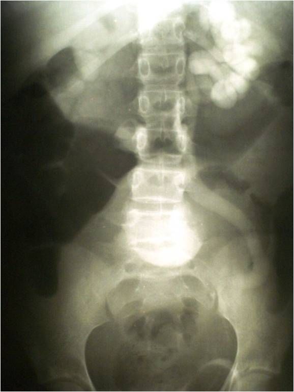
keeping with bladder outlet obstruction (Figure 1).
The anemia was corrected by blood transfusion, the UTI treated with ceftriax-
one. The patient was worked up and prepared for laparotomy. Via a mid-line ab-
dominal incision, the peritoneum and pelvis were explored. The mass was mobi-
lized off the posterior wall of the urinary bladder. Within the mass were embed-
ded the right ovary and the right fallopian tube. There was pelvic and iliac lym-
phadenopathy. A urinary bladder diverticulum was noted. The mass with the
right ovary and right fallopian tube within was resected. Pelvic and iliac lym-
phadenectomy was done. She had an uneventful post-operative recovery. The in-
dwelling urethral catheter was removed on the seventh-day post-operatively. The
DOI: 10.4236/oju.2021.117021 234 Open Journal of UrologyC. A. Odoemene et al.
Table 1. Haematological and other laboratory investigations.
Parameters Results Range
Heamoglobin (HB) 7.2 gm/dL 11.0 - 16.0 gm/dL
Platelet count 165,000/mm3 150,000 - 400,000/mm3
Erythrocyte sedimentation rate 104 mm/hr. 0 - 29 mm/hr (women)
Total White Blood Cell (WBC) Count 11,800/mm3 4000 - 10,000/mm3
Neutrophil—80%
Lymphocyte—19%
Differential Eosinophil—01%
Basophil—0
Monocytes—0
Na+—137 mmol/L (135 - 145)
K+—5.6 mmol/L (3.5 - 5)
1) Serum Electrolytes, Urea Cl−—98 mmol/L (96 - 110)
and creatinine (Pre-catheterization) HCO3—24 mmol/L (22 - 30)
Urea—168 mmol/L (10 - 40)
Creatinine—3.2 mmol/L (0.4 - 1.6)
Na+—140 mmol/L (135 - 145)
K+—3.2 mmol/L (3.5 - 5)
2) Serum Electrolytes 1 week Cl−—100 mmol/L (96 - 110)
post catheterization HCO3− —26 mmol/L (22 - 30)
Urea—35 mg/dL (10 - 40)
Creatinine—1.0 mg/dL (0.4 - 1.6)
Billirubin total 0.5 mg/dl (C. A. Odoemene et al.
patient was discharged on the tenth-day post-operatively to the outpatient de-
partment. Histology report showed ovarian dysgerminoma. The sections from
the ovary and retroperitoneal lymph nodes are similar and show a malignant germ
cell tumour made up of broad and thin trabeculae of pleomorphic malignant
cells. These cells have abundant amphophilic cytoplasm and large vesicular nuc-
lei with a prominent eosinophilic nucleolus. Between the tumour trabeculae, there
are fibrous connective tissue columns that contain many lymphocytes. Within the
tumour masses, there are multinucleated giant tumour cells of trophoblastic type.
Some of the blood vessels within the tumour show intravascular tumour disse-
mination (Figure 2, Figure 3).
Patient was referred to the oncologist for chemotherapy and she did well after
the course of chemotherapy and resumed her academic activities. She was lost to
follow up after 10 months.
Figure 2. H&E × 150 magnification ovarian dysgerminoma. There are anastomosing tra-
beculas of tumor cells separated by fibrous bands that contain lymphocytes.
Figure 3. H&E × 300 magnification ovarian dysgerminoma. Note the frequent multinuc-
leated tumor giant cells among the other tumor cells.
DOI: 10.4236/oju.2021.117021 236 Open Journal of UrologyC. A. Odoemene et al.
3. Discussion
Although relatively common in men, bladder outlet obstruction is relatively un-
common in women in clinical practice and the etiological factors more diverse
in females than in males [3]. The mechanism of obstruction could be urethral
compression, bladder neck distortion or luminal occlusion [2]. In the index pa-
tient, the ovarian tumor compressed and distorted the bladder neck causing lower
urinary tract symptoms, impaired detrusor emptying with increasing residual urine
volume. Furthermore, there was associated daytime urinary frequency, nocturia,
urgency, poor urinary stream, intermittency, terminal dribbling and feeling of
incomplete emptying of the urinary bladder. Storage and voiding symptoms can
coexist in bladder outlet obstruction in females making it a challenge in clinical
practice to make an accurate diagnosis and offering the best form of treatment
[6]. Both urgency urinary incontinence and overflow incontinence coexisted in
this patient. Urinary incontinence adversely impacts the patient, family mem-
bers and the health care system with abstinence from routine activities, increased
rate of depression and reduced quality of life [7]. The index patient experienced
all these and dropped out of school with depression. In addition, the patient with
overflow urinary incontinence has high residual urine volume with associated
high pressure within the bladder which at this stage is considered as high-pressure
chronic retention and can cause renal impairment [7]. The index patient at pres-
entation, laboratory and ultrasound studies showed obstructive nephropathy
and uropathy respectively. A total of 350 mL turbid offensive urine was drained
from the urinary bladder on aseptic urethral catheterization and the patient was
admitted and monitored for complications. The patient in the first 72 hours fol-
lowing catheter insertion made a daily average of 3 liters of urine which was
promptly replaced with intravenous fluids to avoid dehydration and shock. Drai-
nage of more than 300 mL of urine from the bladder after voiding suggests uri-
nary retention [1], and the condition is best managed in a hospital setting with
the patient monitored for post obstructive diuresis [7]. Imaging studies like ab-
dominopelvic ultrasound, CT imaging have been advocated in the investigation
of these patients [1] [3] [5] [6] [8] [9]. Abdominopelvic ultrasonography was
utilized to unravel the etiology of this condition. Furthermore, ultrasonography
could be useful in estimating the residual urine volume [3] [5] and detection of
greater than 200 mL of urine in the bladder after voiding is suggestive of over-
flow urinary incontinence [10]. This patient did not experience acute urinary re-
tention. Acute urinary retention in females is due to impacted pelvic masses that
displace the cervix superiorly and anteriorly compressing the lower bladder lead-
ing to obstruction of the internal urethral orifice [11]. The pelvic mass was mo-
bile in the index patient and not impacted with the internal urethral orifice par-
tially obstructed. The lower abdominal pain the patient had was due to pressure
from the mass and urinary tract infection caused by E. coli.
While lactate dehydrogenase (LDH) levels are known to be elevated in some
patients with dysgerminoma, our patient had normal values of LDH and β-cho-
DOI: 10.4236/oju.2021.117021 237 Open Journal of UrologyC. A. Odoemene et al.
rionic gonadotrophin (βHCG). She had complete resection of the mass with
right salpingo-oophorectomy followed by chemotherapy with bleomycin, etopo-
side and cisplatin (BEP). Even patients with incompletely resected dysgermino-
ma can be rendered disease-free with a combination of cisplatin, vinblastin, and
bleomycin (PVB) [12]. A number of patients had one or more successful preg-
nancies following unilateral salpingo-oophorectomy [13]. However, our patient
was lost to follow up after 10 months of having resumed her academic activities
and in excellent health.
4. Conclusion
Although relatively common in men with voiding dysfunction, bladder outlet
obstruction is relatively rare in women. In this patient right ovarian cancer was
the cause of complications of obstructive uropathy and nephropathy, the patient
and her relative concern was the disabling lower urinary tract symptoms obli-
vious of the underlying pathology. The onus thus lies on the attending clinician
to evaluate the patient diligently and unravel the pathology and in this case, a
right ovarian dysgerminoma which responded excellently to chemotherapy with
the patient resuming her academic activities.
Author Contributions
Study design: Dr. Charles A. Odoemene, Dr. Mrs. Ijeoma Ezeome, Dr. Oke-
chukwu Charles Okafor.
Data acquisition: Charles A. Odoemene, Dr. Mrs. Ijeoma Ezeome, Dr. Oke-
chukwu Charles Okafor.
Data analysis: Charles A. Odoemene, Dr. Mrs. Ijeoma Ezeome.
Drafting of the manuscript: Charles A. Odoemene, Dr. Mrs. Ijeoma Ezeome,
Dr. Okechukwu Charles Okafor.
Critical revision of the manuscript: Charles A. Odoemene, Dr. Mrs. Ijeoma
Ezeome, Dr. Okechukwu Charles Okafor.
Parents Informed Consent
We attest that the patient’s parents gave consent for the publication of this case.
Conflicts of Interest
The authors have no conflicts of interest.
References
[1] Dougherty, J.M. and Rawla, P. (2021) Female Urinary Retention [Updated 2021
Mar. 29]. Stat Pearls [Internet] Treasure Island (FL), Stat Pearls Publishing.
https://www.ncbi.nlm.nih.gov/books/NBK538497/
[2] Mevcha, A. and Drake, M.J. (2010) Etiology and Management of Urinary Retention
in Women. Indian Journal of Urology, 26, 230-235.
https://doi.org/10.4103/0970-1591.65396
DOI: 10.4236/oju.2021.117021 238 Open Journal of UrologyC. A. Odoemene et al.
[3] Yande, S. and Joshi, M. (2011) Bladder Outlet Obstruction in Women. Journal of
Mid-Life Health, 2, 11-17. https://doi.org/10.4103/0976-7800.83257
[4] Dmochowski, R.R. (2005) Bladder Outlet Obstruction: Etiology and Evaluation. Re-
views in Urology, 7, S3-S13.
[5] Shaih, N.A., Hindu, K.A., Shaikh, G.S. and Soornro, A.A. (2015) Bladder Outlet
Obstruction (BOO) in Female: Etiology and Management. Rawal Medical Journal,
40, 289-291.
[6] Raheem, A.A. and Madersbacher, H. (2013) Voiding Dysfunction in Women; How
to Manage It Correctly. Arab Journal of Urology, 11, 319-330.
https://doi.org/10.1016/j.aju.2013.07.005
[7] Mangir, N. and Chapple, C. (2020) Management of Urinary Incontinence in Men.
Trends in Urology & men’s Health, 11, 18-22. https://doi.org/10.1002/tre.740
[8] Selcuk, I., Boyraz, G. and Tuncer, Z.S. (2013) Acute Urinary Retention Due to a Giant
Ovarian Tumour in Reproductive Ages: Case Report. Turkiye Klinikleri Jinekoloji
Obstetrik, 23, 207-210.
[9] Basson, J., Van der Walt, C.I.E. and Heyns, C.F. (2013) Urinary Retention in Women.
CME, 31, 182-184.
[10] Tran, L.N. and Puckett, Y. (2021) Urinary Incontinence [Updated 2021 Apr. 13].
Stat Pearls [Internet] Treasure Island (FL), Stat Pearls Publishing.
https://www.ncbi.nlm.nih.gov/books/NBK559095/
[11] Yang, J.M. and Huang, W.C. (2002) Sonographic Findings of Acute Urinary Reten-
tion Secondary to an Impacted Pelvic Mass. Journal of Ultrasound in Medicine, 21,
1165-1169. https://doi.org/10.7863/jum.2002.21.10.1165
[12] Williams, S.D., Blessing, J.A., Hatch, K.D. and Homesley, H.D. (1991) Chemothe-
rapy of Advanced Dysgerminoma: Trails of the Gynecologic Oncology Group. Journal
of Clinical Oncology, 9, 1950-1955. https://doi.org/10.1200/JCO.1991.9.11.1950
[13] Thomas, G.M., Dembo, A.J., Hacker, N.F. and Depetrillo, A.D. (1987) Current
Therapy for Dysgerminoma of the Ovary. Obstetrics & Gynecology, 70, 268-275.
DOI: 10.4236/oju.2021.117021 239 Open Journal of UrologyYou can also read