Isolated bilateral thumb arthritis: A bizarre initial presentation of common arthritis
←
→
Page content transcription
If your browser does not render page correctly, please read the page content below
In tern a tio n a l
         Sch o la rs
         Jo u rn a ls
International Journal of Medicine and Medical Sciences ISSN: 2167-0404 Vol. 6 (2), pp. 311-316, February,
2016. Available online at www.internationalscholarsjournals.org © International Scholars Journals
                                     Author(s) retain the copyright of this article.

Case Report

        Isolated bilateral thumb arthritis: A bizarre initial
                presentation of common arthritis
                          Hussein Abdullah*1, Khalifa Hamad1 and Al-Sabah Fahd2
                     1
                    Rheumatology Department, King AbdulAziz Medical City, Jeddah, Saudi Arabia.
                 2
                  Rheumatology Department, King AbdulAziz University Hospital, Jeddah, Saudi Arabia.
                                                Accepted 04 October, 2015

    Dactylitis is a known manifestation of seronegative spondyloarthropathy. Here, an unusual case of
    psoriatic arthritis presented initially with isolated erosive metacarpophalengeal joint arthritis of both
    thumbs for 2 years without obvious etiology was reported. Later on, dactylitis and skin psoriasis were
    developed.

    Key words: Erosive arthritis, dactylitis, metacarpophalangeal joint arthritis.

INTRODUCTION

Early arthritis may develop into established rheumatoid          regular medications apart from non steroidal anti-
arthritis or into another definite arthropathy, which may        inflammatory drugs (NSAIDs). Her family history was
resolve spontaneously, or may remain undifferentiated.           negative for arthritis, psoriasis, connective tissue
Intensive interventions early in the course of persistent        diseases or spondyloarthropathy. Upon further clinical
arthritis may profoundly affect long term radiographic           examination, there was tenderness and swelling over
progression (Combe et al., 2007).                                both thumbs’ metacarpophalangeal (MCP) joints with
                                                                 normal range of motion. Other joints’ examination was
CASE REPORT                                                      unremarkable, though there were no skin rashes and nail
                                                                 changes.
A 52-year-old female patient was referred to the                   Laboratory tests revealed that the C-reactive protein
Rheumatology Service with a history of bilateral thumb           (CRP) levels were elevated to 12. Other laboratory
swelling for 1 year. Her problem started 7 months ago            complete blood count, renal and liver functions were
with pain and swelling in the right thumb, then few              normal. Hands X-rays showed bilateral sublaxation and
months after she started to have the same problem in the         erosions with overhanging edges involving the head of
left thumb.                                                      the first metacarpal and the base of proximal phalanx.
   Among the symptoms observed, she had only swelling            Joints’ space was still preserved, with soft tissue swelling,
and tenderness, but she had no redness, trauma or other          but no soft tissue calcification was observed (Figure 1).
joints involvement. There was no indication of morning           These findings were reported by our musculoskeletal
stiffness, skin rashes, history of connective tissue             radiologist.
diseases, skin rashes, constitutional symptoms or history          It was observed that the patient’s chest x-ray was
of spondyloarthropathy. More so, she was not known to            normal. At that time, we started the patient on colchicine
have any medical illness. However, she did not take any          0.5 mg orally two times daily as a case of gouty arthritis
                                                                 since she did not improve on NSAIDs over the past year.
                                                                 Serum uric acid level as well as further workups was
                                                                 requested.
Corresponding author. E-mail: hussein_abdullah@gmail.com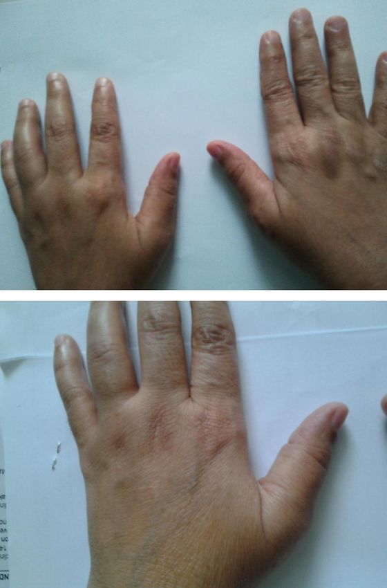
Abdullah et al.       311

       Figure 1. Hands X-rays showed bilateral sublaxation and erosions with overhanging edges involving the head the of first
       metacarpal and the base of proximal phalanx.
Int.J.M ed.Med.Sci.R es. 029
312 Int. J. Med. Med. Sci.
Figure 2. MRI synovitis with marginal erosions and subluxation of the proximal interphalengeal
joint of the right thumb.
Further follow up showed that there was no The authors of this study requested the MRI of the
improvement in the patient’s symptoms. Serum uric acid patient’s right hand to further delineate the condition
was normal as well as serum ferritin, calcium, magnesium showing synovitis with marginal erosions and subluxation
and parathyroid hormone (anti-nuclear antibody). ANA of the proximal interphalengeal joint of the right thumb
and rheumatoid factor (RF) were both negative. However, which could represent erosive arthropathy or
anti cyclic citrullinated peptide (ACPA) was not available inflammatory changes (Figure 2). Since all laboratory
in this study’s lab at that time. workups were negative and the patient did not improveAbdullah et al. 313
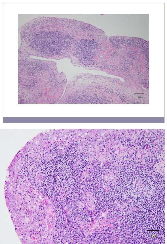
Figure 3. Reactive hyperpalstic synovial tissue with chronic inflammation. No granuloma or
necrosis. No giant cells, No eosinophills or histicytes seen.
on colchicines, we decided to get synovial biopsy which One month after the biopsy, she presented with new
showed reactive hyperpalstic synovial tissue with chronic left fourth finger pain and swelling in addition to pain in
inflammation. However, no granuloma or necrosis was both thumbs. No other joint was affected according to the
observed, and no giant cells, or eosinophills or histicytes patient. Clinical examination showed dactylitis of the left
were seen. Thus, the malignancy of the bacterial and fourth and fifth fingers (Figure 4).
fungal culture was negative (Figure 3). ACPA was strongly positive. We discontinuedInt.J.M ed.Med.Sci.R es. 031
314 Int. J. Med. Med. Sci.
Figure 4. Swelling in both thumbs and dactylitis of left fourth and fifth fingers.
colchicine and started methotrexate (10 mg orally weekly) the patient’s symptoms improved, and no other joint
and folic acid as rheumatoid arthritis (RA). Further follow involvement was observed except for only two swollen
up was done for 2 months. After starting methotrexate, joints (thumbs MCP).Abdullah et al. 315
As regards CRP 17, DAS 28 low disease activity predisposing metabolic disease (including
(Eisuke et al., 2007), the dose of methotrexate was haemochromatosis, primary hyperparathyroidism,
increased to 15 mg weekly and the patient felt clinically hypomagnesaemia) can be found (Zhang et al., 2011a).
better with improvement of thumb swelling as well. It was We screened our patient for these conditions and she
observed that the patient, after the last clinical visit she was normal. The patient did not improve on NSAID or
made before the case was reported, was seen in colchicine which is the recommended management of
dermatology clinic as she developed multiple scaly skin CPP disease (Zhang et al., 2011b).
rash on the hands like psoriasis. Finally, after all the aforementioned workups and
therapeutic measures, our patient did not improve, as she
DISCUSSION had positive ACPA and MRI suggestive of inflammatory
arthritis. We make the diagnosis of rheumatoid arthritis
Here, we report a difficult challenging case to diagnose (RA) as per the American College of Rheumatology/
as the patient presented with symmetrical isolated thumb European League Against Rheumatism (ACR/EULAR)
MCP involvement. She initially has wide differential criteria for RA, and she scored 7 which makes the
diagnosis rather than simple osteoarthritis giving the bony diagnosis of RA (Daniel et al., 2010). We started the
erosions in x-rays. Arthritis of the MCP joint can result in patient on methotrexate (Jasvinder et al., 2012) and she
considerable disability and pain. Inflammatory, improved on it. However, the presence of dactylitis which
posttraumatic, crystalline, and osteoarthritis are common is a manifestation of seronegative spondyloarthropathy
etiologies (Rizzo, 2011). makes the diagnosis uncertain (Olivieri et al., 1996).
Special attention must be paid in this case to erosive Dactylitis is associated with Reiter’s syndrome,
osteoarthritis, a clinically uncommon subset of gene- psoriatic arthritis, sarcoidosis, flexor tendon sheath
ralized osteoarthritis (OA) characterized by a clinical infections, and gout. The presence of dactylitis eliminated
course, which is frequently aggressive. The diagnosis of rheumatoid arthritis from the differential diagnosis (Bruce,
EOA is accepted only for patients that meet American 1998). ACPA as well may be positive in psoriatic arthritis
College of Rheumatology’s clinical criteria for OA of the (Vander Cruyssen et al., 2005). Oligoarticular
hand (Altman et al., 1990). The diagnosis must show presentation is common in psoriatic arthritis (Wright and
radiographic aspects of articular surface erosions which Moll, 1971), though arthritis may precede skin
is central erosion in the proximal plate and marginal manifestations of psoriasis. Our patient later on
proliferation in the distal plate of the distal interphalengeal developed scaly plaques likely psoriasis. So, the
joint (DIP) and proximal interphalengeal joint (PIP) with a diagnosis of psoriatic arthritis was made as the clinical
‘gull wing’ appearance. The commonly involved joints are picture fulfills CASPAR criteria (Taylor, 2006), and
DIP, PIP, and bilateral CMC (Leonardo, 2004). methotrexate was continuously used as treatment for
This study’s patient neither fulfilled The American psoriatic arthritis (Gossec et al., 2012).
College of Rheumatology’s criteria for hand osteoarthritis
nor the typical erosion of erosive osteoarthritis. In this CONCLUSION
difficult case in a middle aged female, a rare differential of
erosive OA comes in mind which is multicentric The patient used in this study finally developed a picture
reticulohistiocytosis (MCRH). It is an uncommon disease that is typical of psoriatic arthritis with dactylitis and skin
with joint and cutaneous manifestations most commonly psoriasis. Her initial presentation was strange with
affecting women in middle age. The diagnosis must be isolated destructive thumbs MCP arthritis. The picture
confirmed by the histological evidence of typical was confusing with the positivity of RF and ACPA which
mononuclear histiocytes and multinucleated giant cells. may be positive as well in psoriatic arthritis.
Arthritis tends to be symmetrical, maximally affecting
interphalangeal joints of the hands. Without the REFERENCES
accompanying skin lesions, the arthritis is commonly
misdiagnosed. Our patient did not have the typical joint Altman R, Alarcón G, Appelrouth D, Bloch D, Borenstein
involvement, neither did she have skin lesions nor the D, Brandt K, Brown C, Cooke TD, Daniel W, Gray
diagnostic histopathology (Trotta et al., 2004). (1990). The American College of Rheumatology criteria
Given the radiological erosions, it is characteristically for the classification and reporting of osteoarthritis of
punched out with overhanging edges for gout the hand. Arthritis. Rheum., 33(11): 1601-1610.
(Schlesinger and Thiele, 2010). We started the patient on Bruce MR (1998). Dactylitis: Implications for clinical
colchicines (Hamburger et al., 2011). As we mentioned practice. Seminars Arthritis Rheum., 28(1): 41-47.
earlier that our patient is not fulfilling ACR criteria for Combe B, Landewe R, Lukas C, Bolosiu HD, Breedveld
hand OA, then we must think of a cause of OA in a typical F, Dougados M, Emery P, Ferraccioli G, Hazes JMW,
location such as calcium pyrophosphate (CPP) disease Klareskog L, Machold K, Martin-Mola E, Nielsen H,
as it may present as pseudo-osteoarthritis, and a Silman A, Smolen J, Yazici H (2007). EULAR316 Int. J. Med. Med. Sci.
recommendations for the management of early arthritis: Rizzo M (2011). Metacarpophalangeal joint arthritis. J.
report of a task force of the European Standing Hand. Surg. Am., 36(2): 345-53.
Committee for International Clinical Studies Including
Schlesinger N, Thiele RG (2010). The pathogenesis of
Therapeutics (ESCISIT). Ann. Rheum. Dis., 66: 34-45.
bone erosions in gouty arthritis. Ann. Rheum. Dis.,
Daniel A, Tuhina N, Alan JS, Julia F, David TF, Clifton 69(11): 1907-1912.
OB, Neal SB, Gerd RB, Vivian PB, Marc DC, Bernard Taylor W, Gladman D, Helliwell P (2006). Classification
C, Karen HC, Maxime D, Paul E, Gianfranco F, criteria for psoriatic arthritis: development of new
Johanna MWH, Kathryn H, Tom WJH, Arthur K, criteria from a large international study. Arthritis
Jonathan K, Tore KK, Timothy L, Philip M, Henri AM, Rheum., 54(8): 2665-2673.
Larry WM, Raymond LN, Theodore P, Josef SS, Ewa
Trotta F, Castellino G, Lo Monaco A (2004). Multicentric
S, Deborah S, Paul PT, Katherine SU, Jiří V, Frederick
reticulohistiocytosis. Best Pract. Res. Clin. Rheumatol.,
W, Gillian H (2010). Rheumatoid Arthritis Classification 18(5): 759-772.
Criteria. Arthritis Rheum., 62(9): 2569-2581.
Vander Cruyssen B, Hoffman IEA, Zmierczak H, Van den
Eisuke I, Hisashi Y, Masako H, Taisuke T, Naoyuki K Berghe M, Kruithof E, De Rycke L, Mielants H, Veys
(2007). Comparison of Disease Activity Score (DAS)28‐ EM, Baeten D, De Keyser F (2005). Anti-citrullinated
erythrocyte sedimentation rate and DAS28‐ C‐reactive peptide antibodies may occur in patients with psoriatic
protein threshold values. Ann. Rheum. Dis. 66(3): 407- arthritis. Ann. Rheum. Dis., 64: 1145-1149.
409. Wright V, Moll JM (1971). Psoriatic arthritis. Bull. Rheum.
Gossec L, Smolen JS, Gaujoux-Viala C, Ash Z, Marzo- Dis., 21(5): 627-632.
Ortega H, van der Heijde D (2012). European League Zhang W, Doherty M, Bardin T, Barskova V, Guerne P-A,
against Rheumatism recommendations for the Jansen TL, Leeb BF, Perez-Ruiz F, Pimentao J, Punzi
management of psoriatic arthritis with pharmacological
L, Richette P, Sivera F, Uhlig T, Watt I, Pascual E
therapies. Ann. Rheum. Dis., 71(1): 4-12.
(2011a). European League Against Rheumatism
Hamburger M, Baraf HS, Adamson TC, Basile J, Bass L, Recommendations. for calcium pyrophosphate
Cole B (2011). Recommendations for the diagnosis and deposition. Part I: terminology and diagnosis. Ann.
management of gout and hyperuricemia. Phys. Rheum. Dis., 70: 563–570
Sportsmed., 39(4): 98-123.
Zhang W, Doherty M, Pascual E, Barskova V, Guerne P-
Jasvinder AS, Daniel EF, Aseem B, Jeffrey RC, Arthur A, Jansen TL, Leeb BF, Perez-Ruiz F, Pimentao J,
FK, Joel MK, Larry WM, James O'D, Kevin LW, Punzi L, Richette P, Sivera F, Uhlig T, Watt I, Bardin T
Timothy B, Louis Bridges Jr. S, Winn Chatham W, (2011b). EULAR recommendations for calcium
Harold EP, Maria S-A, Claire B, Maxime D, Dinesh K, pyrophosphate deposition. Part II: Management. Ann.
Charles MK, Amye L. Leong13, Eric LM, John TS, Rheum. Dis., 70: 571-575.
Eileen M, Karen SK, Archana J, Elizabeth RV, Harsh A,
Sangmee B, Amy SM, Nivedita MP, Kenneth GS
(2012). Update of the 2008 American College of
Rheumatology Recommendations for the Use of
Disease-Modifying Antirheumatic Drugs and Biologic
Agents in the Treatment of Rheumatoid Arthritis.
Arthritis Care Res., 64(5): 625-639.
Leonardo P (2004). Erosive osteoarthritis. Best Pract.
Res. Clin. Rheumatol., 18(5): 739-758.
Olivieri I, Barozzi L, Favaro L, Pierro A, de Matteis M,
Borghi C, Padula A, Ferri S, Pavlica P (1996). Dactylitis
in patients with seronegative spondylarthropathy.
Assessment by ultrasonography and magnetic
resonance imaging. Arthritis Rheum., 39(9): 1524-1528.You can also read