Prostate biopsy free system for laparoscopic radical prostatectomy in a pituitary dwarfism: a case report
←
→
Page content transcription
If your browser does not render page correctly, please read the page content below
Case Report

Prostate biopsy free system for laparoscopic radical prostatectomy
in a pituitary dwarfism: a case report
Jiatong Zhou, Baoling Zhang, Shuai Xia, Tao Li, Ranlu Liu

Department of Urology, The Second Hospital of Tianjin Medical University, Tianjin, China
Correspondence to: Ranlu Liu. Department of Urology, The Second Hospital of Tianjin Medical University, No. 23, Pingjiang Road, Hexi District,
Tianjin 300211, China. Email: 16622080858@163.com.

                 Abstract: Prostate biopsy is the gold standard for the diagnosis of prostate cancer. However, not all
                 patients are suitable for prostate biopsy. For example, some patients have anal stenosis, some patients are
                 too old to withstand the pain caused by puncture, patients who are unwilling to undergo prostate biopsy.
                 We found that there was currently no literature report on a specific solution to this problem. This is the
                 first report of a laparoscopic radical prostatectomy (LRP) in a pituitary dwarfism who didn’t have a prostate
                 biopsy before LRP due to anal stenosis. And this report added a new method to diagnose prostate cancer. We
                 present a case of a 61-year-old pituitary dwarfism who had a prostate specific antigen (PSA) of 32.13 ng/mL
                 by physical examination and didn’t perform prostate biopsy due to anal stenosis. Preoperative prostate MRI
                 suggests a low-signal mass on the left side of the prostate and 68Ga PSMA-11 PET/CT demonstrated that
                 Abnormally high PSMA and CHO uptake on the left side of the prostate. Therefore, combined with the
                 patient’s PSA, MRI and 68Ga PSMA-11 PET/CT, our clinical diagnosis was prostate cancer. Surgery was
                 difficult due to narrow pelvic space, but achievable through LRP. Histological analysis revealed multifocal
                 prostate cancer, with negative surgical margins and no extraprostatic extension. Postoperative patient had no
                 serious complications and was discharged. Based on this case, For the first time, we proposed to make full use
                 of the results of clinical tests and imaging examinations for the diagnosis and treatment of diseases without
                 prostate biopsy.

                 Keywords: Prostate cancer; prostatectomy; pituitary dwarfism; case report

                 Submitted Jan 28, 2020. Accepted for publication Sep 18, 2020.
                 doi: 10.21037/tau-20-489
                 View this article at: http://dx.doi.org/10.21037/tau-20-489

Introduction                                                                   term ADT treatment, the patient’s bone pain symptoms
                                                                               eased and PSA decreased significantly (2) which showed this
Although the current diagnosis of prostate cancer mainly
                                                                               patient got prostate cancer.
depends on prostate biopsy, there were still some special
                                                                                  Therefore, there are still some special cases for the
reports demonstrated that prostate biopsy might be not                         diagnosis of prostate cancer. This is the first report of
a significant diagnosis of prostate cancer. For example, a                     laparoscopic radical prostatectomy (LRP) in pituitary
case report showed that a patient underwent twice prostate                     dwarfism who with no preoperative prostate biopsy due to
biopsies, and the results were all negative, and the metastatic                anal stenosis. We highlighted a case of significantly clinical
lesion of the tumor appeared on the imaging. The biopsy                        diagnosis of prostate cancer treated with LRP, and outline
of metastatic lesion finally confirmed prostate cancer (1),                    surgical and anesthetic considerations before proceeding in
and another case reported that patient similarly underwent                     patients with pituitary dwarfism. We present the following
twice prostate biopsies and the results were negative, and                     article in accordance with the CARE reporting checklist
they also showed distant metastasis on imaging. After long-                    (available at http://dx.doi.org/10.21037/tau-20-489).

© Translational Andrology and Urology. All rights reserved.                             Transl Androl Urol 2020 | http://dx.doi.org/10.21037/tau-20-489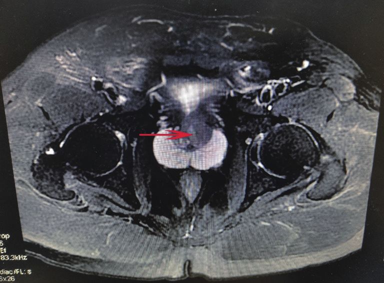
2 Zhou et al. Biopsy free for LRP in pituitary dwarfism
and stable vital signs after LRP. Post-operatively, patients
received adjuvant hemotherapy, followed by PSA test. No
major complication was reported after 3 months. The full
timeline was presented shown in Figure 3.
Discussion
Patients with pituitary dwarfism, who have a human
growth hormone (hGH) deficiency, appear child-like
even as adults, with normally proportioned limbs and
trunk, adenohypophysary hypofunction occurs if there is
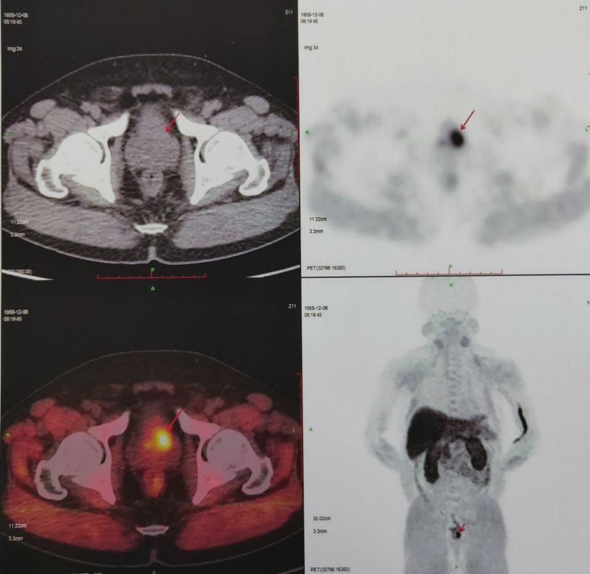
Figure 1 Prostate MRI in the pituitary dwarfism a narrow pelvis parenchymal loss of 75% or more. The causes of these
and short Rectal diameter. Red arrow indicates a prostate mass. patients may be congenital or acquired (3). The diameter of
pelvis is narrow in patients with dwarfism. For women with
dwarfism, the pelvic diameters of persons with pituitary
Case presentation dwarfism are usually only adequate for delivery of a preterm
A 61-year-old pituitary dwarfism presented with moderate infant (4). Therefore, for patients with dwarfism, the narrow
lower urinary tract symptoms. 4 years ago, prostate specific pelvic space greatly increases the difficulty of surgery,
antigen (PSA) level in this patient was 15 ng/mL by especially laparoscopic surgery.
physical examination, and the patient rechecked PSA level Related health problems for consideration for anesthetics
one month ago and the PSA was 31.19 ng/mL. During include cardiac and respiratory difficulties, cervical vertebral
hospitalization, his PSA level was 32.13 ng/mL. There instability in achondroplastic dwarf (5,6). Hence, we
was no history of genetic disease and prostate cancer in his thought that pituitary dwarfism has increased mortality
family. His digital rectal exam (DRE) was failed to perform. compared to the normal population in surgery. Franco et al.
This brought us a big problem that the patient’s anus is too demonstrated that pituitary dwarfism there may be various
narrow to perform prostate biopsy. Because of the unusual anomalies of the dental apparatus, from the morphological
anatomy and prostate biopsy failed, prostate MRI (Figure 1) profile and in terms of development (7). Patients with
and 68Ga PSMA-11 PET/CT (Figure 2) were undertaken dwarfism due to various reasons need to be more cautious
and the results were negative for metastases. And we only during anesthesia, and for patients with pituitary dwarfism,
found abnormal signal in central zone of prostate and no most people have a proportionately smaller airway without
other abnormality, multidisciplinary review occurred. Our anatomic abnormalities (3,8).
radiation oncologists and physicists demonstrated that it In this case, patient with dwarfism had certain risks in
was consistent with the performance of prostate cancer the process of anesthesia, so we should assess the risk of
according to the PSA of the patient and imaging findings. anesthesia well before surgery. Because of the congenital
Therefore, in the absence of a clear histopathology, after pelvic stenosis, there was the possibility of damaging the
multidisciplinary discussions, we combined PSA, MRI, pelvic blood vessels during the operation, which brought
PET-CT to diagnose patients with prostate cancer. From us great difficulty in laparoscopic surgery. On the other
the image, the mass was confined to the prostate tissue hand, due to the patients with congenital anal stenosis
and did not break through the capsula. At present, radical who could not perform prostate biopsy successfully, which
prostatectomy (RP) is one of the main treatment methods brought us great difficulty in clinical diagnosis. Therefore,
for localized prostate cancer. Our radiation oncologists and we proposed a concept of prostate biopsy free. Through
physicists suggested that RP to be the most appropriate imaging diagnosis such as prostate MRI, 68Ga PSMA-11
treatment. Finally, we successfully removed the prostate PET/CT, we could carry out clinical diagnosis of prostate
and bilateral seminal vesicles by LRP. Histological analysis cancer. After consultation with the patient and their
revealed unifocal adenocarcinoma (Gleason 3+5), with families, we decided to carry out LRP for this patient, and
negative surgical margins, no lymphovascular invasion, the operation was successful. Finally, histological analysis
and no extraprostatic extension. No obvious discomfort confirmed that our preoperative diagnosis was accurate. But
© Translational Andrology and Urology. All rights reserved. Transl Androl Urol 2020 | http://dx.doi.org/10.21037/tau-20-489Translational Andrology and Urology, 2020 3 Figure 2 68Ga PSMA-11 PET/CT in the pituitary dwarfism. Red arrow indicates a prostate mass. because of the lack of pathological diagnosis before surgery, Therefore, for these patients with negative biopsy and this method was not suitable for all patients. We also found high suspicion of prostate cancer with distant metastasis, some rare case reports showing patients with bone pain and we could also diagnose prostate cancer through clinical very high PSA who experienced twice prostate biopsy were diagnosis and used tentative ADT treatment to confirm the negative results, and finally through the diagnosis method disease. For our case, we did not find distant metastases, and for bone metastases or lymph node biopsy to confirm the tumor was inside the prostate by imaging examination and diagnosis (1,9,10). Compared with their cases, we found the no enlarged lymph nodes were found, so there was a risk of difference that our patients did not have symptoms of bone misdiagnosis. We needed to combine the indicators of this pain, PSA was not high, and there was no tumor metastasis patient to improve the accuracy of diagnosis. For localized or lymphadenopathy on the MRI, and prostate biopsy could prostate cancer, radical prostatectomy (RP) is the preferred not be completed. Iwamura et al. proposed a case that a treatment. At present, the surgical methods of RP are LRP 64-year-old man with PSA level of 2,036 ng/mL underwent and robotic-assisted LRP. For this patient, we used LRP twice prostate biopsies, and the results were negative. And to excision of prostate. Although we knew that for patients the Imaging showed bone metastases, but no matestatic with pelvic stenosis, RALP was superior to LRP in the lesion biopsy was performed. After ADT, the patient’s bone choice of surgery, but due to the limitations of our surgical pain was alleviated, and PSA level was reduced. Therefore, equipment, we still decided to complete the surgery through they clinically diagnosed patients with prostate cancer (2). LRP. There was no lymph node metastasis was found in © Translational Andrology and Urology. All rights reserved. Transl Androl Urol 2020 | http://dx.doi.org/10.21037/tau-20-489
4                                                                           Zhou et al. Biopsy free for LRP in pituitary dwarfism

Figure 3 Timeline of examination and treatment.

the pathology of the patient, and the image did not show         Technology Committee (19ZXDBSY00050).
systemic metastasis at the same time. The postoperative
pathology showed high-risk prostate cancer, so it was
                                                                 Footnote
necessary to receive endocrine therapy and monitor PSA
levels.                                                          Reporting Checklist: The authors have completed the CARE
   For this case, our diagnosis was successful, and the          reporting checklist. Available at http://dx.doi.org/10.21037/
tumor was removed in time to improve patient survival.           tau-20-489
The strength of this case is that for patients who cannot
undergo prostate biopsy and there is no metastasis and           Conflicts of Interest: All authors have completed the ICMJE
lymphadenopathy on imaging, we combined PSA, MRI, and            uniform disclosure form (available at http://dx.doi.
PET-CT to make a diagnosis to avoid missing the presence         org/10.21037/tau-20-489). The authors have no conflicts of
of prostate malignant tumors. The biggest limitation in          interest to declare.
this case was the risk of misdiagnosis. In this case, no clear
clinical information was obtained to indicate malignant          Ethical Statement: The authors are accountable for all
tumors such as metastatic lesion or suspicious lymph nodes.      aspects of the work in ensuring that questions related
we did not have specific data to enhance the accuracy of our     to the accuracy or integrity of any part of the work are
diagnosis of prostate cancer in this case. Therefore, we hope    appropriately investigated and resolved. All procedures
to provide this case to solve the clinical problems. At the      performed in studies involving human participants were in
same time, we also hope to be able to design a scoring system    accordance with the ethical standards of the institutional
to perform data analysis of laboratory data and subjective       and/or national research committee(s) and with the Helsinki
performance of different imaging studies, so that some           Declaration (as revised in 2013). Written informed consent
patients can avoid unnecessary biopsy. And some patients         was obtained from the patient.
who cannot be biopsied can also be clinically diagnosed.
   All procedures performed in studies involving human           Open Access Statement: This is an Open Access article
participants were in accordance with the ethical standards of    distributed in accordance with the Creative Commons
the institutional and/or national research committee(s) and      Attribution-NonCommercial-NoDerivs 4.0 International
with the Helsinki Declaration (as revised in 2013). Written      License (CC BY-NC-ND 4.0), which permits the non-
informed consent was obtained from the patient.                  commercial replication and distribution of the article with
                                                                 the strict proviso that no changes or edits are made and the
                                                                 original work is properly cited (including links to both the
Conclusions
                                                                 formal publication through the relevant DOI and the license).
Through this case, we can better use laboratory data and         See: https://creativecommons.org/licenses/by-nc-nd/4.0/.
imaging tests to diagnose prostate cancer. Although it
cannot replace prostate biopsy, for patients who are unable
                                                                 References
to perform prostate biopsy or are unwilling to perform
prostate biopsy, we hope to apply the concept of prostate        1.   Ueda Y, Higuchii Y, Hashimoto T, et al. Prostate cancer
biopsy free in clinic.                                                diagnosed through the biopsy of the bone metastatic
                                                                      lesion; a case report. Hinyokika Kiyo 2007;53:327-30.
                                                                 2.   Mitsui Y, Sadahira T, Maruyama Y, et al. Burned-out
Acknowledgments
                                                                      Prostate Cancer ? Primary Metastatic Cancer Not Detected
Funding: The study was supported by Tianjin Science and               on Repeat Biopsy. Acta Med Okayama 2018;72:605-9.

© Translational Andrology and Urology. All rights reserved.               Transl Androl Urol 2020 | http://dx.doi.org/10.21037/tau-20-489
Translational Andrology and Urology, 2020 5
3. Ratner EF, Hamilton CL. Anesthesia for cesarean section in Pituitary Dwarfism: A Case Report and Review of the
in a pituitary dwarf. Anesthesiology 1998;89:253-4. Literature. Case Rep Dent 2017;2017:5849173.
4. Tyson JE, Barnes AC, McKusick VA, et al. Obstetric and 8. Berkowitz ID, Raja SN, Bender KS, et al. Dwarfs:
gynecologic considerations of dwarfism. Am J Obstet pathophysiology and anesthetic implications.
Gynecol 1970;108:688-704. Anesthesiology 1990;73:739-59.
5. Matsui Y, Kawabata H, Ozono K, et al. Skeletal 9. Shimizu K, Nakano S, Okada Y, et al. Hinyokika Kiyo
development of achondroplasia: analysis of genotyped 2019;65:75-80.
patients. Pediatr Int 2001;43:361-3. 10. Iwamura H, Hatakeyama S, Tanaka Y, et al. A case of
6. Wynn J, King TM, Gambello MJ, et al. Mortality in metastatic cancer with markedly elevated PSA level that
achondroplasia study: a 42-year follow-up. Am J Med was not detected by repeat prostate biopsy. BMC Res
Genet A 2007;143A:2502-11. Notes 2014;7:64.
7. Ferrante F, Blasi S, Crippa R, et al. Dental Abnormalities
Cite this article as: Zhou J, Zhang B, Xia S, Li T, Liu R.
Prostate biopsy free system for laparoscopic radical
prostatectomy in a pituitary dwarfism: a case report. Transl
Androl Urol 2020. doi: 10.21037/tau-20-489
© Translational Andrology and Urology. All rights reserved. Transl Androl Urol 2020 | http://dx.doi.org/10.21037/tau-20-489You can also read