Aggressive nasopalatine duct cyst with nasal involvement - Medresearch.in
←
→
Page content transcription
If your browser does not render page correctly, please read the page content below
March, 2018/ Vol 6/Issue 03                                        Print ISSN: 2321-127X, Online ISSN: 2320-8686
                                                                                       Case Report

Aggressive nasopalatine duct cyst with nasal involvement
Pandya D.1, Dey S.2, Bhattacharya M.3, Singh P.4

1
Dr. Divya Pandya, Assistant Professor, Department of Oral Medicine and Maxillofacial Radiology, 2Dr. Soumadip Dey,
Assistant Professor, Department of Oral and Maxillofacial Surgery, 3Dr. Maumita Bhattacharya, Assistant Professor,
Department of Oral Pathology and Microbiology and Forensic Odontology, 4Dr. Pooja Singh, Assistant Professor,
Department of Pedodontic and Preventive Dentistry, all authors are affiliated with Kusum Devi Sundar Lal Dugar Jain
Dental College and Hospital, Kolkata, India.

Corresponding Author: Dr. Divya Pandya, Assistant Professor, Department of Oral Medicine and Radiology, Kusum
Devi Sundar Lal Dugar Jain Dental College and Hospital, Kolkata, India. Dr. Divya Pandya, L 30/1 Bose Para Road,
Kamdahari, Garia, Kolkata, West Bengal, India, E-mail: divyapandya854@gmail.com
………………………………………………………………………………………………………………………………...

Abstract
Nasopalatine duct cyst is a non-odontogenic developmental cyst typically located in the maxillary midline between the
tooth roots of central incisors, these cysts are infrequent and can often be misdiagnosed as periapical lesion or cyst. In
this article, we present a case of which was clinically and radio graphically provisionally diagnosed as nasopalatine duct
cyst in a 62 year old male patient with complaint of swelling in midline of palate. The lesion was surgically removed, and
histo pathologically confirmed the provisional diagnosis, thus concluding that it can be a diagnostic dilemma in clinical
and radiological examinations.

Keywords: Cysts, Developmental, Naso palatine, Non-odontogenic
………………………………………………………………………………………………………………………………...

Introduction
Nasopalatine duct cyst (NPDC) is the most common                 formed between the two maxillae. World Health
non-odontogenic, developmental cyst of non-neoplastic            Organization in 1998 classified these lesions as non-
nature. Its location is peculiar and specific in that it         odontogenic developmental cyst along with naso-
affects the midline anterior maxilla.1Thenasopalatine            alveolar and naso-labial cyst. Although they are most
duct communicates the nasal cavity with the anterior             common non-odontogenic jaw cysts, they accountfor
region of the upper maxilla. During fetal development            only 1% of all maxillary cysts[1,3].
the duct gradually narrows until one or two central
clefts are finally formed on the midline of the upper            NPDC is commonly seen in 4th and 5th decade of life
maxilla. The nasopalatine neurovascular bundle is                with slight male predilection. Clinically it may be
located within the duct and emerges from its intra-bony          asymptomatic and get discovered during routine
trajectory through the nasopalatine foramen [1,2].               radiographic examination. Larger lesion presents as a
                                                                 swelling over anterior maxilla, located between the
NPDC was first described by Meyer in 1914, who                   tooth roots of maxillary central incisors. As the
wrongly identified it as a paranasal sinus. In the past,         presentation of NPDC mimics common periapical
NPDC, was termed as anterior palatine cyst or incisive           lesion like radicular cyst, it is not uncommon to see
canal cyst arising from embryologic remnants of                  evidence of endodontic treatment of associated teeth
nasopalatine duct and was regarded as fissural cysts             due to previous misdiagnosis of the pathology[1,3-6].

Case Report
A 62 year old male patient reported with the chief complaint of a swelling in upper front tooth region of jaw since 2
months. Swelling was insidious in onset and gradually increased to present size with no pain or discharge. Patient
revealed a history of trauma few years back in same region. There was no relevant extraoral finding or lymphadenopathy.
The patient’s medical history was non-contributory.
Manuscript received: 2nd March 2018
Reviewed: 10th March 2018
Author Corrected: 17th March 2018
Accepted for Publication: 21st March 2018

International Journal of Medical Research and Review                    Available online at: www.ijmrr.in 196 | P a g e
March, 2018/ Vol 6/Issue 03 Print ISSN: 2321-127X, Online ISSN: 2320-8686
Case Report
Intraoral examination revealed a well-defined round to oval solitary nodular swelling in mid-line hard palate in
nasopalatine region just beyond palatine rugae, involving both sides of mid-palatine raphe, measuring approximately 1.5
cm x 1.5 cm extending posteriorly to the mesial aspect of maxillary 1stpremolars. On palpation, swelling was soft in
consistency, non tender, compressible, non-reducible and non-fluctuant (Figure 1).
Pulp vitality testing showed vital 11 and 21. Intraoral periapical radiograph of maxillary anterior region revealed a diffuse
radiolucency between roots of central incisors, which was not too appreciable, so a Cone Beam Computed Tomographic
(CBCT) image of maxilla was recommended for 3-dimensional evaluation of lesion.
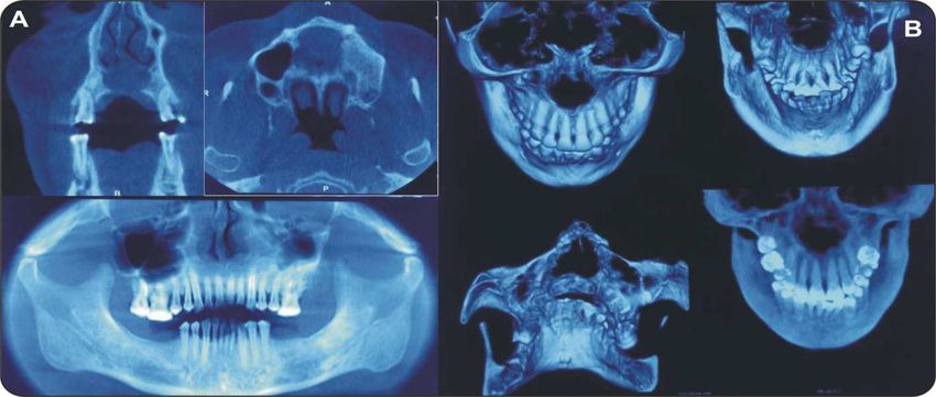
Axial, sagittal and coronal sections with 3-dimensional reformatted images were obtained and assessed to make the
following report – CBCT revealed a well-defined unilocularhypodense lesion in anterior maxilla in 11, 21 regions.
The lesion extended from incisive canal opening to the floor of nasal fossa superinferiorly and from labial to palatal
cortical plate labiopalatally from 13 to 23 region, causing expansion, thinning and destruction of labial cortical plate with
perforation of floor of nasal fossa.
The lesion measured 1.5 cm mesiodistally and supero-inferiorly and 1.4 cm laterally. The lesion was bordered by very
thin irregular sclerotic margin except few areas (Figure 2).
Figure-1: A solitary well-defined swelling in midpalatine region
Figure-2: Multiplanar imaging and reformatting (A) Coronal section (B) Axial section C(C) 3-dimensional reformatted
images showing well defined radiolucency in mid palatine region involving nasal floor and destructing labial cortical
plates.
International Journal of Medical Research and Review Available online at: www.ijmrr.in 197 | P a g e
March, 2018/ Vol 6/Issue 03                                       Print ISSN: 2321-127X, Online ISSN: 2320-8686
                                                                                       Case Report

               Figure-3: Cyst enucleation                      Figure-4: H & E stained histopathologic section

On the basis of history, clinical and radiographic evidence, provisional diagnosis of NPDC was made, with differential
diagnosis of median palatine cyst, periapical cyst and giant cell granuloma. All routine preliminary investigations were
within normal range. Cyst enucleation was done under local anesthesia, crevicular incision was given and palatal
mucoperiosteal flap was raised, complete cystic lining and contents were removed and repositioning of mucosal lining
was performed (Figure 3). Interrupted sutures (3-0 silk) were placed and specimen was sent forhistopathological
examination. The patient was prescribed routine antibiotics and analgesics and was recalled after 1 week for follow up
and reported no complications.

The histopathological report revealed cystic epithelial lining composed of mainly stratified squamous epithelium with
very few areas of simple cuboidal epithelium, with epithelial lining being 2-3 layers thick. The underlying connective
tissue stroma was fibrous with bundles of interlacing collagen fibers, interspersed fibroblasts and chronic inflammatory
cell infiltrate chiefly lymphocytes and plasma cells with few nerves and blood vessels in connective tissue, suggesting a
final diagnosis of NPDC (Figure 4).

Discussion
The NPDC is one of the most common developmental,               Radiographically, it can be detected on routine
non-neoplastic, non-odontogenic cysts of the oral               periapical and occlusal radiographs. It appears as a
cavity, occurring in about 1% of population. They are           round or ovoid radiolucency between the roots of the
believed to develop from remnants of paired embryonic           central incisors. Due to superimposition of the nasal
nasopalatine ducts. NPDC is unique in that it develops          spine, a heart shaped appearance may be seen. Most of
in only a single location, which is the midline of the          the lesions have a well defined sclerotic border. In some
anterior maxilla. It can arise at any age, but is seen most     individuals, a prominent incisive canal can appear as a
often in patients between 30 and 60 years of age. It is         radiolucent area and mimic NPDC. Most authors agree
slightly more common in males than in females, the              that 6 mm should be considered the upper limit for
ratio being 3:1[1,2,5].                                         normal incisive canal radiolucencies larger than this
                                                                should be considered potentially pathologic and merit
Nasopalatine ducts ordinarily undergoes progressive             further investigation[3,4,7-9].
degeneration however, the persistence of the epithelial
remnants may later become the source of epithelia that          Multimodal tomography, can be used as an additional
gives rise to NPDC, from either spontaneous prolife-            imaging modality as it exposes the patient to lesser
ration or proliferation following trauma, bacterial             radiation doses, employs crossed and sectional
infection or mucous retention. Genetic factors have also        tomographic acquisitions in the sagittal plane to yield
been suggested. The cyst is usually asymptomatic and            three dimensional images. Magnetic resonance imaging
discovered on routine radiographs. The most common              may also prove useful in establishing the diagnosis, and
presenting symptoms are swelling of the anterior palate,        particularly contrast the interior of the NPDC with a
drainage and pain. Burning sensation and numbness               high signal intensity[1,6].
may be experienced due to pressure on the nasopalatine
nerve. Occasionally they cause intermittent discharge           A differential diagnosis must be established in order to
with a salty taste. Root resorption and displacement of         avoid unnecessary treatments such as endodontic
teeth is a rare finding[5].                                     procedures in vital permanent upper central incisors.

International Journal of Medical Research and Review                    Available online at: www.ijmrr.in 198 | P a g e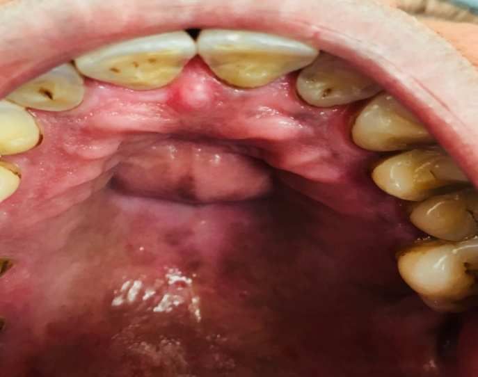
March, 2018/ Vol 6/Issue 03                                     Print ISSN: 2321-127X, Online ISSN: 2320-8686
                                                                                      Case Report
List of differential diagnosis includes radicular cyst,        2. EscodaFrancolí J, Almendros Marqués N, Berini
lateral periodontal cyst, odontogenic cyst, central giant      Aytés L, Gay Escoda C. Nasopalatine duct cyst: report
cell granuloma, ameloblastoma, odontogenicmyxoma               of 22 cases and review of the literature. Med Oral Patol
and central hemangioma. A correct tentative diagnosis          Oral Cir Bucal. 2008 Jul 1;13(7):E438-43.
should be based on positive dental vitality testing and
negative percussion findings of the permanent upper            3. Nilesh K, Vande AV, Suresh KV, Pramod RC,
central incisors provided these teeth do not have pulp or      Suryavanshi P, Kadam NA. A Case of Nasopalatine
periodontal problems[4].                                       Duct Cyst: Presentation, diagnosis and Management. J
                                                               Dent App. 2016;3(2):325-27.
Histopathological examination reveals a cavity lined by
epithelium and surrounded by connective tissue wall. A         4. Shylaja S, Balaji K, Krishna A. Nasopalatine duct
reported 71.8% of NPDCs have squamous, columnar,               cyst: report of a case with review of literature. Indian J
cuboidal or some combination of these epithelial types;        Otolaryngol Head Neck Surg. 2013 Dec;65(4):385-8.
respiratory epithelium is seen in 9.8%. The cyst wall          doi: 10.1007/s12070-011-0242-6. Epub 2011 Apr 11.
may contain a chronic inflammatory reaction consisting
of lymphocytes and plasma cells. Also helpful in the           5. McCrea SJ. Nasopalatine Duct Cyst, a Delayed
diagnosis of NPDC are the presence of neurovascular            Complication to Successful Dental Implant Placement:
bundles, mucous glands and adipose tissue.Treatment of         Diagnosis and Surgical Management. JOI. 2014;XL (2):
NPDC is surgical excision[4,10]. The present case              189-94.
showed typical clinical, radiological and histopatho-
logical features of NPDC.                                      6. Pavankumar K, Sholapurkar AA, Joshi V. Surgical
                                                               Management of Nasopalatine Duct Cyst: case report.
                                                               Rev ClinPesqOdontol. 2010;6(1):81-6.
Conclusion
Clinical and radiological presentation of NPDC can             7. Allmendinger A, Gabe M, Destian S. Median palatine
mimic common periapical lesion like radicular cyst,            cyst. J Radiol Case Rep. 2009;3(7):7-10. doi: 10.3941/
leading to its misdiagnosis. CBCT easily visualizes the        jrcr. v3i7. 269. Epub 2009 Jul 1.
radio-transparency on the midline, with well-defined
sclerotic margins, and informs of the exact location of        8. Salgado H, Felino A, Mesquita P. Extensive naso-
the lesion. In addition, it facilitates planning of the best   palatine cyst with nasal involvement. RPEMD. 2014; 55
surgical approach. Definitive diagnosis is established         (3);171-76.
only after histological evaluation of the excised lining.
                                                               9. Yogesh TL, Shetty A, Nagaraj T, Sinha P.
Funding: Nil, Conflict of interest: None                       Nasopalatine duct cyst: A case report. JCRI. 2014; 1(2):
Permission of IRB: Yes                                         57-60.

References                                                     10. Agarwal S, Jagade M, Thorawade V, Mishra A,
1. Bezalwar A, Tiwari A, Maheshwari V, Patel N.                Joshi S, Ahire D. Palatal Cyst: An Unusual Case
Nasopalatine Duct Cyst: A Case Report. JDMS. 2015;             Report. IJOHNS. 2013;2(1):39-41.
14 (6):60-3.

……………………………
How to cite this article?

Pandya D, Dey S, Bhattacharya M, Singh P. Aggressive nasopalatine duct cyst with nasal involvement. Int J Med Res
Rev 2018;6 (03):196-199. doi:10.17511/ijmrr. 2018.i03.11.
.………………………………………………………………………………………………………………………………..

International Journal of Medical Research and Review                  Available online at: www.ijmrr.in 199 | P a g e
You can also read