Diagnostic Accuracy of Computed Tomography in Predicting Primary Aldosteronism Subtype According to Age
←
→
Page content transcription
If your browser does not render page correctly, please read the page content below
Original                           Endocrinol Metab 2021;36:401-412
                                                                                           https://doi.org/10.3803/EnM.2020.901
                                                        Article                            pISSN 2093-596X · eISSN 2093-5978

Diagnostic Accuracy of Computed Tomography in Predicting
Primary Aldosteronism Subtype According to Age
Seung Hun Lee1, Jong Woo Kim2, Hyun-Ki Yoon2, Jung-Min Koh1, Chan Soo Shin3, Sang Wan Kim3,4,
Jung Hee Kim3

Division of Endocrinology and Metabolism, Department of Medicine, 2Department of Radiology and Research Institute of
1

Radiology, Asan Medical Center, University of Ulsan College of Medicine; 3Department of Internal Medicine, Seoul National
University College of Medicine; 4Department of Internal Medicine, Seoul Metropolitan Government Seoul National University
Boramae Medical Center, Seoul, Korea

Background: Guidelines by the Endocrine Society Guideline on bypassing adrenal vein sampling (AVS) in patients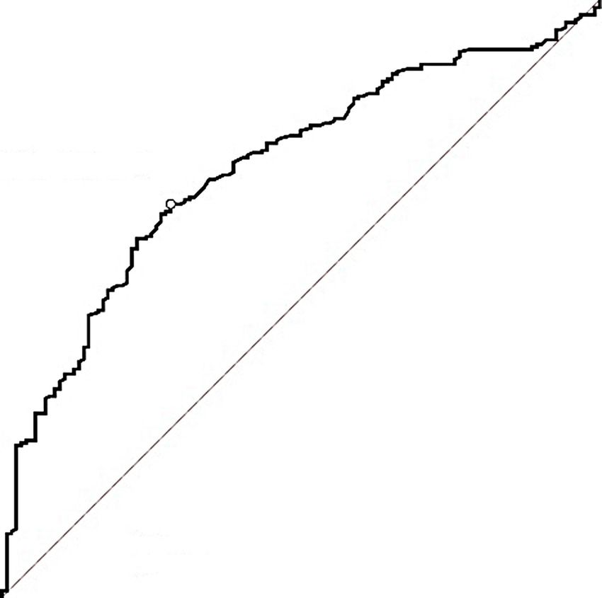
Lee SH, et al.
antagonists [1]. Adrenal venous sampling (AVS) is the standard METHODS
procedure for diagnosing PA subtype [1,5]. However, its use is
limited due to its invasive nature, high cost, and the need for Subjects
technical expertise [5]. Thus, indications for AVS must be opti- This retrospective study enrolled 676 PA patients in two centers
mized by predicting the subtype using an alternative procedure. in Korea; one center from 2000 to 2018 (n=363) [20], and the
Although diagnostic accuracy of adrenal computed tomogra- other from 2007 to 2016 (n=313) [21] using the de-identified
phy (CT) for subtype diagnosis is inadequate [6-11], adrenal CT clinical database [22]. The present study was approved by the
is widely available and less expensive than AVS. Furthermore, Institutional Review Board of Seoul National University Hospi-
score-based algorithms combining CT findings with clinical and tal and Asan Medical Center (no. H-1801-010-911, and no.
biochemical parameters such as age, sex, serum potassium, 2016-0254), and was conducted observing guidelines from the
plasma aldosterone concentration (PAC), PAC to plasma renin Declaration of Helsinki. The requirement for obtaining an in-
activity (PRA) ratio (aldosterone-to-renin ratio [ARR]), esti- formed consent was waived due to the retrospective nature of
mated glomerular filtration rate (eGFR), and the results of con- the study.
firmatory tests have been developed to predict the PA subtype
[8,12-19]. The 2016 Endocrine Society clinical practice guide- Assessment of anthropometric and biochemical
lines published recommended use of adrenal CT as initial work parameters
up to determine PA subtype and exclude adrenal carcinomas [1]. Data regarding age, sex, body mass index (BMI), blood pres-
Moreover, patients aged 15.9 ng/dL, and unilateral inter-assay coefficients of variation were 0.5% and 1.6%.
lesion on CT in the study of JPAS could not lead to the conclu-
sion that AVS can be avoided in these patients. Therefore, JPAS Diagnosis of PA
suggested further validation was needed in the subgroup of pa- Confirmatory testing was performed in patients with HTN with
tients aged 35 to 40 years [10]. high ARR ≥20 and a PAC of >15 ng/dL. PA was confirmed us-
We aimed to investigate the accuracy of adrenal CT in deter- ing saline infusion test [1]. Treatment with diuretics and miner-
mining PA subtype and validate it in patients with marked PA alocorticoid receptor antagonists was discontinued >6 weeks
and unilateral lesion on CT according to age to search the sub- prior to confirming the diagnosis, while treatment with beta-ad-
group of PA patients for bypassing AVS. renergic receptor blockers was discontinued >2 weeks prior.
After shifting from these drugs to calcium channel blockers or
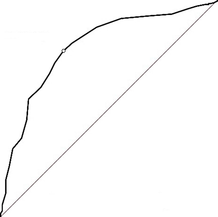
402 www.e-enm.org Copyright © 2021 Korean Endocrine SocietyPredicting the Diagnosis of PA Subtype by CT alpha-adrenergic receptor blockers, BP monitoring was per- act test for categorical variables. Analysis of group difference formed. If BP was unsatisfactorily controlled with the previous was performed using the two afore mentioned tests with post two classes of drug, the addition of angiotensin II receptor hoc Bonferroni correction; α15.9 ng/dL indicated marked PA. to identify independent predictors of concordance among the All patients underwent thin-slice (1 to 3 mm thick) adrenal CT. variables were significant in the univariate analysis. The findings were evaluated by radiologists in each institution To evaluate the ability of age, serum potassium, PAC, and who classified them into unilateral lesion, bilaterally normal, nodule size predictive of concordance of diagnosis between CT and bilateral lesions. Adrenal lesion of CT was defined as a nod- and AVS findings for PA patients with marked PA and unilateral ule or hyperplasia if adrenal gland thickness measured ≥7 mm lesion on CT, receiver-operating characteristics (ROC) curve in diameter [25]. Appearance was considered normal if nodule analysis with the area under the ROC curve (AUC) was per- size or adrenal gland thickness was 30.0 ng/dL, and unilateral lesion on CT by der adrenocorticotropic hormone stimulation [1,5]. Successful age (
Lee SH, et al.

age was 51 years. Approximately 51.1% (228/466) of patients                         and ARR (P=0.012) among patients with unilateral lesion, bi-
had hypokalemia; 79.8% (372/466) had unilateral lesion on CT,                       lateral normal results, and bilateral lesions on CT.
while 11.8% (55/466) had bilateral normal results on CT. Sig-                          Overall prevalence of unilateral PA on AVS was 66.3%
nificant differences were observed in age (P=0.003), DDD of                         (309/466 patients) (Table 2). When including only PA patients
antihypertensive drugs (P=0.039), serum potassium levels (P<                        with SI >5, diagnostic accuracy of CT was 63.7% (279/438), so
0.001), prevalence of hypokalemia (P
Predicting the Diagnosis of PA Subtype by CT

between SI >3 and SI >5 (P=0.851). Prevalence of unilateral           those aged
Table 3. Clinical Findings According to Diagnostic Concordance Rate between CT and Adrenal Vein Sampling Findings in Primary Aldosteronism Patients with Unilateral
                                               Lesion by CT (n=372)
                                                                                        Concordance                 Discordance                                           Univariate analysis                              Multivariate analysis
                                               Variable                                                                                        P value
                                                                                         (n=248)                     (n=124)                                        OR (95% CI)                 P value                OR (95% CI)            P value
                                               Age, yr                                49.0 (42.0–57.0)            53.0 (46.0–60.0)              0.002            0.971 (0.950–0.991)
Predicting the Diagnosis of PA Subtype by CT
100 100
KTable 5. Diagnostic Concordance Rate between CT and AVS Findings in Patients with Hypokalemia, Plasma Aldosterone Concentration >30.0 ng/dL, and Unilateral Le-
sion on CT (n=136), Stratified by Age
Total Age, yr (Predicting the Diagnosis of PA Subtype by CT However, diagnostic accuracy of CT was not statistically signif- As diagnostic concordance rate between CT and AVS findings icant in 139 PA patients with hypokalemia, PAC >30.0 ng/dL, was 11 of 13 (84.6%) PA patients with hypokalemia, PAC and unilateral lesion on CT (91.9% vs. 87.7%); suggesting that >15.9 ng/dL, and unilateral lesion on CT aged 30.0 ng/dL, and unilateral results could not lead to the conclusion that the cut-off value for lesion on CT had a higher risk of unilateral PA, regardless of age to bypass AVS before proceeding to unilateral adrenalecto- age. Using the criteria of hypokalemia, PAC >30 ng/dL, and my was 30.0 ng/dL ings, 105 of 372 (28.2%) bilateral PA would be operated inap- in the previous study by Lim et al. [8] Furthermore, there was propriately by misclassification as unilateral PA. CT findings no significant difference in the diagnostic accuracy of CT in pa- alone would result in adrenalectomies on the wrong side in 19 tients with hypokalemia, PAC >30.0 ng/dL, and unilateral le- of 372 PA patients (5.1%) by showing unilateral hyperaldoste- sion on CT according to age (15.9 ng/dL, ther validation is needed to provide conclusive evidence to sup- and unilateral lesion on CT, there was also a significant differ- port the recent suggestion from the Endocrine Society guide- ence in diagnostic accuracy of CT in patients aged 35 to 39 lines on the age cut-off value of 30 ng/dL, and unilateral lesion on CT aged 15.9 ng/dL, and unilateral lesion on CT aged 30.0 ng/dL, hypokalemia, and unilateral racy of CT in those aged
Lee SH, et al.

on AVS was higher in patients with unilateral disease than in         al study, which is well-associated with a risk of bias from resid-
those with bilateral normal results on CT (50.8% vs. 14.6%, re-       ual confounders. Third, we excluded cases with intermediate LI
spectively; P
Predicting the Diagnosis of PA Subtype by CT

      Iannaccone A, et al. Long-term cardio- and cerebrovascular       13. Nanba K, Tsuiki M, Nakao K, Nanba A, Usui T, Tagami T,
      events in patients with primary aldosteronism. J Clin Endo-            et al. A subtype prediction score for primary aldosteronism.
      crinol Metab 2013;98:4826-33.                                          J Hum Hypertens 2014;28:716-20.
 3.   Reincke M, Fischer E, Gerum S, Merkle K, Schulz S, Pal-          14.   Kupers EM, Amar L, Raynaud A, Plouin PF, Steichen O. A
      lauf A, et al. Observational study mortality in treated prima-         clinical prediction score to diagnose unilateral primary aldo-
      ry aldosteronism: the German Conn’s registry. Hypertension             steronism. J Clin Endocrinol Metab 2012;97:3530-7.
      2012;60:618-24.                                                  15.   Kocjan T, Janez A, Stankovic M, Vidmar G, Jensterle M. A
 4.   Savard S, Amar L, Plouin PF, Steichen O. Cardiovascular                new clinical prediction criterion accurately determines a sub-
      complications associated with primary aldosteronism: a con-            set of patients with bilateral primary aldosteronism before
      trolled cross-sectional study. Hypertension 2013;62:331-6.             adrenal venous sampling. Endocr Pract 2016;22:587-94.
 5.   Monticone S, Viola A, Rossato D, Veglio F, Reincke M, Go-        16.   Kobayashi H, Haketa A, Ueno T, Ikeda Y, Hatanaka Y,
      mez-Sanchez C, et al. Adrenal vein sampling in primary al-             Tanaka S, et al. Scoring system for the diagnosis of bilateral
      dosteronism: towards a standardised protocol. Lancet Dia-              primary aldosteronism in the outpatient setting before adre-
      betes Endocrinol 2015;3:296-303.                                       nal venous sampling. Clin Endocrinol (Oxf) 2017;86:467-
 6.   Kempers MJ, Lenders JW, van Outheusden L, van der Wilt                 72.
      GJ, Schultze Kool LJ, Hermus AR, et al. Systematic review:       17.   Kobayashi H, Abe M, Soma M, Takeda Y, Kurihara I, Itoh H,
      diagnostic procedures to differentiate unilateral from bilat-          et al. Development and validation of subtype prediction
      eral adrenal abnormality in primary aldosteronism. Ann In-             scores for the workup of primary aldosteronism. J Hypertens
      tern Med 2009;151:329-37.                                              2018;36:2269-76.
 7.   Ladurner R, Sommerey S, Buechner S, Dietz A, Degenhart           18.   Kamemura K, Wada N, Ichijo T, Matsuda Y, Fujii Y, Kai T,
      C, Hallfeldt K, et al. Accuracy of adrenal imaging and adre-           et al. Significance of adrenal computed tomography in pre-
      nal venous sampling in diagnosing unilateral primary aldo-             dicting laterality and indicating adrenal vein sampling in
      steronism. Eur J Clin Invest 2017;47:372-7.                            primary aldosteronism. J Hum Hypertens 2017;31:195-9.
 8.   Lim V, Guo Q, Grant CS, Thompson GB, Richards ML,                19.   Burrello J, Burrello A, Pieroni J, Sconfienza E, Forestiero V,
      Farley DR, et al. Accuracy of adrenal imaging and adrenal              Rabbia P, et al. Development and validation of prediction
      venous sampling in predicting surgical cure of primary aldo-           models for subtype diagnosis of patients with primary aldo-
      steronism. J Clin Endocrinol Metab 2014;99:2712-9.                     steronism. J Clin Endocrinol Metab 2020;105:dgaa379.
 9.   Mulatero P, Bertello C, Rossato D, Mengozzi G, Milan A,          20.   Park KS, Kim JH, Yang YS, Hong AR, Lee DH, Moon MK,
      Garrone C, et al. Roles of clinical criteria, computed tomog-          et al. Outcomes analysis of surgical and medical treatments
      raphy scan, and adrenal vein sampling in differential diag-            for patients with primary aldosteronism. Endocr J 2017;64:
      nosis of primary aldosteronism subtypes. J Clin Endocrinol             623-32.
      Metab 2008;93:1366-71.                                           21.   Kim BJ, Kwak MK, Ahn SH, Kim H, Lee SH, Koh JM.
10.   Umakoshi H, Ogasawara T, Takeda Y, Kurihara I, Itoh H,                 Lower trabecular bone score in patients with primary aldo-
      Katabami T, et al. Accuracy of adrenal computed tomogra-               steronism: human skeletal deterioration by aldosterone ex-
      phy in predicting the unilateral subtype in young patients             cess. J Clin Endocrinol Metab 2018;103:615-21.
      with hypokalaemia and elevation of aldosterone in primary        22.   Shin SY, Park YR, Shin Y, Choi HJ, Park J, Lyu Y, et al. A
      aldosteronism. Clin Endocrinol (Oxf) 2018;88:645-51.                   de-identification method for bilingual clinical texts of vari-
11.   Williams TA, Burrello J, Sechi LA, Fardella CE, Matrozova              ous note types. J Korean Med Sci 2015;30:7-15.
      J, Adolf C, et al. Computed tomography and adrenal venous        23.   Levey AS, Bosch JP, Lewis JB, Greene T, Rogers N, Roth D.
      sampling in the diagnosis of unilateral primary aldosteron-            A more accurate method to estimate glomerular filtration
      ism. Hypertension 2018;72:641-9.                                       rate from serum creatinine: a new prediction equation. Mod-
12.   Riester A, Fischer E, Degenhart C, Reiser MF, Bidlingmaier             ification of Diet in Renal Disease Study Group. Ann Intern
      M, Beuschlein F, et al. Age below 40 or a recently proposed            Med 1999;130:461-70.
      clinical prediction score cannot bypass adrenal venous sam-      24.   WHO Collaborating Centre for Drug Statistics Methodolo-
      pling in primary aldosteronism. J Clin Endocrinol Metab                gy, Norwegian Institute of Public Health, World Health Or-
      2014;99:E1035-9.                                                       ganization. ATC/DDD index 2021. [Internet]. Oslo: World

Copyright © 2021 Korean Endocrine Society                                                                         www.e-enm.org       411
Lee SH, et al.
Health Organization Collaborating Centre for Drug Statis- 29. Satoh F, Abe T, Tanemoto M, Nakamura M, Abe M, Uruno
tics Methodology; 2020 [cited 2021 Feb 1]. Available from: A, et al. Localization of aldosterone-producing adrenocorti-
https://www.whocc.no/atc_ddd_index/. cal adenomas: significance of adrenal venous sampling. Hy-
25. Vincent JM, Morrison ID, Armstrong P, Reznek RH. The pertens Res 2007;30:1083-95.
size of normal adrenal glands on computed tomography. 30. Young WF, Stanson AW, Thompson GB, Grant CS, Farley
Clin Radiol 1994;49:453-5. DR, van Heerden JA. Role for adrenal venous sampling in
26. Perkins NJ, Schisterman EF. The Youden index and the opti- primary aldosteronism. Surgery 2004;136:1227-35.
mal cut-point corrected for measurement error. Biom J 31. Williams TA, Lenders JWM, Mulatero P, Burrello J, Rotten-
2005;47:428-41. kolber M, Adolf C, et al. Outcomes after adrenalectomy for
27. Umakoshi H, Tsuiki M, Takeda Y, Kurihara I, Itoh H, Kata- unilateral primary aldosteronism: an international consensus
bami T, et al. Significance of computed tomography and se- on outcome measures and analysis of remission rates in an
rum potassium in predicting subtype diagnosis of primary international cohort. Lancet Diabetes Endocrinol 2017;5:
aldosteronism. J Clin Endocrinol Metab 2018;103:900-8. 689-99.
28. Omura M, Sasano H, Saito J, Yamaguchi K, Kakuta Y, Ni- 32. Rossi GP, Barisa M, Allolio B, Auchus RJ, Amar L, Cohen
shikawa T. Clinical characteristics of aldosterone-producing D, et al. The Adrenal Vein Sampling International Study
microadenoma, macroadenoma, and idiopathic hyperaldo- (AVIS) for identifying the major subtypes of primary aldo-
steronism in 93 patients with primary aldosteronism. Hyper- steronism. J Clin Endocrinol Metab 2012;97:1606-14.
tens Res 2006;29:883-9.
412 www.e-enm.org Copyright © 2021 Korean Endocrine SocietyYou can also read