CONGENITAL PULMONARY AIRWAY MALFORMATION (CPAM): A CASE REPORT - FI-Admin
←
→
Page content transcription
If your browser does not render page correctly, please read the page content below
ISSN Versión Online: 2308-0531
                Rev. Fac. Med. Hum. January 2021;21(1):217-221.
                                                                                                                                  Facultad de Medicina Humana URP
                DOI 10.25176/RFMH.v21i1.3488
                CLINICAL CASE

                  CONGENITAL PULMONARY AIRWAY MALFORMATION
                             (CPAM): A CASE REPORT
                                 MALFORMACIÓN CONGÉNITA DE LA VÍA AÉREA PULMONAR (MCVAP), REPORTE DE CASO
                                                                                José Luis Medina Valdivia1,2

                  ABSTRACT
                  Pulmonary malformations include different abnormalities of the respiratory system, including congenital
                  pulmonary airway malformation (MCVAP), formerly known as cystic adenomatous malformation, which
                  is a rare disease with an incidence of 1 in 8,300 to 35,000 live births. Five classification patterns have been
                  described according to the number and size of the cyst, in addition to their histological characteristics,
                  with type 1 MCVAP being the most frequent, showing displacement of adjacent structures according to
CLINICAL CASE

                  size, associated with brochioalveolar carcinoma, and good prognosis after resection surgical. We present
                  the case of a four-year-old female patient with recurrent hospitalizations for pneumonia and bronchial
                  obstructive syndrome. The thorough anamnesis and physical examination supplemented with the chest
                  x-ray and tomography allowed the diagnosis to be suspected. Later, the patient underwent surgery,
                  there were no complications and the respiratory symptoms disappeared. The histopathological study
                  confirmed the diagnosis.
                  Key words: Pulmonary disease; congenital anomalies; cysts; pneumonia; hospitalized child (source:
                  MeSH NLM).

                  RESUMEN
                  Las malformaciones pulmonares comprenden distintas anomalías del sistema respiratorio, entre ellas
                  la malformación congénita de la vía aérea pulmonar (MCVAP), antes conocida como malformación
                  adenomatosa quística, que es una enfermedad rara con una incidencia de 1 por 8 300 a 35 000 nacidos
                  vivos. Se ha descrito cinco patrones de clasificación de acuerdo al número y tamaño de quiste, además
                  de sus características histológicas, siendo la MCVAP tipo 1 la más frecuente, presenta desplazamiento de
                  estructuras adyacentes según tamaño, asociados a carcinoma broquioalveolar, y de buen pronóstico tras
                  resección quirúrgica. Presentamos el caso, de una paciente de sexo femenino de cuatro años de edad con
                  hospitalizaciones recurrentes por neumonía y síndrome obstructivo bronquial. La acuciosa anamnesis y
                  examen físico complementando con la radiografía de tórax y tomografía permitió la sospecha diagnóstica.
                  Posteriormente la paciente fue intervenida quirúrgicamente, no se presentaron complicaciones y los
                  síntomas respiratorios desaparecieron. El estudio histopatológico confirmó el diagnóstico.
                  Palabras clave: Enfermedad pulmonar; anomalías congénitas; quistes; neumonía; niño hospitalizado
                  (fuente: DeCS BIREME).

                 1 Servicio de Pediatría Hospital Regional de Moquegua, Moquegua-Perú.
                 2
                   Pediatrician.
                 Cite as: José Luis Medina-Valdivia. Congenital pulmonary airway malformation (CPAM): a case report. Rev. Fac. Med. Hum. January 2021;
                 21(1):217-221. DOI 10.25176/RFMH.v21i1.3488

                Journal home page: http://revistas.urp.edu.pe/index.php/RFMH

                  Article published by the Magazine of the Faculty of Human Medicine of the Ricardo Palma University. It is an open access article, distributed under the terms of the
                  Creative Commons License: Creative Commons Attribution 4.0 International, CC BY 4.0 (https://creativecommons.org/licenses/by/4.0/), that allows non-commercial
                  use, distribution and reproduction in any medium, provided that the original work is duly cited. For commercial use, please contact revista.medicina@urp.pe

                                                                                                                                                                             Pág. 217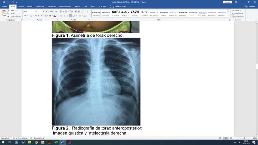
INTRODUCTION CASE REPORT
Congenital anomalies of the respiratory system include A 4-year-old female patient was admitted to the
a large number of pathologies which can affect the Pediatric Services of Hospital Regional de Moquegua
development of the larynx, trachea and bronchi, lung because of dry cough, respiratory distress and
parenchyma, diaphragm and thoracic wall. Congenital fever the previously 5 days. No significant history
pulmonary malformations are divided into: upper at birth, but she was hospitalized several times
and lower respiratory tract congenital malformations. because of bronchiolitis, bronchial obstructive
Congenital pulmonary airway malformation (CPAM), syndrome, recurrent pneumonia (all of them
formerly known as congenital cystic adenomatous without complications) and presumptive diagnosis
malformation, pulmonary sequestration, congenital
of pulmonary tuberculosis since the first month of
lobar emphysema and bronchial atresia(1), appear in
life. The patient showed stable progression during
the lower respiratory tract congenital malformations.
the first two days of hospitalization. Thereafter, she
CPAM is a rare disease with an incidence of 1 per 8,300 presented oxygen desaturation up to 87%, heart rate
to 35,000 live births. It represents approximately of 100/minute and temperature of 37°C. On physical
25% of all congenital lung disease, it has equal examination, nasal flaring, asymmetry of anterior right
CLINICAL CASE
frequency in the right lung than in the left one and chest (Figure 1), subcostal and intercostal retractions
slight predominance in males. Unilobular disease were detected. On auscultation, vesicular sound in
is much more common (85-95%) than multilobular right hemithorax was decreased. Complementary
disease(1,2). examination related to the complete blood count
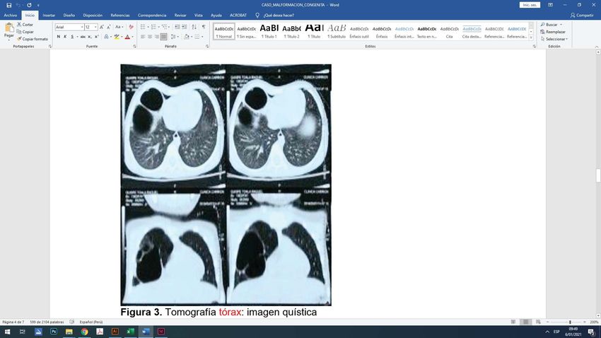
CPAM may present at birth as an incompatible with with leukocytosis, neutrophilia, CRP (+++), the
life syndrome. Prematures, stillbirths and neonates images of the chest radiograph and chest CT (Figure
are frequently affected. They promptly die from 2 and 3 respectively) revealed bilobed cystic image
respiratory insufficiency and anasarca. Only 10% in right lung coupled with basal right atelectasis
of the cases shows up during the first year of life. suggestive of a congenital pulmonary malformation.
There are cases described in older children, and even Patient received oxygen, antibiotic and respiratory
adults; however, those cases are very uncommon and physiotherapy. After stable condition, she was
generally suspected of recurrent infections(2). referred to Instituto Nacional de Salud del Niño San
A case of 4-year-old girl who was admitted to the Borja where she underwent surgery (right upper
Pediatric Services of Hospital Regional de Moquegua and middle lobectomy) confirming the diagnostic
with history of repeated hospitalizations because of suspicion based on histopathological study (CPAM
lower respiratory infections and suspected diagnosis type I: two cystic cavities, the biggest 7x4x4 cm in size
of congenital pulmonary airway malformation type I and the smallest 4x3x3 in diameter, along with upper
is presented. This is due to a careful anamnesis, good and middle lobe ectomy of focal Bronchiectasis,
physical examination, auxiliary imaging examinations atelectasis, diffuse vascular congestion, focal nodular
such as X-ray and tomography, with a subsequent bronchiolitis, and presence of foamy macrophages
diagnostic confirmation through histopathological and foreign-body giant cell reaction of the cyst
study. epithelium).
Pág. 218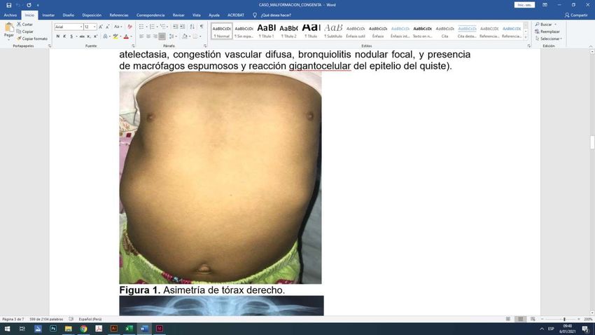
CLINICAL CASE
Figure 1. Asymmetry of right chest.                   Figure 2. Anteroposterior chest radiograph:
                                                       cystic image and right atelectasis.

Figure 3. CT chest: bilobed cystic image and right atelectasis.

                                                                                                Pág. 219
DISCUSSION
CPAM comprise a cysts range with various sizes of now classified into five groups, based on the cysts
variable histology. In 1977, the early Stocker et al.’s amount and size, as well as their histopathological
classification included three types. However, it is origin (Table 1)(2,3).
Table 1. New Stocker et al.’s classification of CPAM.
Type 0 Acinar dysgenesis and dysplasia of the airways. Incompatible with life
Type 1 One or multiple cysts over 2 cm in diameter of bronchus or bronchiole
Type 2 One or multiple cysts under 2 cm in diameter of bronchiole
Type 3 Solid lesion with some cyst under 0,5 cm of bronchiole and alveolar duct
CLINICAL CASE
Type 4 Acinar origin multiple cysts
Among CPAM Type I, is the most common. The fact diagnosis(6).
of lesions being located and can only affect a part of
Surgical resection is intended to prevent further
one lobe makes it the type with the best prognosis.
lung complications such as recurrent infection,
It can also be multilocated(4). Most of the cases
compression of adjacent structures and malignant
occur in the neonatal period or intrauterine period,
transformation(7). Thoracoscopic excision has been
but in rare cases they may show later. The clinical
suggested in literature of having apparently good
presentation is variable and relies directly on the
results(8). The definitive diagnosis is determined by
size of the lung mass. Hydrops fetalis development
histopathological study as it is showed that cyst
is associated with the worst prognosis and high
wall is covered by the bronchial epithelium(8,10). In
mortality during prenatal stage. Clinical picture is
conclusion, prenatal diagnosis of the congenital
variable and is related to the size lung lesion too(5).
malformation of the pulmonary tract allows the earlier
Microscopically, there is a marked boundary between
postnatal diagnosis confirmation. This contributes in
the lesion and the adjacent normal lung, but there is
taking timely therapeutic action to prevent further
no capsule. The larger cystic spaces are covered by the
complications. Uncommonly this pathology is
pseudostratified ciliated columnar epithelium and the
diagnosed in infants, preschoolers, school children
lung between the cysts may reveal overgrowth and
and/or adolescents. Early surgical treatment prevents
underdeveloped alveolar parenchyma. Historically,
from possible future pulmonary complications.
mucous cell hyperplasia is described in 35 to 50%
of the cases. Malignant transformation is rare, but In Colombia, a similar case of a 4-year-old girl who
areas of consolidation that are not resolved during presented recurrent respiratory infections and even
CPAM imaging should be considered (mucinous received treatment for tuberculosis was reported by
adenocarcinoma). After complete resection the Guzmán et al.(11). In 2018, Ventura et al.(12) reported
prognosis is favourable, although recurrence of the the first three cases of congenital pulmonary airway
disease has been described if it is completely resected, malformation with prenatal diagnosis intrauterinally
sometimes decades after the original cyst resection. treated at Instituto Nacional Materno Perinatal
It may also show signs of additional infection when located in Lima, Peru. In Argentina a huge CPAM case
resected, including an aspergilloma. that simulates tension pneumothorax(13) was reported
by Wang(13) in 2019.
Sometimes the diagnosis of congenital pulmonary
airway malformations can be difficult. Whether We reported a case of CPAM Type I at the Pediatric
clinically or radiologically it can simulate other Services of Hospital Regional de Moquegua. This is a
diseases. Therefore, health care providers should rarely diagnosed disease in our environment, perhaps
be aware of such fact in order to furnish an early due to the difficulty of carrying out high resolution
Pág. 220gestational ultrasounds.The patient, with a history of aid, taking a detailed and complete medical history,
frequent respiratory infections, had to wait until she as in the case of this patient, made it possible to
was four years old to be diagnosed and treated. observe the recurrence of hospitalizations with
inconclusive respiratory diagnosis, to propose
CONCLUSION presumptive and differential diagnosis as well as to
In developing countries where it is difficult to access refer the patient for timely surgical treatment.
sophisticated technological resources as a diagnostic
Authorship contributions: The author participated Conflict of interest: The author declares no conflicts
in the conception and design of the work; data of interest in the publication of this case report.
collection, analysis and interpretation; critical review Received: December 13, 2020
and writing of the final version.
Approved: December 13, 2020
Financing: Self-financed.
CLINICAL CASE
Correspondence: José Luis Medina Valdivia
Address: Urbanización los Damascos C-1, Moquegua, Mariscal Nieto-Perú
Telephone number: 953951080
E-mail: jlzf29@hotmail.com
BIBLIOGRAPHIC REFERENCES
1. Zhu H, Liu D, Jia H. Analysis of Wnt7B and BMP4 expression patterns Quística. Presentación de caso. Fac Tecnol la salud. 2018; 9(1):173–8.
in congenital pulmonary airway malformation. Pediatr Pulmonol.
marzo de 2020; 55(3):765-70. 8. Ramenofsky ML, Leape LL, McCauley RGK. Broncogenic cyst. J Pediatr
Surg. 1979; 14: 219-24.
2. Stocker JT. Congenital Pulmonary airway malformation : a new name
for an expanded classification of congenital cysticadenomatoid 9. Salcedo M, Alva LF, Sotelo R, Peña ES, Lule MS, Falcón V. Quiste
malformation of the lung .Histopatology.2002;41:424-31 broncogénico: Reporte de dos casos y revisión de la literatura. Rev
Inst Nal Enf Resp Mex. 2004; 17: 35-41.
3. Posada Saldarriaga Ricardo Neumología Pediátrica :Asociación
Colombiana de Neumología Pediátrica .Bogotá Colombia :Editorial 10. Kumar A, Aggarwal S, Halder S, Kumar S, Khilnani GC.Thoracoscopic
Medica Distribuna 2016 excision of mediastinal bronchogenic cyst: A case report and review
of literature. Indian J Chest Dis Allied Sci. 2003; 45: 199-201.
4. Barazzone-Argiroffo C, Lascano Maillard J, Vidal I, Bochaton-Piallat
ML, Blaskovic S, Donati Y, et al. New insights on congenital pulmonary 11. Guzmán-Vélez JE, Ossa-Galvis MM. Malformación congénita de la vía
airways malformations revealed by proteomic analyses. Orphanet J aérea pulmonar. Rev CES Med 2014; 28(2): 283-292
Rare Dis. 28 de 2019; 14(1):272.
12. Ventura Laveriano W, Chang Wong K, Lacunza Paredes R, Nazario
5. Fajardo-Escolar AP, Díaz-Bohada L. Case report Anesthetic Redondo C, Saldaña Montes P, Moreno Gonzales R, Alvarado Zelada
management in two infants with cystic adenomatoid malformation J, Castillo Urquiaga W, Zárate Girao M, Limay Ríos A. Manejo prenatal
– Case report. Colomb J Anesthesiol [Internet]. 2017; 45(S1):76– 80. de la malformación adenomatoidea quística pulmonar, variedad
Available from: http://dx.doi.org/10.1016/j. rcae.2016.11.014 macroquística. Reporte de los primeros casos tratados intraútero
en el Perú y revisión de la literatura. Rev Peru Ginecol Obstet. 2018;
6. Chaparro A, María. Resultado postnatal de fetos con malformación 64(1):91-97
adenomatoidea quística pulmonar en la unidad maternofetal clínica
Colombia. [Colombia]: Universidad del Rosario; 2013 13. Wang N, Wu X Zhang S, Li N. Quiste pulmonar congénito gigante que
imita neumotórax a tensión en un niño. Arch Argent Pediatr 2019;
7. González Y, Ortiz J, Silva A, Romero A. Enfermedad Adenomatoidea 117(4):e416-e419.
Pág. 221You can also read