Rheumatoid Arthritis Related Interstitial Lung Disease: Patterns of High-resolution Computed Tomography - Cureus
←
→
Page content transcription
If your browser does not render page correctly, please read the page content below

Open Access Original
                                         Article                                                  DOI: 10.7759/cureus.6875

                                         Rheumatoid Arthritis Related Interstitial
                                         Lung Disease: Patterns of High-resolution
                                         Computed Tomography
                                         Mahesh Gautam 1 , Mah Jabeen Masood 2 , Sadaf Arooj 2 , Mufazzal-e-Haque Mahmud 3 ,
                                         Muhammad Umer Mukhtar 4

                                         1. Radiology, Nobel Medical College, Biratnagar, NPL 2. Radiology, King Edward Medical University,
                                         Lahore, PAK 3. Rheumatology and Immunology, Shaikh Zayed Hospital, Lahore, PAK 4. Medicine, King
                                         Edward Medical University, Lahore, PAK

                                         Corresponding author: Muhammad Umer Mukhtar, m.umermukhtar@hotmail.com

                                         Abstract
                                         Background and aim
                                         Rheumatoid arthritis (RA) is a chronic inflammatory systemic disease characterized by bilateral
                                         involvement of mostly small joints of hands and feet. There can be various extra-articular
                                         manifestations of the disease including lung parenchymal disease. Pulmonary involvement in
                                         RA patients leads to increased morbidity and mortality. The overall burden of RA related
                                         pulmonary disease is underestimated due to the limitation of resources in underdeveloped
                                         countries. High-resolution computed tomography (HRCT) is an important tool used to
                                         diagnose different abnormalities in RA related interstitial lung disease (ILD). The objective of
                                         the study was to evaluate HRCT findings in patients of RA related ILD and categorize the
                                         radiological findings according to clinical findings.

                                         Method
                                         This descriptive prospective observational study was conducted at Mayo Hospital, Lahore from
                                         June 2014 to June 2015. Patients of RA suspected of lung disease after selection underwent
                                         HRCT chest on 128-slice Hitachi CT scanner (Hitachi Global, Tokyo, Japan) in the radiology
                                         department. Images were reconstructed and evaluated by experienced radiologists. Findings
                                         were recorded on a questionnaire. Data was analyzed on SPSS version 21 (IBM Corp, Armonk,
                                         US).

                                         Results
                                         Out of the 54 patients scanned, interlobular septal thickening was the most common finding
                                         found in 22 of the patients. Ground-glass opacification was recognized in 21 patients,
Received 01/14/2020
                                         honeycombing in nine and bronchiectasis in two patients. Regarding zonal predilection of
Review began 01/25/2020
Review ended 01/31/2020                  disease pattern, lower zones of lungs were found involved in most of the cases. The disease was
Published 02/04/2020                     found to be bilateral in 15 patients. Based on these findings, usual interstitial pneumonitis
                                         (UIP) was diagnosed in six patients and non-specific interstitial pneumonitis (NSIP) in 14
© Copyright 2020
Gautam et al. This is an open access     others.
article distributed under the terms of
the Creative Commons Attribution
License CC-BY 4.0., which permits        Conclusion
unrestricted use, distribution, and
                                         This study concluded that HRCT images are very useful in diagnosing interstitial lung disease
reproduction in any medium, provided
the original author and source are       related to rheumatoid arthritis.
credited.

                                         How to cite this article
                                         Gautam M, Masood M, Arooj S, et al. (February 04, 2020) Rheumatoid Arthritis Related Interstitial Lung
                                         Disease: Patterns of High-resolution Computed Tomography. Cureus 12(2): e6875. DOI
                                         10.7759/cureus.6875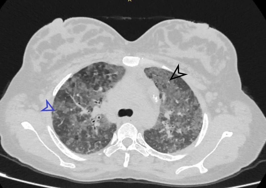
Categories: Radiology, Allergy/Immunology, Rheumatology
                                   Keywords: rheumatoid arthritis, interstitial lung disease, hrct

                                   Introduction
                                   Rheumatoid arthritis (RA) is a chronic inflammatory systemic disease exhibiting clinical signs
                                   and symptoms of predominantly joint disease [1-3]. This disease is characterized by
                                   symmetrical bilateral involvement of mostly small joints of hand and feet; however as it leads
                                   to chronic synovitis, all joints may be involved [4-5]. There can be various extra-articular
                                   manifestations (EAM) of the disease e.g. upper airway, lower airway, pleural, vascular and lung
                                   parenchymal disease, etc. [6-8]. The overall burden of RA related pulmonary disease is
                                   underestimated due to the limitation of resources in underdeveloped countries [9]. Prevalence
                                   of interstitial lung disease (ILD) was found to be 97.9% per 100000 with more of the secondary
                                   ILDs than primary ILD. is around 19-44% [10]. Pulmonary involvement seen in RA patients has
                                   high clinical significance as it leads to increased morbidity and mortality [11]. The most
                                   important characteristic of RA related pulmonary disease is that almost all anatomical parts of
                                   the lung are prone to RA related tissue injury [12]. The overall risk of having ILD in RA patients
                                   is 19.2% as compared to the risk of having ILD in the common population [13]. ILD is a
                                   spectrum of pulmonary diseases that involve all parts of pulmonary bronchovascular units,
                                   including alveolar epithelium, capillary endothelium, alveoli, perivascular connective tissue,
                                   and perilymphatic tissues. RA is only second to systemic sclerosis as far as the incidence of ILD
                                   in connective tissue disorders is concerned [14]. The natural history of ILD is not very well
                                   defined in patients. Complications may arise themself or secondary to immunosuppressive drug
                                   treatment. High-resolution computed tomography (HRCT), that emerged during the past
                                   decade, is an important tool to diagnose the different abnormalities in RA related ILD.

                                   Materials And Methods
                                   From June 2014 to June 2015, 54 patients were selected from the outpatient department of
                                   Mayo Hospital, Lahore, with RA related pulmonary disease, according to American College of
                                   Radiology (ACR) criteria 2010 [15]. The complete history was taken, and patients with co-
                                   morbidities like pulmonary tuberculosis, chronic obstructive pulmonary disease (COPD), and
                                   lung masses were excluded after evaluating chest X-rays. HRCT chest was performed using a
                                   multislice multidetector scanner. Axial images were acquired in a supine position, taking
                                   complete deep inspiration. Images were taken using a 0.5 m slice thickness with at least 1 cm
                                   slice interval. Image reconstruction was done using a bone algorithm.

                                   Results
                                   Altogether 54 cases were studied to complete the sample size of the project. Out of the 54
                                   patients, 18 (33.33%) were male, while 36 (66.67%) were female. The mean age of the patient
                                   was 44.17± 11.315 years, with the minimum age being 15 years and the maximum age being 65
                                   years. Patients had a variable presentation, and the commonest presentation to the hospital
                                   was exertional dyspnea (Table 1).

2020 Gautam et al. Cureus 12(2): e6875. DOI 10.7759/cureus.6875                                                                 2 of 12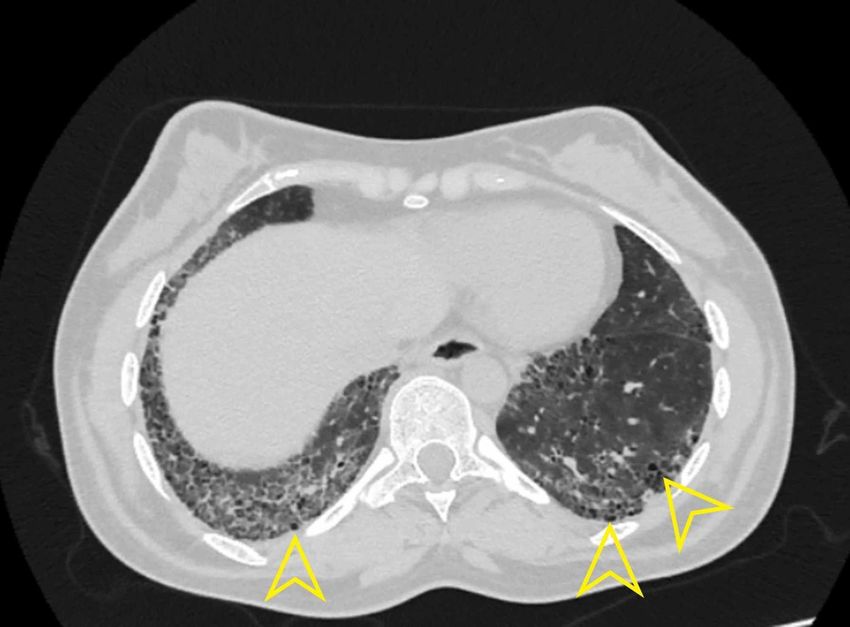
Symptoms                                           Present                  Duration in months

     Cough                                              7 (13%)                  3-6 months

     Exertional dyspnea                                 20 (37%)                 1-3 months

     Wheezing                                           0                        0

     Phlegm                                             3 (5.6%)                 1 month

    TABLE 1: Frequency distribution of the respiratory symptoms

                                      Regarding the findings of HRCT, interlobular septal thickening was the most common finding,
                                      present in 22 (40.7%) patients. Similarly, ground-glass opacity (GGO) was present in 21 (38.9%)
                                      patients, while nine (16.7%) patients had honeycombing (Table 2).

     HRCT Findings                                                                         Frequency %

     Ground-glass attenuation                                                              21 (38.9%)

     Honeycombing                                                                          9 (16.7%)

     Interlobular septal thickening                                                        21 (38.9%)

     Nodule                                                                                0

     Air trapping                                                                          1 (1.9%)

     Bronchiectasis                                                                        1 (1.9%)

     Traction bronchiectasis                                                               15 (27.8%)

     Reticular shadowing                                                                   22 (40.7%)

     Mosaic perfusion                                                                      2 (3.7%)

     Architectural distortion                                                              3 (5.6%)

    TABLE 2: Descriptive statistics of the patients with early rheumatoid arthritis using
    HRCT
    HRCT - high-resolution computed tomography

                                      Bronchiectasis was present in two (3.7%) patients, whereas traction bronchiectasis was present
                                      in 17 (31.5%). Air trapping was present in one (1.9%), mosaic perfusion in two (3.7%), and
                                      architectural distortion in three (5.6%) patients. GGO, interlobular septal thickening and
                                      reticular shadowing, honeycombing, air trapping, mosaic perfusion, and air trapping were
                                      found to involve both the lungs. Fifteen (27.8%) patients had bilateral symmetrical traction
                                      bronchiectasis, whereas one (1.9%) patient had traction bronchiectasis involving right lung and

2020 Gautam et al. Cureus 12(2): e6875. DOI 10.7759/cureus.6875                                                                 3 of 12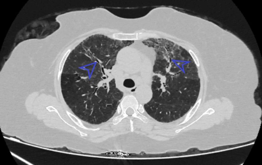
one (1.9%) patient had traction bronchiectasis involving left lung only (Table 3). Similarly, one
                                      (1.9%) had unilateral bronchiectasis involving the right lung, and one (1.9%) patient had
                                      bronchiectasis involving the left lung.

                                            Unilateral right lung              Unilateral left lung            Bilateral
     Findings
                                            involvement                        involvement                     involvement

     Ground-glass attenuation               0                                  0                               21 (38.9%)

     Honeycombing                           0                                  0                               9 (16.7%)

     Interlobular septal thickening         0                                  0                               22 (40.7%)

     Nodule                                 0                                  0                               0

     Air trapping                           0                                  0                               1 (1.9%)

     Bronchiectasis                         1 (1.9%)                           1 (1.9%)                        0

     Traction bronchiectasis                1 (1.9%)                           1 (1.9%)                        15 (27.8%)

     Reticular shadowing                    0                                  0                               22 (40.7%)

     Air space opacity                      0                                  0                               0

     Emphysema                              0                                  0                               0

     Cysts                                  0                                  0                               0

     Tree-in-bud appearance                 0                                  0                               0

     Crazy-paving appearance                0                                  0                               0

     Mosaic perfusion                       0                                  2 (3.7%)                        0

     Architectural distortion               0                                  3 (5.6%)                        0

     Thickening of bronchovascular
                                            0                                  0                               0
     bundle

    TABLE 3: Descriptive statistics of the HRCT findings in unilateral and bilateral
    involvement of the right and left lung
    HRCT- high-resolution computed tomography

                                      Regarding the zone involvement, the lower zone was found to be more frequently involved
                                      (Table 4). GGO was found to involve lower zone in 14 (25.9%) patients, one (1.9%) patient had
                                      involvement of mid zone, five (9.3%) patients had involvement of the middle and lower zone
                                      and one (1.9%) patient had involvement of all three zones, i.e., upper, mid and lower.
                                      Honeycombing was present in the lower zone in six (11.1%) patients, two (3.7%) patients had
                                      honeycombing in the middle and lower zone, and one (1.9%) had honeycombing involving all
                                      three zones. Thirteen (24.1%) patients had interlobular septal thickening involving mid and
                                      lower zone, eight (14.8%) had interlobular septal thickening in the lower zone, and one (1.9%)

2020 Gautam et al. Cureus 12(2): e6875. DOI 10.7759/cureus.6875                                                                    4 of 12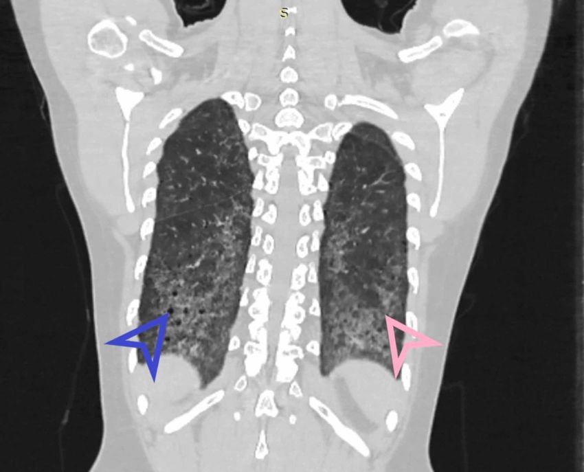
had interlobular septal thickening involving all three zones.

                           Right lung                                          Left lung

     HRCT finding
                                                      mid and     upper mid                                  mid and    upper mid
                           upper   mid      lower                              upper    mid        lower
                                                      lower       and lower                                  lower      and lower

     Ground-glass                  1        14        5                        1        14         5
                           0                                      1 (4.8%)                                   1 (4.8%)
     Opacity                       (4.8%)   (66.7%)   (23.8%)                  (4.8%)   (66.7%)    (23.8%)

                                            6         2                                            6         2
     Honeycombing          0       0                              1 (11.1%)    0        0                               1 (11.1%)
                                            (66.7%)   (22.2%)                                      (66.7%)   (22.2%)

     Interlobular septal                    8         13                                           8         13
                           0       0                              1 (4.0%)     0        0                               1 (4.0%)
     thickening                             (36.0%)   (59.0%)                                      (36.0%)   (59.0%)

     Nodule                0       0        0         0           0            0        0          0         0          0

                                            1                                                      1
     Air trapping          0       0                  0           0            0        0                    0          0
                                            (100%)                                                 (100%)

                                                                                                   1
     Bronchiectasis        0       0        0         0           1 (100%)     0        0                    0          0
                                                                                                   (100%)

     Traction                      1        1                                           1          1
                           0                          0           0            0                             0          0
     bronchiectasis                (6.7%)   (100%)                                      (6.7%)     (100%)

                                            16        6                                            16        6
     Reticular shadowing   0       0                              1 (4.5%)     0        0                               1 (4.5%)
                                            (68.2%)   (27.3%)                                      (68.2%)   (27.3%)

    TABLE 4: Descriptive statistics of the HRCT findings in term of zone involved among
    patients with early rheumatoid arthritis
    HRCT - high-resolution computed tomography

                                   Based on these patterns of involvement of the lung, usual interstitial pneumonia (UIP) was
                                   diagnosed in six (11.1%) patients (Table 5). Similarly, non-specific interstitial pneumonia
                                   (NSIP) was diagnosed in 14 (25.9%) of the patients. Findings of HRCT that did not fall under
                                   any defined category were labeled as others, as were present in five (9.3%) patients.

2020 Gautam et al. Cureus 12(2): e6875. DOI 10.7759/cureus.6875                                                                     5 of 12
Diagnosis Frequency Percent Valid percent Cumulative percent
Normal 29 53.7 53.7 53.7
Usual interstitial pneumonia 6 11.1 11.1 64.8
NSIP 14 25.9 25.9 90.7
Others 5 9.3 9.3 100.0
Total 54 100.0 100.0
TABLE 5: Frequency distribution of the HRCT diagnosis in patients with early
rheumatoid arthritis
NSIP - non-specific interstitial pneumonia
Discussion
The present study indicates that females are at increased risk of RA with the statistical odds of
(66.67% vs. 33.3%). The results of this study are comparable with the study by Gabbay E et al.
that demonstrated females to be at an increased risk of RA (69.44%) [16]. El Khalik KA et al.
found that RA was more common in females than in males (79.4% vs. 20.58%) [17]. This
disparity may be due to bias in the selection of women. The present study provides evidence
that RA patients were older individuals of 44.17 ±11.315 years. Gabbay E et al. demonstrated
that RA was more commonly found in older patients 51.8 ±16.0 years [16]. Affara NK et al.
examined RA patients to be 52.6 ± 5.1 years old [18]. The discrepancy in the mean age of the
patients in our study may be present due to the small sample size.
Affara NK et al. examined HRCT abnormalities in their study, i.e., GGO was present in 30%, air
space opacification (AS) was present in 13.3%, mixed AS/GGO was present in 13.3%,
honeycombing in 10%, septal thickening in 26.7% and traction bronchiectasis in 11.7% of the
subjects [18]. In contrast with these studies, our study reported interlobular septal thickening
as the most common HRCT finding, present in 22 (40.7%) patients and illustrated in Figures 1
and 2.
2020 Gautam et al. Cureus 12(2): e6875. DOI 10.7759/cureus.6875 6 of 12FIGURE 1: Axial section revealing bilateral basal intralobular
septal thickening with sparing of subpleural areas
Black arrowhead shows Interlobular septal thickening.
FIGURE 2: Patchy areas of GGO and interlobular septal
thickening involving bilateral upper lobes
Black arrowhead indicates the ground-glass opacity (GGO). Blue arrowhead shows interlobular
2020 Gautam et al. Cureus 12(2): e6875. DOI 10.7759/cureus.6875 7 of 12septal thickening.
Similarly, GGO was present in 21 (38.9%) patients (Figure 2), nine (16.7%) patients had
honeycombing. Bronchiectasis was present in two (3.7%), whereas traction bronchiectasis was
present in 17 (31.5%) such as shown in Figure 3. Air trapping was present in one (1.9%), two
(3.7%) patients had mosaic perfusion, and three (5.6%) patients had architectural distortion.
FIGURE 3: Axial section showing bronchiectasis
Blue arrowhead indicates bronchiectasis.
Metafratzi ZM et al. .used normal healthy individuals as control against known patients of RA
[19]. They used a semi-quantitative grading system, which has also been described in the past.
The presence and extent of findings on HRCT chest were coded according to lung zones
bilaterally, making a total of six zones. Only air trapping was given a score of eight on its
presence on paired inspiratory and expiratory images. The control subjects showed minimal
findings with scores of less than 3.6. The most common findings were air trapping and
bronchiectasis. The abnormalities noticed in patients were equal to a score of 5.2 (moderate in
severity) with only air trapping having a score of 14 (maximum severity). Other findings were
bronchiectasis, bronchial wall thickening, macro nodules, and GGO.
Chansakul TN, in their study, concluded that traction bronchiectasis and the extent of
honeycombing (as our earlier findings illustrated in Figures 4-6) were strongly associated with
morbidity and mortality [20]. RA related ILD carried a bad prognosis when they compared HRCT
findings to pulmonary function tests. They also completely evaluated patients’ intrathoracic
noncardiac findings on CT scans in terms of pleural, parenchymal, vascular disease as well as
drug-related complications and opportunistic infections. They found that UIP (illustrated in
Figure 5) was commoner than NSIP. There was more overlap between NSIP and UIP patterns.
2020 Gautam et al. Cureus 12(2): e6875. DOI 10.7759/cureus.6875 8 of 12FIGURE 4: Coronal HRCT showing basal predominance of
fibrosis and honeycombing in UIP
HRCT - high-resolution computed tomography; UIP - usual interstitial pneumonia
Blue arrowhead indicates honeycombing. Pink arrowhead shows fibrosis.
2020 Gautam et al. Cureus 12(2): e6875. DOI 10.7759/cureus.6875 9 of 12FIGURE 5: Axial section high-resolution computed tomography
showing UIP
Yellow arrowhead indicating honeycombing in usual interstitial pneumonia (UIP).
FIGURE 6: Coronal view HRCT shows bilateral symmetrical
2020 Gautam et al. Cureus 12(2): e6875. DOI 10.7759/cureus.6875 10 of 12fibrosis in lower lobes
HRCT - high-resolution computed tomography
Blue arrowhead shows honeycombing.
Conclusions
Multiple pulmonary manifestations of RA are well known. Investigation for ILD is mandatory
for evaluating the clinical progression of RA. HRCT detects subtle abnormalities in RA patients
even without respiratory symptoms. Thus, HRCT is the investigation of choice to delineate in
detail the bronchovascular involvement and disease patterns of the lungs in patients of RA.
Additional Information
Disclosures
Human subjects: Consent was obtained by all participants in this study. Institutional Review
Board of King Edward Medical University, Lahore issued approval 476/RC/KEMU. The Synopsis
of Dr. Mahesh Gautam, PG Trainee - MD Diagnostic Radiology, Department of Radiology, King
Edward Medical University was presented before the Institutional Review Board for ethical
approval. The committee after discussion approved the synopsis and found it appropriate and
awarded the study ethical approval. Animal subjects: All authors have confirmed that this
study did not involve animal subjects or tissue. Conflicts of interest: In compliance with the
ICMJE uniform disclosure form, all authors declare the following: Payment/services info: All
authors have declared that no financial support was received from any organization for the
submitted work. Financial relationships: All authors have declared that they have no
financial relationships at present or within the previous three years with any organizations that
might have an interest in the submitted work. Other relationships: All authors have declared
that there are no other relationships or activities that could appear to have influenced the
submitted work.
References
1. Akhter E, Bilal S, Kiani A, Haque U: Prevalence of arthritis in India and Pakistan: a review .
Rheumatol Int. 2011, 31:849-855. 10.1007/s00296-011-1820-3
2. Johnson C: Recent advances in the pathogenesis, prediction and management of rheumatoid
arthritis associated with interstitial lung disease. Curr Opin Rheumatol. 2017, 29:254-259.
10.1097/BOR.0000000000000380
3. Shaw M, Collins BF, Ho LA, Raghu G: Rheumatoid arthritis-associated lung disease . Eur
Respir Rev. 2015, 24:1-16. 10.1183/09059180.00008014
4. Bongartz T, Nannini C, Medina-Velasquez YF, et al.: Incidence and mortality of interstitial
lung disease in rheumatoid arthritis: a population-based study. Arthritis Rheum. 2010,
62:1583-1591. 10.1002/art.27405
5. Norton S, Koduri G, Nikiphorou E, Dixey J, Williams P, Young A: A study of baseline
prevalence and cumulative incidence of comorbidity and extra-articular manifestations in RA
and their impact on outcome. Rheumatology. 2013, 52:99-110. 10.1093/rheumatology/kes262
6. Hamblin MJ, Horton MR: Rheumatoid arthritis associated interstitial lung disease: diagnostic
dilemma. Pulm Med. 2011, 872120. 10.1155/2011/872120
7. Pappas DA, Giles JT, Connors G, Lechtzin N, Bathon JM, Danoff SK: Research article
Respiratory symptoms and disease characteristics as predictors of pulmonary function
abnormalities in patients with rheumatoid arthritis: an observational cohort study. Arthritis
Res Ther. 2010 , 12:R104. 10.1186/ar3037
8. Dokwal CP: Pulmonary manifestations of collagen vascular disease . Pulse. 2010, 4:16-21.
10.3329/pulse.v4i1.6958
2020 Gautam et al. Cureus 12(2): e6875. DOI 10.7759/cureus.6875 11 of 129. Yunt ZX, Solomon J: Lung disease in rheumatoid arthritis . Rheum Dis Clin. 2015, 41:225-236.
10. Duchemann B, Annesi-Maesano I, Jacobe de Naurois C, et al.: Prevalence and incidence of
interstitial lung diseases in a multi-ethnic county of Greater Paris. Eur Respir J. 2017,
50:1602419. 10.1183/13993003.02419-2016
11. Assayag D, Elicker BM, Urbania TH, et al.: Rheumatoid arthritis-associated interstitial lung
disease: radiologic identification of usual interstitial pneumonia pattern. Radiology. 2014,
270:583-588. 10.1148/radiol.13130187
12. Zou YQ, YS, Ding XN, Ying ZH: The clinical significance of HRCT in evaluation of patients
with rheumatoid arthritis-associated interstitial lung disease: a report from China. Rheumatol
Int. 2012, 32:669-673. 10.1007/s00296-010-1665-1
13. Salaffi F, Carotti M,Di Carlo M,Tardella M, Giovagnoni A: High-resolution computed
tomography of the lung in patients with rheumatoid arthritis: prevalence of interstitial lung
disease involvement and determinants of abnormalities. Ann Rheum Dis. 2019, 78:350.
14. Bhattacharya P, Ghosh S, Sengupta S, Dasgupta A, Ghosh, K, Ghosh B: Clinicoradiological
profile of interstitial lung disease in rheumatoid arthritis. Asian J Med Sci. 2018, 10:66-71.
10.3126/ajms.v10i1.20123
15. Aletaha D, Neogi T, Silman AJ, et al.: 2010 rheumatoid arthritis classification criteria: an
American College of Rheumatology/European League Against Rheumatism collaborative
initiative. Arthritis Rheumatol. 2010, 62:2569-2581. 10.1002/art.27584
16. Gabbay E, Tarala R, Will R, Carroll G, Adler B, Cameron D, Lake FR: Interstitial lung disease in
recent onset rheumatoid arthritis. Am J Respir Crit Care Med. 1997, 156:528-535.
10.1164/ajrccm.156.2.9609016
17. Abd el-khalik KA, El-Sayed ZA, Faheem MS, Fouda E, Abdurrahman L, Abd El-Ghany S: High
resolution computed tomography and pulmonary function tests in childhood systemic lupus
erythematosus and juvenile rheumatoid arthritis. Egypt J Pediatr Allergy Immunol. 2004, 2:8-
15.
18. Affara NK, Refaat AM, Elgawish MH, Zakaria MA, Dashti KA: .High-resolution CT and
pulmonary function tests in rheumatoid arthritis patients with subclinical interstitial lung
disease in Kuwait. Egypt Rheumatol. 2015, 38:77-83. 10.1016/j.ejr.2015.06.003
19. Metafratzi ZM, Georgiadis AN, Ioannidou CV, et al.: Pulmonary involvement in patients with
early rheumatoid arthritis. Scand J Rheumatol. 2017, 36:338-344.
10.1080/03009740701393957
20. Chansakul T, Dellaripa PF, Doylec TJ, Madan R: Intra-thoracic rheumatoid arthritis: Imaging
spectrum of typical findings and treatment related complications. Eur J Radiol. 2015, 84:1981-
1991. 10.1016/j.ejrad.2015.07.008
2020 Gautam et al. Cureus 12(2): e6875. DOI 10.7759/cureus.6875 12 of 12You can also read