INTRA LESIONAL TRIAMCINOLONE ACETONIDE INJECTION IN THE TREATMENT OF CHRONIC RECALCITRANT PLAQUE PSORIASIS
←
→
Page content transcription
If your browser does not render page correctly, please read the page content below
I.J.A.B.R., VOL. 2(4) 2012: 750-755                                                                               ISSN 2250 - 3579

INTRA LESIONAL TRIAMCINOLONE ACETONIDE INJECTION IN THE
  TREATMENT OF CHRONIC RECALCITRANT PLAQUE PSORIASIS
                         a
                         Galawish A. Abdullah, bAmmar T. Aliwe & cMohammed Y. Abbas
                     a
                      Department of Medicine. Al-Kindy College of Medicine. University of Baghdad. Iraq.
                                      b
                                        Al-Sader General Hospital. Ministry of Health.
                     c
                      Department of Medicine. Al-Kindy College of Medicine. University of Baghdad. Iraq.

ABSTRACT
Psoriasis is a common, chronic, distressing skin disease of unknown etiology that affects 2-3% of the population. Until
now, unfortunately, there is no unique curative systemic or topical treatment. Intra lesional injection of triamcinolone
acetonide (3-10 mg/ml) is one of the known treatment option recommended for small resistant lesions for many years. The
aim of this study was to induce atrophy by increasing the concentration of triamcinolone acetonide(15-20 mg/ml) for
chronic recalcitrant hypertrophic plaque psoriasis. Forty patients with 167 lesions enrolled in this study, 84 lesions used as
a control side and 83 lesions as a treatment side. Follow up was performed monthly for 6 months. The results of the
injections were statistically significant except for trunkal lesions. Atrophy occurred in only 25% of the patients and in most
of the cases were temporary resolving in few months.

KEY WORDS: psoriasis, intralesional, triamcinolone acetonide.

INTRODUCTION                                                          dye laser, and Excimer laser –generated 308-nm UVB
Psoriasis is a common chronic, relapsing and disfiguring,             radiation).5       Intralesional triamcinolone acetonide
inflammatory and proliferative disorder of the skin, in               10mg/ml and triamcinolone hexacetonide (5mg/ml) can be
which both genetic and environmental influences have a                infiltrated intradermally into localized psoriatic lesion by
critical role.1 It affects about 2% of the U.S. population, 2         needle injection. This is a valuable technique in
and in Iraq the incidence of psoriasis was 1.5%.3 Psoriasis           troublesome, small, resistant lesions on the back of the
is considered to be a disorder of keratinocyte proliferation          hands, especially the knuckles, intensely pruritic small
in the epidermis secondary to activated lymphocytes in the            plaques or lichenoid lesions. The effect is long-lasting &
dermis; however the precise mechanism and sequence of                 repetition of the injection may be unnecessary for several
interaction between keratinocytes and immune cells is not             months.1
yet fully understood.1
The main pathogenic changes in psoriasis are:1                        PATIENTS & METHODS
                                                                      Study design
1-Epidermal hyper proliferation with loss of                          An open labeled clinical trial was performed on patients
differentiation.                                                      diagnosed clinically as having chronic recalcitrant plaque
2-Dilatation & proliferation of dermal blood vessels.                 psoriasis attending the out-patient department of
3-Accumulation of inflammatory cells, particularly                    Dermatology and Venereology at Al-Kindy teaching
neutrophils & T-lymphocytes.                                          hospital during the period from January 2007 through
                                                                      august 2011. A total of 40 patients were included in the
Chronic stable plaque type psoriasis (psoriasis vulgaris )            study; 22(55%) males and 18(45%) females. Their ages
occurs in the majority of patients as indolent lesions                ranged from 8-50 years with a mean 27.65±13.11.
present for months or years and changing only slowly.                 Patient’s selection
Clinically the classical lesion of psoriasis presents as a            The following criteria were used for selecting patients in
sharply marginated erythematous papule, rich red in color             this study:
with silvery –white scales. Scales are lamellar, loose and            1. Paitients with bilateral symmetrical distribution of
easily removed by scratching. Papules coalesce to form                    lesions were chosen; one side was considered as a
plaques with polycyclic and serpiginious patterns                         treatment side and the other side as the control side (to
distributed mainly over the extensor surfaces of knees,                   be injected after finishing the study). All patients had
elbows, lumbosacral areas, umbilicus and retro auricular                  between 2-8 lesions except one patient who had seven
areas .4 Most stable plaque psoriasis should first be                     lesions in the trunk; three of them were injected and the
approached with topical therapy, which disrupts the                       other four lesions were used as a control.
patient’s routine as little as possible. Tar preparations,            2. Only patients with chronic recalcitrant, stable plaque
vitamin D 3 analogues (calcipotriene), topical                            psoriasis who were treated for two months with topical
corticosteroids, anthralin and tazarotene are the mainstays               calcipotriol ointment and clobetasol propionate, and
of topical treatments. 2 Recalcitrant plaques of psoriasis                either showed no improvement or relapsed
have been treated by lasers (CO2 laser resurfacing, pulsed                immediately after discontinuation of therapy were

                                                                750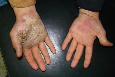
Acetonide injection in the treatment of chronic recalcitrant plaque psoriasis
enrolled in the study after stopping therapy for two MATERIALS & TECHNIQUES
months. All patients received one single session of triamcinolone
3. Patients with preexisting chronic illness such as acetonide intralesional treatment.
diabetes and hypertension and patients on systemic Triamcinolone acetonide suspension was used in a dilution
immunosuppressive medications were excluded from of 15-20 mg/ml (20 mg/ml were used only for very thick
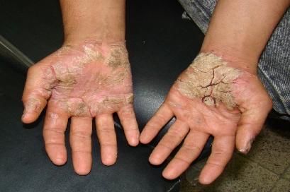
the study. hyperkeratotic lesions in palms and soles) using a small
4. A written consent and agreement was taken from the insulin syringe with a needle gauge 29, firmly locked so
patients or their parents (if they were children) that one that it won't disengage. For the pediatric age group (8-16
side will be injected during the study period and the years old) a total dose not exceeding 1mg/kg is used; for
other side will be injected after finishing the study. The adult patients a total dose not exceeding 80mg is used.
developments of side effects (atrophy, pigmentary Lesions were injected intradermally with 0.1-0.2ml of the
changes and infection) were also discussed with the diluted material in adjacent areas so as the whole lesion
patients or their parents. was infiltrated. Blood pressure and fasting blood sugar
Clinical assessment: The surface area of each plaque was levels were measured before initiation of therapy and after
measured separately pre and post injection by measuring one week of treatment. Follow up on a monthly basis for
the maximum length and width. up to 6 months was done to monitor the clinical response,
Statistical analysis: for determination of the statistical development of complications and to report side effects.
significance among different variables descriptive
statistics (like mean and standard deviation) were used RESULTS
together with analytic statistics which is t-test between two Of the 40 patients enrolled in this study; 22(55%) were
dependent means. males and 18(45%) were females. Their age ranged from
8-50 years with a mean of 27.65±13.17 [ table 1]. All
patients continued the 6 months period time of the study.
Most of the patients had 4 lesions (16 patients) [table2].
TABLE 1: showing age and gender distribution of patients.
No %
Age (years) 50years 1 2.5
Mean±SD (Range) 27.65±13.17 8-50
Gender Male 22 55.0
Female 18 45.0
TABLE 2: showing distribution of lesions
Site of lesion No %
Ear 1 1.2
Elbow 13 15.5
Dorsum of hand 5 6.0
Palm 15 17.9
Trunk 7 8.3
Knee 19 22.6
Ankle 9 10.7
Dorsum of foot 6 7.1
Sole 9 10.7
TABLE 3: showing total number of lesions.
Total number of lesions No. of patients %
2 12 30.0
4 16 40.0
6 8 20.0
7 1 2.5
8 3 7.5
The total number of lesions was 167, distributed mainly in 13.00±10.52 (1-40cm) and, after 6 months was
palms and on knees [table3] and only one patient had 13.00±10.52 (1-40cm). The mean lesion size in the
lesions in both external ears (choncae). Eighty four lesions treatment group was 12.05±10.05 (1-42cm) and 6 months
were used as a control group and 83 lesions as a treatment after treatment was 1.97±5.09 (0-30), with a P value
group. The mean lesions size for the control group was 0.0001 which is highly significant. The P value between
751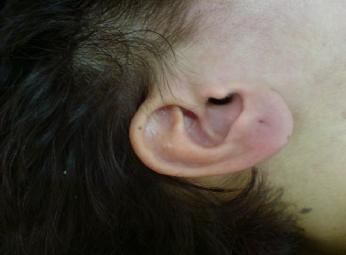
I.J.A.B.R., VOL. 2(4) 2012: 750-755                                                                              ISSN 2250 - 3579

the control and treatment groups in the beginning of the             was not significant between the two groups (treatment and
study was 0.076 which is not significant & it became                 control), where as it was highly significant for each site in
0.0001 after 6 months which is highly significant [table4            the treatment group pre and post injections for lesions on
and figure1]. The mean and the standard deviation were               the elbows, knees, palms and soles, dorsum of the hands
also measured for each site alone [table 5, figure 2 and             and ankles.
3].The P value for each site prior to the injection session

                TABLE 4: showing means lesion size of the control and treated groups pre and post injection.
                 P value          After 6 months                Before                    Size of lesion (cm)
                 -                13.00±10.52 (1-40)            13.00±10.52 (1-40)        Control group
                 0.0001*          1.97±5.09 (0-30)              12.05±10.05 (0-42)        Treated group
                                  0.0001*                       0.076                     P value
                                          -Data were presented as Mean±SD (Range)
                        *Significant using Students-t-test for difference between two dependent means

                   FIGURE1: showing mean lesion size of control and treated group pre and postinjection.

                  TABLE 5: showing mean lesion size of each site pre and post injection and control group.
       P value                         Size of lesion (cm)                                       Site of lesion
       Treat before X   Cont X treat   treated after      treated before      Control
       treat after      before         6 months
      -                -               0±                 7.00±               1.00±               Ear
      0.0001*          0.053           0.92±3.33          7.86±2.98           9.58±4.53           Elbow
      0.007*           0.629           0±                 2.14±0.95           2.48±1.43           Dorsum of hand
      0.0001*          0.247           1.33±5.16          17.41±8.17          19.79±11.65         Palm
      0.356               0.547             8.03±10.48          9.10±9.89         7.83±5.72         Trunk
      0.0001*             0.073             2.77±5.82           13.97±9.87        16.03±8.91        Knee
      0.048*              0.674             0.22±0.44           5.04±6.68         4.22±2.91         Ankle
      0.070               0.415             1.18±1.59           2.83±2.14         3.33±3.20         Dorsum of foot
      0.0001*             0.999             1.72±1.46           26.65±7.52        26.64±7.53        Sole
                                              -Data were presented as Mean±SD
                        *Significant using Students-t-test for difference between two dependent means

However, the P value was not significant for lesions on the          temporary and resolved in a few months. Only two
trunk and dorsum of the foot. There was only one patient             patients (5%) developed secondary infection in form of
who had lesions in both external ears (ear conchae) and it           abscess formation in the sole region 10-14 days after the
showed complete resolution after 6 months of one                     injection. Hypersensitivity reactions or panniculitis were
injection session. [Figure 4and 5]. All patients complained          not recorded [table6]. Most of the patients were satisfied
from pain and discomfort at the site of injections mainly in         with the outcome of the injection; (60%) showed full
the palms and soles. Ten patients (25%) and seven patients           satisfaction, (27.5%) were partially satisfied and only
(17.5%) developed atrophy and hypo pigmentation                      (12.5%) were not satisfied [table7].
respectively. Atrophy and hypo pigmentation were

                                                               752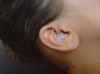
Acetonide injection in the treatment of chronic recalcitrant plaque psoriasis

   FIGURE 2: mean lesion size of each site of the control and treated pre and postinjection.

FIGURE 3: mean lesion size of each site of the control and treated group pre and postinjection.

                                TABLE 6: showing side effect
              %               No            Local side effects after 1 month
              100             40                  Pain
              25              10                   atrophy
              17.5            7                   Hypopigmentation
              5               2                   Abscess

                           TABLE 7: showing patient's satisfaction
                 %             No            Patient satisfaction
                60.0           24                    Fully satisfied
                27.5           11                    Partially satisfied
                12.5            5                    Not satisfied

   FIGURE 4: preinjection                            FIGURE 5: postinjection 2 months

                                             753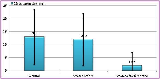
I.J.A.B.R., VOL. 2(4) 2012: 750-755                                                                                 ISSN 2250 - 3579

                  FIGURE 6: preinjection.                                         FIGURE 7: postinjection 4 months.

DISCUSSION                                                              and rapid relapsing rate, make it a last choice when other
Psoriasis is a common, chronic, relapsing, distressing skin             modalities fail, or the first choice when the patient desires
disease of unknown etiology that affects 2-3% of the                    to have it.
population1. Recently psoriasis is thought to be an                     Macgugan & co workers (1963) showed that a single
immunologically mediated disease where T-cells play an                  injection of triamcinolone acetonide induced adrenal
important role in its pathogenesis.6 until now;                         suppression as measured by a decrease in plasma cortisol
unfortunately, there is no unique curative systemic or                  concentration which persisted for up to 4 days; but in
topical treatment. Psoriasis for most patients is more                  contrast an injection of 25 mg triamcinolone diacetate or
emotionally than physically disabling. It is a disease that             50 mg triamcinolone acetonide produced only an
erodes the self image, causing shame and embarrassment                  occasional transient adrenal suppression. It was concluded
and forces the patient into a life of concealment and self              therefore that 25 mg or less is a safe dose.10 So after
consciousness.7Chronic stable psoriasis is “one of the                  discussion with the patients about the possibility of
miseries that beset mankind “; therefore even when the                  development of side effects, it was more acceptable for
patient has only a few asymptomatic, chronic plaques the                them to accept atrophy and hypo pigmentation than the
disease is more serious than it appears. Thus, when                     daily application of topical remedies which they use for
choosing a treatment regimen it is important to reconcile               several months with only partial response and rapid
the extent and measurable severity of the disease with the              relapse after discontinuation of treatment. All 40 patients
patient’s own perception of his or her disease. In this                 who were selected for this study had bilateral chronic
context it is notable that a recent study found that 40                 recalcitrant plaque psoriasis. All were treated for two
percent of patients felt frustrated with the ineffectiveness            months with calcipotriol and clobetasol propionate and,
of their current therapies, and 32%reported that treatment              showed either no response or relapsed immediately after
was not aggressive enough.5 Intralesional injections with               discontinuation of therapy. One side was used as a control
triamcinolone acetonide( 3-10 mg/ml) is one of the known                side and other side as the treatment side. The mean lesion
treatment options recommended for small resistant lesions               size in the treated side after 6 months from a single
for many years.1 The mechanism of action of                             injection was significantly lower when compared with the
corticosteroid has specific and non specific effects. These             control side, and from the mean lesion size in the same
different mechanisms of action, include: anti                           side before injection, with a P value 0.0001. P value for
inflammatory, immunosuppressive, antiproliferative and                  each site was highly significant 0.0001 for lesions on the
vasoconstrictive effects.8                                              palm and sole, elbow, and knee regions. Whereas lesions
Quantitative measurements of surface areas of corneocytes               on the trunk usually relapsed within 6 months of the
from desequemative portion of psoriatic lesion showed                   injection. Atrophy occurred in 25% of the patients and in
that the surface areas of corneocyte gradually increase in              most was temporary and resolved within few months.
size following one intralesional injection of triamcinolone             Most of the patients were satisfied with the injection and
acetonide.9 From these facts and the fact that most patients            they always asked about the next injection. Controlled
accept atrophy rather than the hyperkeratotic disfiguring               trials with Excimer- laser treatment also showed effective
and disabling lesions, we decided to increase the                       and promising results for localized plaques1, but are much
concentration of the triamcinolone and so increase these                more costly are require repeated sessions of treatment. So
pharmacological and clinical effects with possible                      intralesional triamcinolone acetonide 15-20 mg/ml is an
increased atrophy. Usually dermatologist use10 mg/ml to                 ideal way to treat chronic plaque psoriasis resistant to
avoid or minimize atrophy, and since the goal of our study              other form of therapy except for trunkal lesions which
was to induce atrophy so we increased the concentration of              showed a high relapse rate.
triamcinolone acetonide to (15-20) mg/ml injected at the
thickened hyperkeratotic lesions mostly palms and soles,                REFERENCES
in order to induce rapid clinical improvement at these                   [1]. Griffiths C.E.M, Barker J. N. W. N. Psoriasis, In:
sites. The rapid, long lasting effects of intralesional steroid               Burns DA, Breathnach SM, Cox NH and Griffiths
in comparison to other topical modalities like tar,                           CEM ( eds). Rook's Textbook of Dermatology, eight
anthraline, topical steroids…etc, which need daily                            edition, London; Blackwell L td; 2010, 20: 20.1-
application with their messy, clothes discoloring effects                     20.60.

                                                                  754
Acetonide injection in the treatment of chronic recalcitrant plaque psoriasis
[2]. Asha G pardasani, Steven R. Feldman: Treatment immunomodulators and systemic therapies;
of psoriasis: An Algorithm-based primary care U.S.Experience. Br J Dermatol 2004,151(1):3-15.
physicians. American Family Physicians.2000; 1;
16(3):725-733. [7]. HabifTP,.Psoriasis and Other Papulosquamous
Diseases, In:Habif T.P(eds). Clinical Dermatology,
[3]. Al Nuamy S. Epidemiology and HLA typing of fifth edition, Mosby –Elsevier inc;2010,8:264-308.
psoriasis in Iraq. A study submitted to University of
Baghdad, College of Medicine. Diploma Thesis [8]. Isabel C. Valencia, Francisco A. Kerdel. Topical
1988; 45. corticosteroid, In: Wolff, Klaus; Gold Smith,
Lowell A.; Katz, Stephen I.; Gilchrest, Barbara A,;
[4]. Wolff K., Johnson R.A., Psoriasis, In: Wolff K., Paller, Amy S; Leffell, David J(eds). Fitzpatrick's
Johnson R.A., (Eds). Fitzpatrick’s color Atlas & Dermatology in General Medicine. Seventh
Synopsis of Clinical Dermatology, sixth edition, edition;Mc Graw Hill. Philadelphia, 2008,
New York ;Mc Graw Hill;2009,4: 53-71. 216:2103-2106.
[5]. Gudjonsson J E, Elder JT ,.psoriasis In: Wolff [9]. Herbert Goldschmidt: Surface area measurements
Klaus, Gold Smith, Lowell A.; Katz, Stephen I.; of psoriatic corneocytes: effects of intralesional
Gilchrest, Barbara A,; Paller Amy S; Leffell David, steroid therapy. Journal of investigative
J. (Eds). Fitzpatrick's Dermatology in General dermatology1979, 73:558-560.
Medicine. Seventh edition; Mc Graw Hill.
Philadelphia, 2008; 18:169-193. [10]. Teik K.O, Poh F.W:Treatment of various
dermatoses by intralesional injection of
[6]. T Kormeili N J, Lowe p s, Yamauchi. Psoriasis : Triamcenolone acetonide.A clinical trial of 23
Immunopathogenesis and Evolving cases. Singapore Medical journal 1965;4: 107-109.
755You can also read