Accuracy of Scheimpflug Holladay equivalent keratometry readings after corneal refractive surgery
←
→
Page content transcription
If your browser does not render page correctly, please read the page content below
ARTICLE

   Accuracy of Scheimpflug Holladay equivalent
        keratometry readings after corneal
                 refractive surgery
   Qiongyan Tang, MD, Kenneth J. Hoffer, MD, Michael D. Olson, OD, PhD, Kevin M. Miller, MD

                 PURPOSE: To determine the accuracy of Pentacam Scheimpflug system Holladay equivalent kera-
                 tometry (K) readings (EKRs) in calculating intraocular lens (IOL) power after corneal refractive sur-
                 gery, including laser in situ keratomileusis (LASIK), photorefractive keratectomy (PRK), and radial
                 keratotomy (RK).
                 SETTING: Jules Stein Eye Institute, David Geffen School of Medicine at University of California, Los
                 Angeles, Los Angeles, California, USA.
                 METHODS: In this combined retrospective and prospective clinical study, patients who had cataract
                 surgery after corneal refractive surgery were recalled to have Scheimpflug imaging of the operated
                 cornea and Holladay EKR determination. The Holladay EKR was compared with a gold-standard K
                 value, which was the back-calculated value using the original Hoffer formula based on the actual
                 surgical outcomes. Eyes without a history of refractive surgery served as controls.
                 RESULTS: Twenty-seven patients (41 eyes) were evaluated; 26 eyes had previous LASIK or PRK and
                 15, previous RK. Forty-one eyes served as controls. The mean error of the Holladay EKR in eyes with
                 previous LASIK or PRK was C1.84 diopters (D) (range C0.66 to C4.94 D). The mean error in eyes
                 with previous RK was C2.17 D (range C0.48 to C3.09 D). In the control eyes, the mean EKR error
                 was C1.38 D (range 0.17 to C2.54 D).
                 CONCLUSIONS: The Holladay EKR calculated using version 1.16r04 of the Scheimpflug system
                 software was inaccurate in virgin corneas and in those with a history of LASIK, PRK, or RK using
                 current IOL power calculation formulas. The Scheimpflug power measurements were consistently
                 steeper than the true corneal power.
                 J Cataract Refract Surg 2009; 35:1198–1203 Q 2009 ASCRS and ESCRS

Precise intraocular lens (IOL) power calculation after                estimation accuracy to an extent, postoperative refrac-
laser in situ keratomileusis (LASIK), photorefractive                 tive surprises still occur.
keratectomy (PRK), and radial keratotomy (RK) is                         The ideal method for measuring corneal K is directly
a worldwide challenge for cataract surgeons. The pri-                 using a device that works independently of refractive
mary problem is obtaining the true corneal or kera-                   surgery information. Standard topography and kera-
tometry (K) power after the cornea has been                           tometry are generally accurate in virgin eyes but inac-
surgically altered. In the setting of previous myopic                 curate after refractive surgery because they are blind to
keratorefractive surgery, eyes usually end up hyper-                  the center of the cornea and do not measure the contri-
opic after cataract surgery unless specific measures                  bution of the posterior corneal surface. Most topogra-
are taken to compensate for surgical changes in cor-                  phers and keratometers also depend on a standard
neal curvature. Various methods for K estimation                      corneal index of refraction that may have been altered
have been developed1–3; these include the clinical his-               by refractive surgery.
tory method,4,5 contact lens method,4,6 vertexed IOL                     The Pentacam (Oculus, Optikgera GmbH) is a rotat-
power method,7 and Ianchulev intraoperative autore-                   ing single Scheimpflug camera system. It measures an-
fraction method.8 There are also many methods for ad-                 terior and posterior corneal surface elevations and
justing the IOL power calculation to make up for the                  corneal thickness and computes anterior and posterior
error in K, such as the Aramberri double-K technique.9                curvatures to obtain the net corneal power. In the
Although most of these methods improve K                              system’s software version 1.16r04, the Holladay

1198   Q 2009 ASCRS and ESCRS                                                                             0886-3350/09/$dsee front matter
       Published by Elsevier Inc.                                                                            doi:10.1016/j.jcrs.2009.02.030
SCHEIMPFLUG HOLLADAY K READINGS AFTER REFRACTIVE SURGERY                                               1199

equivalent K reading (EKR) report, developed in coop-
                                                                           Table 1. Clinical and surgical characteristics of the study
eration with Jack T. Holladay, MD, uses data from the                      patients.
4.5 mm optical zone. Theoretically, this Scheimpflug
system seems promising for measuring the true cor-                         Characteristic                                            Value
neal power after keratorefractive surgery. The purpose
                                                                           Sex, n (%)
of this study was to determine the accuracy of the sys-                      Female                                                 19 (80.5)
tem’s Holladay EKR in calculating IOL power after                            Male                                                    8 (19.5)
LASIK, PRK, and RK.                                                        Age (y)
                                                                             Mean                                                     64.4
                                                                             Range                                                   37–79
PATIENTS AND METHODS                                                       Refractive surgery, n (%)
                                                                             Myopic
After institutional review board approval, consecutive pa-
                                                                                LASIK                                               18 (43.9)
tients who had cataract surgery performed by the same sur-
                                                                                PRK                                                  2 (4.9)
geon (K.M.M.) after they had keratorefractive surgery at the
Jules Stein Eye Institute or elsewhere were recalled for addi-                  RK                                                  15 (36.6)
tional testing. Consecutive patients who had cataract sur-                   Hyperopic
gery without previous refractive surgery were also recalled                     LASIK                                                6 (14.6)
to serve as controls.                                                      Time from refractive surgery
   To be included in the study, patients had to be 21 years or             to cataract surgery (mo)
older and have a postoperative corrected distance visual acu-                Mean                                                     65.9
ity of 20/40 or better so that an accurate postoperative refrac-             Range                                                   23–239
tion could be obtained. The LASIK and PRK groups were                      Time from cataract surgery
combined in the analysis because the procedures change
                                                                           to Scheimpflug measurement (mo)
the anterior cornea without affecting the posterior cornea.
                                                                             Mean                                                     23.8
In contrast, RK changes both surfaces simultaneously.
   Cataract surgery was by phacoemulsification. In most                      Range                                                  0.5–70.5
cases, peripheral corneal relaxing incisions were created at               LASIK Z laser in situ keratomileusis; PRK Z photorefractive keratec-
the time of cataract surgery. In the practice of the surgeon,              tomy; RK Z radial keratotomy
a phacoemulsification incision placed on the steep axis is
considered to be a relaxing incision. Relaxing incisions re-
duce or modify astigmatism but have little effect on the
spherical equivalent power of the cornea postoperatively.                the corneal vertex to the anterior IOL vertex was measured
   Patients had 3 tests at the time of the recall visit: (1)             with the pachymeter.
Scheimpflug imaging of the operated cornea, (2) manifest re-                The principal plane of the IOL, also known as the effective
fraction in the operated eye using a phoropter and the Jack-             lens position (ELP), was computed by adding 50% of the
son cross-cylinder technique, and (3) anterior chamber depth             manufacturer-reported central thickness of the implanted
(ACD) measurement using an optical pachymeter (Optical                   IOL to the ACD measured by optical pachymetry. Based
Pachymeter II, Haag-Streit International).10 The distance from           on the axial length (AL) measured preoperatively by immer-
                                                                         sion A-scan ultrasound, the measured ELP, the power of the
                                                                         implanted IOL, and the postoperative refraction, a gold-
                                                                         standard K value was back-calculated for each eye using
Submitted: July 28, 2008.                                                the original 1974 Hoffer formula11 (ie, without the Q for-
Final revision submitted: February 18, 2009.                             mula, which manipulates the ACD value.) The mean Holla-
Accepted: February 22, 2009.                                             day EKRs from the Scheimpflug system were compared with
From the Department of Ophthalmology (Tang, Hoffer, Olson,
Miller), David Geffen School of Medicine at UCLA and the Jules
Stein Eye Institute, Los Angeles, California, USA; Department of
                                                                           Table 2. Clinical characteristics of the control patients.
Ophthalmology (Tang), Union Hospital, Tongji Medical College,
Huazhong University of Science and Technology, Wuhan, China.               Characteristic                                            Value
No author has a financial or proprietary interest in any material or       Sex, n (%)
method mentioned.                                                            Female                                                 21 (72.4)
Presented in part at the ASCRS Symposium on Cataract, IOL and                Male                                                    8 (27.6)
Refractive Surgery, Chicago, Illinois, USA, April 2008.                    Age (y)
                                                                             Mean                                                     69.3
Supported by unrestricted gifts from John A. Lyddon and the Carl             Range                                                   50–81
and Roberta Deutsch Foundation.                                            Time from cataract surgery
                                                                           to Scheimpflug measurement (mo)
Corresponding author: Kevin M. Miller, MD, Jules Stein Eye Insti-            Mean                                                      5.0
tute, 100 Stein Plaza, UCLA, Los Angeles, California 90095-7002,             Range                                                  0.5–35.5
USA. E-mail: kmiller@ucla.edu.

                                                 J CATARACT REFRACT SURG - VOL 35, JULY 2009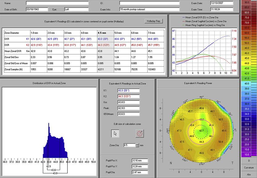
1200 SCHEIMPFLUG HOLLADAY K READINGS AFTER REFRACTIVE SURGERY
Table 3. Intraocular lenses implanted in study and control eyes.
Study Eyes (n)
Center Thickness
IOL Model (Manufacturer) of 20.0 D IOL (mm) IOL Design m-LASIK h-LASIK m-PRK RK Control Eyes (n)
SA60AT (Alcon) 0.625 Equiconvex 2 2 0 3 1
SN60WF (Alcon) 0.590 Asymmetrically biconvex 3 0 0 0 20
SN60D3 (Alcon) 0.550 Asymmetrically biconvex 0 0 0 0 6
SN6AD3 (Alcon) 0.550 Asymmetrically biconvex 0 0 0 0 11
CC4204BF (Staar) 1.251 Equiconvex 13 3 2 12 3
ZA9003 (Abbott Medical Optics*) 0.959 Asymmetrically biconvex 0 1 0 0 0
h-LASIK Z hyperopic laser in situ keratomileusis; IOL Z intraocular lens; m-LASIK Z myopic laser in situ keratomilsusis; m-PRK Z myopic photorefractive
keratectomy
*Formerly Advanced Medical Optics
the back-calculated Hoffer formula gold-standard K values mean Holladay EKR in the 26 eyes that had LASIK
using the paired t test. or PRK showed a difference ranging from C0.66 to
C4.94 D. In the 15 eyes that had RK, the difference
RESULTS ranged from C0.48 to C3.09 D. On average, the
The study group comprised 27 patients (41 eyes); 24 Scheimpflug system overestimated the true power of
eyes were post-LASIK, 2 were post-PRK, and 15 the central cornea in study eyes by approximately
were post-RK. The control group comprised 29 consec- 2.00 D. Subtracting the mean back-calculated K value
utive patients (41 eyes). Table 1 shows the characteris- from the mean Holladay EKR in the 41 control eyes
tics of the study group and Table 2, of the control yielded a difference ranging from 0.17 to C2.54 D.
group. All study eyes and more than 90% of control The differences were statistically significant in all sub-
eyes received peripheral corneal relaxing incisions at groups (P!.001, paired t test).
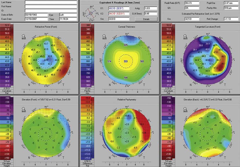
the time of cataract surgery. Figure 1, A, shows a plot of the Scheimpflug Holla-
Table 3 shows the IOL models implanted in study day EKRs for the post-LASIK and post-PRK eyes ver-
eyes and control eyes. All IOLs were biconvex. Equi- sus the back-calculated Hoffer K values. All data in the
convex IOLs were implanted in 90.2% of study eyes study eyes fall below the solid line in the figure, indi-
and 9.8% of control eyes. Asymmetrically biconvex cating overestimation of the true back-calculated cor-
IOLs were implanted in the remainder of eyes. The ac- neal power by the Scheimpflug system. Note that as
curacy of ELP calculation was greatest in the eyes with the K readings become steeper, the distance between
an equiconvex IOL. The maximum error can be no the lines representing the best-fit linear regression
greater than lens thickness divided by 2 for the asym- and the perfect correspondence decreases. As the cor-
metrically biconvex IOLs. nea becomes flatter, the Scheimpflug system error in-
Table 4 shows the Holladay EKR values, back-calcu- creases. Figure 1, B, shows a plot of the same data
lated true corneal powers, and mean differences. Sub- using the Bland-Altman method. Figure 2 shows plots
tracting the mean back-calculated K value from the of the data in the 15 post-RK eyes and Figure 3, in the
Table 4. Holladay EKR values, back-calculated true corneal powers, and mean differences.
Mean (D)
Group/Subgroup Number Holladay EKR Back-Calculated Corneal Power Difference Error Range (D) P Value
Study
LASIK and PRK 26 41.5 39.7 C1.84 C0.66 to C4.94 !.001
RK 15 39.0 36.9 C2.17 C0.48 to C3.09 !.001
Combined 41 40.6 38.6 C1.96 C0.48 to C4.94 !.001
Control 41 44.2 42.8 C1.38 0.17 to C2.54 !.001
EKR Z equivalent keratometry reading; LASIK Z laser in situ keratomileusis; PRK Z photorefractive keratectomy; RK Z radial keratotomy
J CATARACT REFRACT SURG - VOL 35, JULY 2009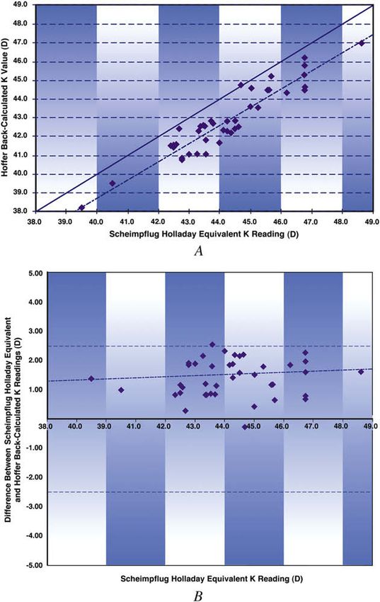
SCHEIMPFLUG HOLLADAY K READINGS AFTER REFRACTIVE SURGERY                                            1201

Figure 1. A: Pentacam Holladay EKRs versus Hoffer back-calculated        Figure 2. A: Pentacam Holladay EKRs versus Hoffer back-calculated
K values in 26 eyes with a history of preoperative LASIK or PRK. The     K values in the 15 eyes with a history of preoperative RK. The solid
diamond-shaped data points represent eyes that had myopic LASIK.         line designates perfect correspondence. The dashed line is a best-fit
The round data points represent eyes that had hyperopic LASIK. The       linear regression. B: The same data as in A shown in a Bland-Altman
solid line designates perfect correspondence. The dashed line is         plot (K Z keratometry).
a best-fit linear regression. B: The same data as in A shown in a
Bland-Altman plot (K Z keratometry).
                                                                         that have had keratorefractive surgery. Given cur-
                                                                         rently available IOL power calculation formulas, the
41 control eyes. The Scheimpflug system overesti-                        measurements it reports are inaccurate.
mated the true power of the cornea in all but 1 eye in                      Lackerbauer et al.12 found the Pentacam system to
the control group.                                                       be more accurate than keratography in estimating cen-
   Figure 4 shows a Holladay report from the                             tral corneal power after myopic LASIK; however, the
Scheimpflug system for a post-LASIK eye. The 6                           authors did not apply their findings to IOL power cal-
maps show typical post-LASIK findings, including                         culation. Borasio et al.13 used anterior and posterior
reduced central pachymetry, flattening of central                        curvature and corneal thickness data from the Penta-
anterior corneal curvature, and normal posterior el-                     cam system in their BESSt formula and found the ap-
evation. The mean EKR (43.8 D) in the 4.5 mm op-                         proach to be more accurate than several other
tical zone is displayed in the upper central panel.                      methods. However, they also found a consistent cor-
Figure 5 shows a detailed Holladay report for the                        neal power error of C1.30 D, similar to our 1.38 D er-
same eye.                                                                ror in control eyes. In their paper, the authors state
                                                                         that ‘‘the Gaussian optics formula consistently under-
DISCUSSION                                                               estimated corneal values by 1.30 D on average, indi-
The Pentacam Holladay report software is intended to                     cating that either K values measured with corneal
improve corneal power estimation, especially in eyes                     topography in virgin eyes overestimate corneal power

                                                 J CATARACT REFRACT SURG - VOL 35, JULY 2009
1202                                SCHEIMPFLUG HOLLADAY K READINGS AFTER REFRACTIVE SURGERY

                                                                           by more than 1.00 D or anterior or posterior corneal
                                                                           curvatures measured with the Pentacam are not
                                                                           correct.’’
                                                                              In this study, we used the Hoffer back-calculated
                                                                           K value as our gold standard because this formula
                                                                           was developed on the basis of the Gullstrand eye
                                                                           model without altering the ACD in the formula,
                                                                           which is in contrast to other third-generation
                                                                           formulas (Haigis, Hoffer Q, Holladay 1 and 2,
                                                                           and SRK/T). The latter formulas alter or ‘‘fudge’’
                                                                           the actual ELP and would produce erroneous
                                                                           results if used for this purpose. The optical pachy-
                                                                           meter we used to measure ACD in our study was
                                                                           also designed according to the optical principles
                                                                           of the Gullstrand model eye and should theoreti-
                                                                           cally be more accurate than ultrasound or other
                                                                           imaging modalities. Our gold-standard K was
                                                                           back-calculated based on surgical outcomes data,
                                                                           including AL, ELP, implanted IOL power, and post-
                                                                           operative refraction. According to the first-order
                                                                           optics Gullstrand eye model, this back-calculated
                                                                           K value should best reflect true postoperative
                                                                           corneal power.
                                                                              If the mean C1.38 D error found in the control eyes
                                                                           is used to offset the measurements obtained in the eyes
                                                                           that had previous refractive surgery, sizable relative
                                                                           errors of C0.46 D for post-LASIK and post-PRK eyes
                                                                           and C0.79 D for post-RK eyes continue to be present,
                                                                           indicating that a quick fix to the Pentacam system is
                                                                           not possible. The inaccuracy of the measurements
                                                                           with the Scheimpflug system increased as the change
Figure 3. A: Pentacam Holladay EKRs versus Hoffer back-calculated          in corneal power induced by keratorefractive surgery
K values in the 41 normal control eyes. The solid line designates per-     increased, as shown by the divergence of the lines in
fect correspondence. The dashed line is a best-fit linear regression.      Figures 1 and 2. Conversely, there was little
B: The same data as in A shown in a Bland-Altman plot
(K Z keratometry).

                                                                                          Figure 4. A Scheimpflug system Holladay report for
                                                                                          a post-LASIK eye (software version 1.16r04).

                                                   J CATARACT REFRACT SURG - VOL 35, JULY 2009
SCHEIMPFLUG HOLLADAY K READINGS AFTER REFRACTIVE SURGERY 1203
Figure 5. A detailed Holladay report showing addi-
tional data for the eye in Figure 4. Upper left panel:
The EKRs in different optical zones. Lower left panel:
A histogram of EKR powers in the 4.5 mm zone.
divergence in the lines as a function of corneal steep- 6. Holladay JT. Cataract surgery in patients with previous kerator-
ness or flatness for the virgin corneas (Figure 3, A). efractive surgery (RK, PRK, and LASIK). Ophthalmic Pract
1997; 15:238–244
In a letter to the editor, Norrby14 suggested that the 7. Feiz V, Mannis MJ, Garcia-Ferrer F, Kandavel G,
‘‘problem’’ with the Pentacam system might lie in IOL Darlington JK, Kim E, Caspar J, Wang JL, Wang W. Intraocular
power formulas that use paraxial (thin and thick lens) lens power calculation after laser in situ keratomileusis for
optics and IOL constants and not with the Scheimp- myopia and hyperopia; a standardized approach. Cornea
flug system itself. He theorizes that the Scheimpflug 2001; 20:792–797
8. Odenthal MTP, Eggink CA, Melles G, Pamayer JH,
measurements may be accurate and that current IOL Geerards AJM, Beekhuis WH. Clinical and theoretical results
power calculation formulas may have to be adjusted of intraocular lens power calculation for cataract surgery after
to accept the system’s more accurate K readings. He photorefractive keratectomy for myopia. Arch Ophthalmol
recommends replacing simple paraxial optics formu- 2002; 120:431–438
las with ray-tracing approaches to improve IOL power 9. Aramberri J. Intraocular lens power calculation after corneal
refractive surgery: double-K method. J Cataract Refract Surg
calculation. 2003; 29:2063–2068
In summary, our study found that the Holladay 10. Hoffer KJ. To the editor [letter regarding lens power calculation
EKR, calculated by using version 1.16r04 of the Penta- and the problem of the short eye]. Ophthalmic Surg 1982;
cam software, was inaccurate in virgin corneas and 13:962
corneas with a history of LASIK, PRK, or RK using cur- 11. Hoffer KJ. The Hoffer Q formula: a comparison of theoretic and
regression formulas. J Cataract Refract Surg 1993; 19:700–712;
rent-generation IOL power calculation formulas. The errata 1994; 20:677
system should be used with caution as a sole instru- 12. Lackerbauer CA, Hartmann L, Frömlich S, Schaumberger M,
ment for determining corneal power in post-refractive Kollias A. Evaluation der zentralen Hornhautbrechkraft nach my-
surgery eyes. The Holladay EKR consistently mea- oper LASIK. [Measurement of the central corneal power after
sured a steeper central power than true corneal power myopic LASIK]. Ophthalmologe 2008; 105:60–65
13. Borasio E, Stevens J, Smith GT. Estimation of true corneal
based on paraxial optics and surgical outcomes data. power after keratorefractive surgery in eyes requiring cataract
surgery: BESSt formula. J Cataract Refract Surg 2006;
32:2004–2014
14. Norrby S. Pentacam keratometry and IOL power calculation
REFERENCES
[letter]. J Cataract Refract Surg 2008; 34:3
1. Hamilton DR, Hardten DR. Cataract surgery in patients with prior
refractive surgery. Curr Opin Ophthalmol 2003; 14:44–53
2. Speicher L. Intra-ocular lens calculation status after corneal re-
fractive surgery. Curr Opin Ophthalmol 2001; 12:17–29 First author:
3. Seitz B, Langenbucher A. Intraocular lens power calculation in Dr. Qiongyan Tang
eyes after corneal refractive surgery. J Refract Surg 2000;
16:349–361 Department of Ophthalmology, David
4. Holladay JT. Consultations in refractive surgery [comment]. Geffen School of Medicine at UCLA and
Refract Corneal Surg 1989; 5:203 the Jules Stein Eye Institute,
5. Hoffer KJ. Intraocular lens power calculation for eyes after Los Angeles, California, USA
refractive keratotomy. J Refract Surg 1995; 11:490–493
J CATARACT REFRACT SURG - VOL 35, JULY 2009You can also read