Dermatoscopic pitfalls in differentiating pigmented Spitz naevi from cutaneous melanomas
←
→
Page content transcription
If your browser does not render page correctly, please read the page content below
British Journal of Dermatology 1999; 141: 788±793.

Dermatoscopic pitfalls in differentiating pigmented Spitz naevi
from cutaneous melanomas
                    G.ARGENZIANO, M.SCALVENZI, S.STAIBANO,* B.BRUNETTI,² D.PICCOLO,³
                    M.DELFINO, G.DE ROSA* AND H.P.SOYER§
                    Departments of Dermatology and *Biomorphological and Functional Sciences, Pathology Section, Federico II University of Naples,
                    Via S. Pansini 5, 80131 Naples, Italy
                    ²The Salerno 2nd Health Care District, Salerno, Italy
                    ³Department of Dermatology, University of L'Aquila, Italy
                    §Department of Dermatology, University of Graz, Austria
                    Accepted for publication 3 June 1999

Summary             Epiluminescence microscopy (ELM, skin surface microscopy, dermoscopy, dermatoscopy) is a
                    valuable method for improving the diagnostic accuracy in pigmented skin lesions. Specific ELM
                    criteria are already recognized for differentiating pigmented Spitz naevi (PSN) from cutaneous
                    melanomas (CM). Our purpose was to describe the ELM appearance of a series of PSN with emphasis
                    on PSN and CM with overlapping features. Thirty-six consecutive patients with PSN, and three
                    cases of CM (selected from a larger database) exhibiting ELM `spitzoid' features, were evaluated
                    clinically, dermatoscopically and histopathologically. Most PSN (27 of 36; 75%) displayed two
                    typical ELM patterns, namely, the starburst (19 of 36; 53%) or the globular pattern (eight of 36;
                    22%), which were correlated to different histopathological findings. In nine of 36 (25%) PSN,
                    atypical ELM features which are more commonly seen in CM were observed. These PSN with an
                    atypical pattern were characterized by an uneven distribution of colours and structures, and an
                    irregular diffuse pigmentation resembling blue±white veil or irregular extensions (black blotches).
                    These atypical lesions mostly occurred in children and showed no history of growth. In contrast, in
                    three examples of CM, the typical ELM criteria of malignancy were less recognizable and either the
                    characteristic starburst or globular pattern usually seen in PSN was present. These three lesions
                    occurred in adults and had a recent history of change in colour, shape or size. The overlap in ELM
                    features of some PSN and CM represents a major diagnostic pitfall when ELM examination is
                    considered alone. In these atypical cases, clinical history including the age of the patient may be the
                    only clue to enable a correct diagnosis.
                    Key words: clinical diagnosis, dermatoscopy, epiluminescence microscopy, melanoma, pigmented
                    Spitz naevi, skin surface microscopy

Epiluminescence microscopy (ELM, skin surface micro-                        cutaneous melanomas (CM).2,3,6 PSN can be easily
scopy, dermoscopy, dermatoscopy, magnified oil immer-                       identified by a prominent symmetrical starburst or
sion diascopy) is an in vivo, non-invasive technique that                   globular pattern, with a central, bizarre or reticular
has disclosed a new dimension of clinical morphology                        depigmentation, and a rim of brown globules at the
in pigmented skin lesions. Different incident light                         periphery, which in some instances may mimic pseudo-
magnification systems with an oil immersion technique                       pods. In contrast, CM are asymmetrical and irregularly
have been used for performing this investigation.1                          pigmented with variable combinations of broadened
Previous studies have demonstrated that use of ELM                          pigment network, blue±white veil, irregular black dots
improves the clinical accuracy in diagnosing pigmented                      or brown globules, peripheral depigmentation, irregu-
skin lesions.2±5 Specific ELM criteria have been described                  lar extensions (black blotches) as well as `true' pseudo-
for differentiating pigmented Spitz naevi (PSN) from                        pods and radial streaming at the edge of the lesion.7,8
                                                                            The purpose of this study was to describe the ELM
Correspondence: Giuseppe Argenziano. E-mail: argenziano@tin.it

788                                                                                                 q 1999 British Association of Dermatologists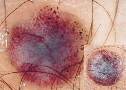
DERMATOSCOPY IN SPITZ NAEVI 789

appearance of a series of PSN in correlation with their
histopathological findings and to focus on PSN and CM
with overlapping features.

Subjects and methods
In this study, 36 consecutive patients (15 men and 21
women aged 5±43 years, mean 21´3) with PSN, and
three patients with CM, were included. These CM were
selected from a larger database of lesions (122 CM
which underwent a complete clinical, dermatoscopic
and histopathological examination) because they
exhibited `spitzoid' ELM features.6 All these lesions
underwent careful clinical evaluation, and all were
also examined with a stereomicroscope (Wild M650,
Heerbrugg AG, Switzerland) using immersion oil and a
glass slide (to render the epidermis translucent). All
lesions were photographed using either Dermaphot                         Figure 1. Starburst pattern of a pigmented spindle-cell naevus (Reed
                                                                         naevus) (original magnification  16). Insert: clinical appearance
equipment (Heine Optotechnik, Herrsching, Germany),
at a fixed magnification of  10, or a Nikon F3 camera
mounted on the stereomicroscope, with six- to 40-fold                    pods at the periphery, was observed. Because of their
magnification.                                                           characteristic dermatoscopic appearance, all these
   Each lesion was carefully examined for the evalu-                     lesions were correctly classified as PSN. On clinical
ation of the standard ELM criteria, most of which were                   examination by naked eye, however, only 12 of 19
listed in the guidelines of the Consensus Meeting held                   lesions were diagnosed as PSN, whereas seven of 19
in Hamburg in 1989.7,8 The evaluation of ELM criteria                    lesions were considered clinically atypical.
was carried out with the consensus of at least two of                       Histopathologically, most of the lesions of this first
three investigators (G.A., M.S., B.B.). Following clinical               group of cases exhibited the morphological findings of
and dermatoscopic examination, all lesions were                          pigmented spindle-cell naevus (Reed naevus), namely,
excised and diagnosed histopathologically. All slides                    symmetrical and well-circumscribed proliferations of
were evaluated by two pathologists (G.D.R., S.S.) using                  spindle-shaped melanocytes involving the epidermis
a double-headed light microscope and the final diag-                     and/or papillary dermis.9±11 The spindle-shaped melano-
nosis was the result of their complete agreement.                        cytes were arranged in fascicles closely packed along
                                                                         the dermoepidermal junction. In addition, large amounts
Results
By ELM examination, the PSN could be divided into
three groups.

Group 1
Nineteen of 36 (53%) cases of PSN showed a typical
starburst pattern with symmetrical, prominent, grey±
blue to black pigmentation, and central, bizarre or
reticular hypopigmentation. Most of these lesions
exhibited a characteristic rim of grey±brown to black
globules regularly distributed at the periphery, mimick-
ing pseudopods or radial streaming (Fig. 1). In addi-
tion, seven cases showed a regular and prominent
pigment network, whereas in three cases, only the                        Figure 2. Globular pattern of a spindle- and/or epithelioid-cell
prominent black±blue pigmentation, with no pseudo-                       naevus (Spitz naevus) (original magnification 16).

q 1999 British Association of Dermatologists, British Journal of Dermatology, 141, 788±793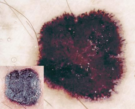
790 G.ARGENZIANO et al.
Table 1 Clinical data, dermatoscopic findings, and preoperative and histopathological diagnoses of pigmented Spitz naevi (PSN) with atypical
epiluminescence microscopy (ELM) patterns
Overall
Patient Sex/age preoperative
no. (years) Location Clinical history ELM features diagnosisa Histopathology
1 F/11 Upper limb No change Uneven distribution of colours and structures and PSN Reed naevus
blue±white veil (Fig. 3)
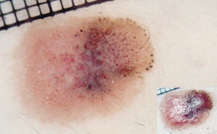
2 F/8 Lower limb No change Blue±white veil and irregular black blotches (Fig. 4) PSN Spitz naevusb
3 F/6 Lower limb No change Asymmetry of colours and structures, PSN Spitz naevus
blue±white veil and irregular black dots
4 M/12 Back No change Blue±white veil, irregular prominent pigment PSN Reed naevus
network and depigmented areas
5 F/13 Upper limb No change Uneven distribution of colours and structures PSN Spitz naevusb
and radial streaming
6 M/29 Lower limb No change Blue±white veil and irregular black dots (Fig. 5) PSN vs. Reed naevus
melanoma
7 M/43 Back No change Irregular pigment network and grey±blue areas Atypical Spitz naevus
naevus vs.
melanoma
8 F/37 Buttock Colour change Irregular black blotches and radial streaming Melanoma Reed naevus
9 F/32 Lower limb Change in size Blue±white veil and irregular brown globules Melanoma Reed naevus
a b
Independently of the preoperative diagnosis, all these lesions were excised because of the atypical ELM appearance. Case with atypical
histopathological features.
of melanin and numerous melanophages were present exhibit a radial (or stellate) appearance. Brown to
in the papillary dermis. grey±blue globules and dots often extended throughout
the entire surface of the lesion (Fig. 2). In two less
Group 2 pigmented lesions a dotted vascular pattern was also
detectable. By means of clinical examination these
A second group of PSN (eight of 36; 22%) had ELM lesions were classified as PSN (four of eight) or Clark
features of a symmetrical, basically globular pattern naevus (four of eight), whereas on ELM examination all
with regular, discrete, brown to grey±blue pigmenta- these lesions were correctly diagnosed as PSN.
tion in the centre, and a characteristic rim of large The histopathological correlates of this second group
brown globules at the periphery. In contrast to the of lesions were again similar, mostly revealing typical
starburst pattern, the globules at the periphery did not
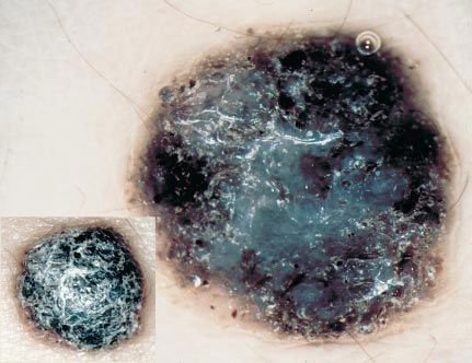
Figure 3. Atypical epiluminescence microscopy features with an Figure 4. Atypical epiluminescence microscopy features character-
uneven distribution of colours and structures, blue±white veil, and ized by blue±white veil and irregular black blotches. The histopatho-
dotted vascular pattern. The histopathological examination revealed logical examination revealed a Spitz naevus (original magnification
a Reed naevus (original magnification 16). 10).
q 1999 British Association of Dermatologists, British Journal of Dermatology, 141, 788±793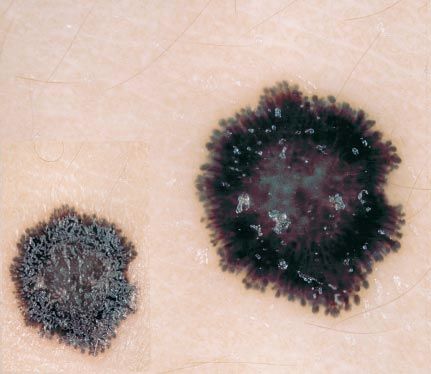
DERMATOSCOPY IN SPITZ NAEVI 791

                                                                         brown globules, black dots and depigmented areas were
                                                                         irregularly distributed throughout the lesions, with
                                                                         pseudopods and radial streaming at the periphery.
                                                                         Occasionally, a dotted vascular pattern was observed
                                                                         (Figs 3±5). All these lesions were clinically atypical.
                                                                         Patient histories showed that seven of these nine
                                                                         atypical PSN had not changed in colour, size or shape.
                                                                         Moreover, five of the nine patients were under 14 years
                                                                         of age (Table 1). Histopathological examination revealed
                                                                         five cases of pigmented spindle-cell naevus (Reed naevus)
                                                                         and four cases of spindle- and/or epithelioid-cell naevus.
                                                                         Two of these lesions (patients 2 and 5, Table 1) showed
                                                                         atypical architectural and cytomorphological features,
                                                                         i.e. a pagetoid spread of single melanocytes, nests of
Figure 5. Reed naevus showing irregular black dots at the periphery      melanocytes varying in size and shape, a tendency of
and a blue±white veil (original magnification 10).
                                                                         nests to become confluent, and a dense lymphocytic
                                                                         infiltrate intermingled with melanophages. In addition,
Spitz naevus (spindle- and/or epithelioid-cell naevus).                  pronounced nuclear atypia, necrotic cells and mitotic
These tumours displayed a symmetrical silhouette and                     figures were present. However, these lesions were
sharp circumscription, with striking nests of spindle                    identified as PSN because of symmetry, sharp circum-
and/or large epithelioid cells involving the epidermis                   scription and maturation of melanocytes with progres-
and/or the papillary and reticular dermis. Maturation                    sive descent into the dermis.
of melanocytes (gradual diminution of nuclear and
cellular sizes) with progressive descent into the dermis
was a constant finding, whereas necrotic cells and                       Cutaneous melanoma
mitotic figures were only found occasionally.                            Three patients with CM that exhibited dermatoscopic
                                                                         criteria in favour of Spitz naevi were observed and are
                                                                         briefly summarized below.
Group 3                                                                     Figure 6 shows a rapidly enlarging 12-mm plaque of
                                                                         1 year duration located on the left leg of a 39-year-old
A third group of PSN (nine of 36; 25%; Table 1)
                                                                         woman. ELM examination revealed an asymmetrical
exhibited an atypical ELM appearance characterized by
                                                                         distribution of colours and structures in one axis,
an asymmetrical silhouette formed by an uneven
distribution of colours and structures, and an irregular
diffuse pigmentation caused by irregular extensions
(black blotches) and a blue±white veil. Pigment network,

Figure 6. Cutaneous melanoma with epiluminescence microscopy             Figure 7. Cutaneous melanoma showing the typical starburst pat-
findings reminiscent of Spitz naevus (original magnification 6).        tern of a Reed naevus (see also Fig. 1) (original magnification 10).

q 1999 British Association of Dermatologists, British Journal of Dermatology, 141, 788±793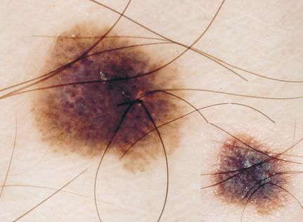
792 G.ARGENZIANO et al.

                                                                       in the dermis. There was a low mitotic activity.
                                                                       However, no maturation of melanocytes was observed.
                                                                       The Breslow thickness was 0´7 mm (Clark level III).
                                                                          Figure 8 shows a 5-mm flat plaque located on the
                                                                       left thigh of a 45-year-old man, with a history of a
                                                                       recent change in colour. Dermatoscopic examination
                                                                       revealed a relatively symmetrical distribution of colours
                                                                       and structures with a rim of brown globules at the
                                                                       periphery and discrete grey pigmentation in the centre.
                                                                       Both the clinical and dermatoscopic diagnoses were in
                                                                       favour of melanocytic naevus but the lesion was
                                                                       biopsied because of the clinical history. Histopatho-
                                                                       logical examination revealed the morphological fea-
                                                                       tures of a typical malignant melanoma with a Breslow
Figure 8. Cutaneous melanoma with epiluminescence microscopy           thickness of 0´5 mm (Clark level II).
features suggesting a Spitz naevus (original magnification 10).

                                                                       Discussion
several brown globules partially disposed at the peri-                 PSN are well-known simulators of CM from a clinical,
phery, and a larger amelanotic portion with a globular                 dermatoscopic and histopathological point of view.
vascular pattern filling the `holes' of a reticular                    Clinically, PSN may be larger than `common' naevi,
depigmentation. A few black dots and discrete grey±                    may have an irregular outline, a scaly, crusted or
blue areas were also detectable. Despite the presence of               eroded surface, and may show variegated colours.12,13
some ELM criteria suggestive of Spitz naevus, the                      Because these clinical features are also characteristic of
clinical features and the clinical data suggested the                  CM, PSN are often difficult to differentiate from CM by
final diagnosis of melanoma. Histopathological exam-                   clinical criteria alone.
ination revealed an asymmetrical melanocytic prolif-                      ELM is a valuable method for improving the diag-
eration of large, atypical spindle and epithelioid cells               nostic accuracy in pigmented skin lesions, and in the
and a prominent vascular and desmoplastic stroma.                      case of PSN an increase in diagnostic accuracy from
The thinned epidermis was characterized by slight                      56% to 93% has been reported previously.6 However, in
hyperkeratosis, hypergranulosis and a minimal page-                    the present study, 25% of PSN revealed atypical ELM
toid spread of melanocytes. Absence of maturation,                     features that did not allow a correct differentiation from
nuclear pleomorphism and mitotic figures were                          CM. Nachbar et al.14 have reported the possibility of
observed. The Breslow thickness was 1´1 mm (Clark                      misclassification of PSN due to an asymmetrical silhou-
level III).                                                            ette and an uneven distribution of colours and struc-
   Figure 7 shows a slowly growing 8-mm flat plaque of                 tures by means of the ABCD rule of dermatoscopy. In
1 year duration on the right thigh of a 46-year-old                    addition, most of our atypical cases displayed an
man. The clinical diagnosis favoured melanoma. ELM                     irregular, diffuse, grey±blue pigmentation resembling
examination revealed a symmetrical starburst pattern                   blue±white veil. This is usually felt to represent a
with prominent pigmentation, and central, bizarre                      specific ELM criterion for the diagnosis of mela-
hypopigmentation. In addition, there was a rim of                      noma.7,15 As in melanoma, the grey±blue pigmenta-
grey±brown to black globules regularly distributed at                  tion could be histopathologically correlated to clusters
the periphery mimicking pseudopods. Because of its                     of melanophages within the mid-reticular dermis.16±18
characteristic appearance, this lesion was dermato-                    Despite the atypical ELM pattern of these PSN, the
scopically classified as PSN. Histopathological exami-                 preoperative diagnosis in most cases favoured a benign
nation showed a hyperpigmented, partially asymmetrical                 lesion because of the clinical situation, namely, a
proliferation of confluent junctional nests of melanocytes             pigmented skin lesion occurring in children and
varying in size and shape. There was also a focal pagetoid             showing no history of growth (Table 1).
spread of melanocytes within the upper layers of the                      The potential for confusion between Spitz naevi and
epidermis. The melanocytes were atypical, mostly                       melanomas exists even at a histopathological level. In
spindled at the dermoepidermal junction and epithelioid                this study, two of the dermatoscopically atypical PSN

                                                q 1999 British Association of Dermatologists, British Journal of Dermatology, 141, 788±793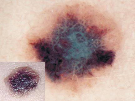
DERMATOSCOPY IN SPITZ NAEVI 793

were extremely difficult to differentiate from melanoma                   2 Pehamberger H, Steiner A, Wolff K. In vivo epiluminescence
because these lesions also displayed histopathological                      microscopy of pigmented skin lesions. I. Pattern analysis of
                                                                            pigmented skin lesions. J Am Acad Dermatol 1987; 17:
findings in common with melanoma. However, PSN                              571±83.
can be usually recognized histopathologically by                          3 Steiner A, Pehamberger H, Wolff K. In vivo epiluminescence
architectural features that often have an ELM counter-                      microscopy of pigmented skin lesions. II. Diagnosis of small
part. PSN are usually dermatoscopically and histo-                          pigmented skin lesions and early detection of malignant
                                                                            melanoma. J Am Acad Dermatol 1987; 17: 584±91.
pathologically symmetrical, and characterized by one                      4 Pehamberger H, Binder M, Steiner A, Wolff K. In vivo
of two different ELM patterns, the first typified by a                      epiluminescence microscopy: improvement of early diagnosis of
prominent, black to blue diffuse pigmentation and                           melanoma. J Invest Dermatol 1993; 100 (Suppl.): 356±62S.
radial streaks (pseudopods) distributed regularly at the                  5 Steiner A, Binder M, Schemper M et al. Statistical evaluation of
                                                                            epiluminescence microscopy criteria for melanocytic pigmented
periphery in a stellate or radiate pattern (starburst                       skin lesions. J Am Acad Dermatol 1993; 29: 581±8.
pattern), and the second revealing a discrete, brown to                   6 Steiner A, Pehamberger H, Binder M, Wolff K. Pigmented Spitz
grey±blue pigmentation and a peripheral rim of large                        nevi: improvement of the diagnostic accuracy by epiluminescence
brown globules which often extend throughout the                            microscopy. J Am Acad Dermatol 1992; 27: 697±701.
                                                                          7 Bahmer FA, Fritsch P, Kreusch J et al. Terminology in surface
entire lesion (globular pattern). These two distinct ELM                    microscopy. J Am Acad Dermatol 1990; 23: 1159±62.
patterns were found to be predominantly associated                        8 Soyer HP, Smolle J, Leitinger G et al. Diagnostic reliability of
with the pigmented spindle-cell naevus (Reed naevusÐ                        dermoscopic criteria for detecting malignant melanoma. Derma-
starburst pattern) and with the spindle- and/or                             tology 1995; 190: 25±30.
                                                                          9 Cochran AJ, Bailly C, Paul E et al. (eds) Melanocytic Tumors. A
epithelioid-cell naevus (Spitz naevusÐglobular pattern),                    Guide to Diagnosis. Philadelphia: Lippincott-Raven Publishers,
respectively. The heavily pigmented nests of melanocytes                    1997; 145±55.
closely packed along the dermoepidermal junction                         10 Elder DE, Murphy GF (eds) Melanocytic Tumors of the Skin. Armed
presumably represent the histopathological correlate                        Forces Institute of Pathology, 1991; 40±57.
                                                                         11 Barnhill RL, Fitzpatrick TB, Fandrey K et al. (eds) Color Atlas and
of the starburst pattern commonly seen in Reed naevi.                       Synopsis of Pigmented Lesions. New York: McGraw-Hill, 1995;
In contrast, the large nests of melanocytes within the                      113±16.
epidermis and/or papillary dermis may be the histo-                      12 Rhodes AR. Neoplasms: benign neoplasias, hyperplasias and
pathological correlate of the globular pattern fre-                         dysplasias of melanocytes. In: Dermatology in General Medicine
                                                                            (Fitzpatrick TB, Eisen AZ, Wolff K et al., eds), 4th edn. New York:
quently seen in spindle- and/or epithelioid-cell naevi.                     McGraw-Hill, 1993: 996±1077.
   Remarkably, we also observed three of 122 CM                          13 Kopf AW, Andrade R. Benign juvenile melanoma. In: Yearbook of
showing very few or even no ELM features suggestive of                      Dermatology, 1965±6 (Kopf A, Andrade R, eds). Chicago: Year
malignancy and exhibiting either the globular (Figs 6                       Book, 1966; 7.
                                                                         14 Nachbar F, Stolz W, Merkle T et al. The ABCD rule of
and 8) or the starburst pattern (Fig. 7) mostly seen in                     dermatoscopy. J Am Acad Dermatol 1994; 30: 551±9.
PSN. Previous studies reported ELM pitfalls in diagnos-                  15 Menzies SW, Ingvar C, McCarthy WH. A sensitivity and specificity
ing CM in heavily pigmented lesions that do not reveal                      analysis of the surface microscopy features of invasive melanoma.
the criteria necessary for ELM pattern analysis,4 and in                    Melanoma Res 1996; 6: 55±62.
                                                                         16 Yadav S, Vossaert KA, Kopf AW et al. Histopathologic correlates of
hypomelanotic or `featureless' CM lacking specific ELM                      structures seen on dermoscopy (epiluminescence microscopy).
criteria of malignancy.19 Our cases demonstrate the                         Am J Dermatopathol 1993; 15: 297±305.
possibility of missing CM due to the presence of ELM                     17 Argenziano G, Fabbrocini G, Carli P et al. Epiluminescence
features typical for PSN. Thus, surgical excision and                       microscopy: criteria of cutaneous melanoma progression. J Am
                                                                            Acad Dermatol 1997; 37: 68±74.
subsequent histopathological examination should be                       18 Soyer HP, Smolle J, Hodl S et al. Surface microscopy: a new
performed for pigmented skin lesions in adult patients                      approach to the diagnosis of cutaneous pigmented tumors. Am J
or showing a history of recent change in colour, shape                      Dermatopathol 1989; 11: 1±11.
or size, even if they have the characteristic ELM                        19 Menzies SW, Ingvar C, Crotty KA, McCarthy WH. Frequency and
                                                                            morphologic characteristics of invasive melanomas lacking
features of Spitz naevi.                                                    specific surface microscopic features. Arch Dermatol 1996; 132:
                                                                            1178±82.

References
 1 Cohen D, Sangueza O, Fass E, Stiller M. In vivo cutaneous surface
   microscopy: revised nomenclature. Int J Dermatol 1993; 32:
   257±8.

q 1999 British Association of Dermatologists, British Journal of Dermatology, 141, 788±793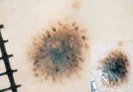
You can also read