Radioiodine Therapy-Induced Conversion of Toxic Adenoma to Graves' Disease - Cureus
←
→
Page content transcription
If your browser does not render page correctly, please read the page content below
Open Access Case
                                         Report                                               DOI: 10.7759/cureus.8683

                                         Radioiodine Therapy-Induced Conversion
                                         of Toxic Adenoma to Graves’ Disease
                                         Anis Rehman 1 , Silvana Obici 2 , Abid Yaqub 3

                                         1. Endocrinology, Southern Illinois University School of Medicine, Springfield, USA 2. Division of
                                         Endocrinology and Metabolism, Stony Brook University, Stony Brook, USA 3. Division of Endocrinology,
                                         Diabetes and Metabolism, University of Cincinnati Medical Center, Cincinnati, USA

                                         Corresponding author: Silvana Obici, silvana.obici@stonybrookmedicine.edu

                                         Abstract
                                         We present a 50-year-old female who was evaluated for the symptoms of thyrotoxicosis. She
                                         had low thyroid stimulating hormone (TSH) 0.02 with normal free thyroxine (FT4) 1.00 (0.61-
                                         1.76 ng/dL) and normal total triiodothyronine (TT3) 1.0 (0.60-2.20 ng/mL) levels. Her
                                         thyrotropin receptor antibody (TRAbs) and thyroid peroxidase antibody (TPOAb) titers were
                                         negative. Thyroid ultrasound revealed an ill-defined, heterogeneous, 1.8 cm x 0.8 cm x 0.7 cm
                                         nodule in the left lower lobe. 123-radioiodine (RAI) thyroid scan revealed 38.5% uptake, which
                                         was concentrated in the lower left thyroid lobe, a finding consistent with a solitary toxic
                                         adenoma of the thyroid.

                                         The patient became clinically and biochemically euthyroid on methimazole (MMI). She then
                                         underwent 131-RAI therapy with 12 mCi, which cured her hyperthyroidism with normalization
                                         of TSH levels for four months. She then developed overt thyrotoxicosis with low TSH of 0.02,
                                         elevated TT3 of 3.2, and normal FT4 of 0.91. Repeat TRAbs and TPOAb were elevated along
                                         with diffusely increased uptake on the I-123 RAI thyroid uptake scan, consistent with Graves’
                                         disease (GD). The patient was then placed on MMI again to bridge to definitive treatment with
                                         total thyroidectomy. Our case is a rare case where the patient with solitary toxic adenoma with
                                         negative TPOAb serology developed GD following I-131 RAI treatment.

                                         Categories: Endocrinology/Diabetes/Metabolism, Family/General Practice, Internal Medicine
                                         Keywords: i-131 radioiodine treatment, graves’ disease, toxic nodular disease, toxic adenoma

                                         Introduction
                                         The pathogenesis of toxic adenoma (TA) and Graves’ disease (GD) is very distinct. TA results
                                         from somatic mutations leading to nodules with autonomous activity and growth [1]. It is more
                                         prevalent in older population. On the contrary, GD is more common among the younger
Received 05/04/2020
Review began 05/31/2020                  population. It is induced by circulating antibodies directed against the thyroid stimulating
Review ended 06/09/2020                  hormone (TSH) receptor, a G-protein-coupled receptor that stimulates growth and stimulates
Published 06/18/2020                     biosynthesis and release of thyroid hormones [2]. Both TA and GD can present with overt or
© Copyright 2020                         subclinical thyrotoxicosis.
Rehman et al. This is an open access
article distributed under the terms of
                                         Graves’ disease commonly presents with signs and symptoms of tachycardia, weight loss,
the Creative Commons Attribution
License CC-BY 4.0., which permits
                                         tremors, anxiety, diarrhea, and heat intolerance. Patients may also develop Graves’
unrestricted use, distribution, and      ophthalmopathy and dermopathy [3]. Its incidence has been found to increase with a genetic
reproduction in any medium, provided     predisposition, particularly with human leukocyte antigen DR3 (HLA DR3), which is associated
the original author and source are       with an increased incidence of autoimmune processes [3-4]. Interestingly, GD has also been
credited.
                                         known to be triggered by viral or bacterial infections [4]. Upon review of literature, several case

                                         How to cite this article
                                         Rehman A, Obici S, Yaqub A (June 18, 2020) Radioiodine Therapy-Induced Conversion of Toxic Adenoma
                                         to Graves’ Disease. Cureus 12(6): e8683. DOI 10.7759/cureus.8683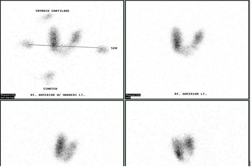
studies have described the onset of GD following I-131 radioiodine (RAI) treatment in toxic
                                  nodular goiter [5-12].

                                  I-131 RAI therapy has thyroid-selective destructive properties, which makes it an effective
                                  treatment for toxic nodular goiter as well as GD [1]. However, I-131 RAI may lead to the
                                  complete destruction of the thyroid gland, resulting in hypothyroidism. Transient
                                  hyperthyroidism within zero to eight weeks after I-131 RAI treatment may occur due to
                                  radiation thyroiditis. I-131 RAI treatment has been reported to trigger autoimmunity in 5%-
                                  5.4% of patients with multinodular goiter and in 0%-5.3% of patients with solitary nodular
                                  thyroid adenoma [13]. The incidence of seroconversion to positive titers for thyrotropin
                                  receptor antibody (TRAbs) after I-131 RAI therapy has been reported to be 5% [8]. Those with
                                  positive thyroid peroxidase antibody (TPOAb) titers before RAI-131 therapy have a much higher
                                  risk of seroconversion, which is reported to be 22% in one case series [6, 8].

                                  Here, we present a rare case of serologically TPOAb negative solitary toxic nodule which turned
                                  into serologically TPOAb and TRAbs positive GD after I-131 RAI treatment. We also review the
                                  medical literature regarding the role of I-131 RAI therapy in triggering an autoimmune
                                  response leading to the development of GD in patients with pre-existing nodular goiter.

                                  Case Presentation
                                  A 50-year-old female was referred to our endocrinology clinic with subacute onset of fatigue,
                                  palpitations, hot flashes, loose stools, dry skin, tremors, anxiety, and insomnia. There was no
                                  prior radiation exposure to head and neck, family history of thyroid or autoimmune disease, or
                                  recent exposure to iodinated contrast. She also denied taking any iodine or thyroid
                                  supplements. Her physical examination was unremarkable with no clinically palpable thyroid
                                  enlargement, Graves’ ophthalmopathy, or dermopathy. She was noted to have slight tremors of
                                  outstretched fingers.

                                  Thyroid function tests revealed a TSH low at 0.02 (0.34-5.60 uIU/mL) with normal free
                                  thyroxine (FT4) 1.00 (0.61-1.76 ng/dL), normal total triiodothyronine (TT3) 1.1 (0.60-2.20
                                  ng/mL), and normal free triiodothyonine (FT3) of 3.1 (2.0-3.6 pg/mL). Her serology titers were
                                  negative for both TRAbs < 0.9 IU/L and TPOAb < 10 IU/mL (see Table 1).

                                  I-123 RAI thyroid scan revealed 38.5% uptake concentrated in the lower portion of the left
                                  thyroid lobe, suggesting the presence of a hot nodule in the left lower thyroid lobe consistent
                                  with a clinical diagnosis of toxic adenoma (Figure 1). We then evaluated the patient with
                                  thyroid ultrasound, which revealed a normal-sized thyroid gland without any hyper-vascularity
                                  on color Doppler flow. However, the ultrasound did show an ill-defined, heterogeneous,
                                  isoechoic to a hypoechoic nodule, measuring 1.8 cm x 0.8 cm x 0.7 cm in size, which was
                                  present in the lower aspect of the left lobe corresponding to the hot area on the thyroid scan.
                                  The nodule did not have high-risk features such as micro-calcifications, irregular borders, or
                                  being taller than wider in shape.

2020 Rehman et al. Cureus 12(6): e8683. DOI 10.7759/cureus.8683                                                                 2 of 8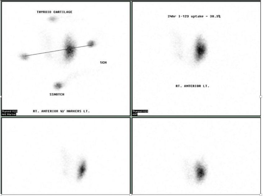
FIGURE 1: 123-RAI thyroid uptake (38.5%) prior to 131-RAI
                                     treatment.
                                     The report suggests the presence of left lower lobe hot nodule as noted above.

                                     RAI, radioiodine

                                  The patient was started on 5 mg of methimazole (MMI) orally per day, which led to both
                                  biochemical and clinical euthyroidism. Her thyroid function test, four months after the
                                  initiation of MMI revealed TSH of 0.99 (0.34-5.60 uIU/mL), FT4 of 0.91 (0.61-1.76 ng/dL), TT3
                                  of 1.0 (0.60-2.20 ng/mL), and FT3 of 3.2 (2.0-3.6 pg/mL) (Table 1).

2020 Rehman et al. Cureus 12(6): e8683. DOI 10.7759/cureus.8683                                                              3 of 8
Timeline
Events TSH FT4 TT3 FT3 TRAbs TPOAb
(months)
0.02
0 Pre-MMI and I-131 RAI treatment 1.00 1.1 3.1FIGURE 2: The repeat I-123 RAI scan after I-131 RAI treatment.
The figure reveals a diffuse uptake (37.7%) without any localized hot or cold areas/nodules.
RAI, radioiodine
Discussion
We describe a case of I-131 RAI treatment causing the transition of the toxic nodular disease to
GD. It is a rare occurrence, initially described by Boddenberg et al. in 1993 [12], and
subsequently reported in a few case reports and small prospective and retrospective studies [5-
12]. Besides 1-131 RAI treatment, other conditions that may lead to damage to thyroid cells
include surgical manipulation for parathyroidectomy, external radiation therapy for
nonthyroidal diseases, subacute thyroiditis, and percutaneous ethanol injections, which have
been reported to cause autoimmunity [5, 10]. Interestingly, our patient did not undergo any of
these procedures.
A few potential mechanisms have been proposed in the literature to explain I-131 RAI
treatment leading to the development of autoimmunity. However, none of them have been
specifically tested or proven. One such mechanism is thought to occur in a two-step process
with an initial phase of necrosis of thyroid cells, causing the release of antigens, which in turn
stimulates autoimmunity against thyroid and production of TSH-stimulating
immunoglobulins [5]. NyGaard et al. investigated this concept by postulating that an increase in
circulating thyroglobulin (tg) could act as a trigger to autoimmunity after I-131 RAI therapy;
however, tg levels were similar in the control group when compared to the patient developing
GD after I-131 RAI treatment [5]. It is possible that there is some other antigen, which is the
culprit rather than tg. An alternative hypothesis is that I-131 RAI therapy may kill the
suppressor T cells in the thyroid, which in turn may cause an imbalance between T helper and
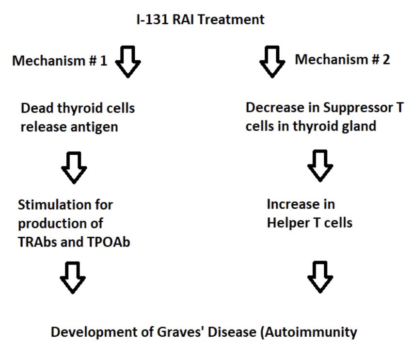
suppressor cells resulting in autoimmunity (Figure 3) [11].
2020 Rehman et al. Cureus 12(6): e8683. DOI 10.7759/cureus.8683 5 of 8FIGURE 3: Proposed mechanisms for development of GD after
I-131 RAI treatment in toxic nodular disease.
GD, Grave’s disease; RAI, radioiodine
A recent systematic review revealed that positive TPOAb titers, glandular hypo-echogenicity,
and I-123 RAI diffuse uptake scan, increase risk of development of autoimmunity up to 10-folds
after I-131 RAI treatment [10, 12]. Interestingly, in our patient, none of these risk factors were
present. Therefore our case is somewhat unusual and unique as the estimated time to onset of
autoimmunity after I-131 RAI exposure has been described to be 3-10 months [10-11, 13-14]. In
our patient, the timeline was four months post I-131 RAI therapy for the transition from TA to
GD. Susceptible genetic make-up along with environmental risk factors is known to contribute
to the onset of GD. However our patient denied prior known family history of autoimmune
disease [2].
In 1911, Marine and Lenhart reported the presence of thyroid nodules and autoimmunity
concomitantly [15]. I-131 RAI therapy leading to the development of autoimmunity is different
than Marine-Lenhart syndrome as in the former thyroid nodular disease precedes the
development of GD following I-131 RAI treatment [8, 11].
Once diagnosed with hyperthyroidism after more than three months of I-131 RAI treatment,
the patients described in the literature have increased uptake on repeat I-123 RAI scan. These
patients have positive TPOAb and TRAbs titers, thus confirming GD [5, 11, 14]. Our patient was
similar to the cases described in the literature in this regard; she developed positive serology
with diffuse uptake on the I-123 RAI scan, a classical presentation of GD, suggesting that I-131
RAI can trigger a de novo autoimmune response. Conversely, not all patients treated with I-131
2020 Rehman et al. Cureus 12(6): e8683. DOI 10.7759/cureus.8683 6 of 8RAI therapy show seroconversion from negative to positive titers for TPOAb and TRAbs, which
suggests that only a subset of predisposed individuals will have this response [5, 14].
Elevated TRAbs titers are detected in more than 90% of patients with GD [2]. However, a small
portion of patients has negative autoimmune serology, which could be ascribed to the detection
limits of our current serological assays. It has been proposed in the literature that, in some
cases, the production of TRAbs titers can be restricted to intra-thyroidal tissue [16]. As a
variable degree of lymphocytes infiltration is present in GD, we speculate that our TRAbs-
negative patient may have had intra-thyroidal lymphocyte infiltration restricted to only a
portion of the thyroid, and thus presenting with I-123 RAI uptake scan typical of the toxic
nodule [2-3]. In this scenario, the subsequent I-131 RAI therapy could have triggered
exacerbation of an autoimmune reaction against the whole thyroid and the conversion from
seronegative to seropositive TRAbs titers.
Conclusions
Hyperthyroid patients who have nodular thyroid disease with no prior autoimmune disease,
and who receive I-131 RAI treatment should be counseled and monitored for GD after 3-10
months of RAI treatment. Positive TPOAb titers, glandular hypo-echogenicity, and diffuse I-
123 RAI uptake scan increase the risk of development of autoimmunity. However, it is essential
to know that a few patients may still transition to GD from toxic nodular disease after I-131 RAI
treatment despite the absence of these risk factors. Although some experts have proposed
potential explanations for this phenomenon, the underlying pathogenetic mechanism remains
elusive to a large extent. More research is needed in this area to clearly understand the
pathophysiologic changes that lead to the development of GD following RAI-131 treatment in
patients with nodular thyroid disease.
Additional Information
Disclosures
Human subjects: Consent was obtained by all participants in this study. Conflicts of interest:
In compliance with the ICMJE uniform disclosure form, all authors declare the following:
Payment/services info: All authors have declared that no financial support was received from
any organization for the submitted work. Financial relationships: All authors have declared
that they have no financial relationships at present or within the previous three years with any
organizations that might have an interest in the submitted work. Other relationships: All
authors have declared that there are no other relationships or activities that could appear to
have influenced the submitted work.
Acknowledgements
The authors would like to acknowledge Jagit Padda MD for her assistance in writing the
manuscript.
References
1. Carlé A, Andersen SL, Boelaert K, Laurberg P: Management of endocrine disease: subclinical
thyrotoxicosis: prevalence, causes and choice of therapy. Eur J Endocrinol. 2017, 176:325-337.
10.1530/EJE-16-0276
2. Morshed SA, Latif R, Davies TF: Delineating the autoimmune mechanisms in Graves’ disease .
Immunol Res. 2012, 54:191-203. 10.1007/s12026-012-8312-8
3. Figueroa-Vega N, Alfonso-Pérez M, Benedicto I, et al.: Increased circulating pro-inflammatory
cytokines and Th17 lymphocytes in Hashimoto's thyroiditis. J Clin Endocrinol Metab. 2010,
95:953-962. 10.1210/jc.2009-1719
2020 Rehman et al. Cureus 12(6): e8683. DOI 10.7759/cureus.8683 7 of 84. Ye XP, Yuan FF, Zhang LL, et al.: ITM2A expands evidence for genetic and environmental
interaction in Graves’ disease pathogenesis. J Clin Endocrinol Metab. 2017, 102:652-660.
10.1210/jc.2016-2625
5. Nygaard B, Knudsen JH, Heqedüs L, et al.: Thyrotropin receptor antibodies and Graves’
disease, a side-effect of 131 I treatment in patients with nontoxic goiter. J Clin Endocrinol
Metab. 1997, 82:2926-2930. 10.1210/jcem.82.9.4227
6. Nygaard B, Faber J, Veje A, Heqedüs L, Hansen JM: Appearance of Graves’-like disease after
radioiodine therapy for toxic as well as non-toxic multinodular goitre. Clin Endocrinol. 1995,
43:129-130. 10.1111/j.1365-2265.1995.tb01904.x
7. Niepomniszcze H, Pitoia F, Goodall C, Manavela M, Bruno OD: Development of Graves’
hyperthyroidism after radioiodine treatment for a toxic nodule: is the hyperthyroidism always
triggered by 1311 therapy?. Thyroid. 2001, 11:991. 10.1089/105072501753211091
8. Nygaard B, Faber J, Veje A, Heqedüs L, Hansen JM: Transition of nodular toxic goiter to
autoimmune hyperthyroidism trigged by 131I therapy. Thyroid. 1999, 9:477-481.
10.1089/thy.1999.9.477
9. Van Leussen JJ, Edelbroek MA, Talsma MA, de Heide LJ: Graves' disease induced by Na(131)
therapy for toxic multinodular goitre. Neth J Med. 2000, 57:194-197. 10.1016/s0300-
2977(00)00068-1
10. Schmidt M, Gorbauch E, Dietlein M, et al.: Incidence of postradioiodine immunogenic
hyperthyroidism/Graves’ disease in relation to a temporary increase in thyrotropin receptor
antibodies after radioiodine therapy for autonomous thyroid disease. Thyroid. 2006, 16:281-
288. 10.1089/thy.2006.16.281
11. Huysmans AK, Hermus RM, Edelbroek MA, et al.: Autoimmune hyperthyroidism occuring late
after radioiodine treatment for volume reduction of large multinodular goiters. Thyroid. 1997,
7:535-539. 10.1089/thy.1997.7.535
12. Boddenberg B, Voth E, Schicha H: Immunogenic hyperthyroidism following radioiodine
ablation of focal autonomy. Nuklearmedizin. 1993, 32:18-22.
13. Yürekli Y, Cengiz A, Güney E: Graves’ disease induced by radioiodine therapy for toxic nodular
goiter: a case report. Mol Imaging Radionucl Ther. 2015, 24:135-137. 10.4274/mirt.74046
14. Roque C, Vasconcelos CA: 131 I-Induced Graves' disease in patients treated for toxic
multinodular goitre: systematic review and descriptive analysis. J Endocrinol Invest. 2018,
41:1019-1028. 10.1007/s40618-018-0827-y
15. Marine D, Lenhart CH: Pathological anatomy of exophthalmic goiter . Arch Intern Med. 1911,
8:265-316.
16. Sugenoya A, Kobayashi S, Kasuga Y, et al.: Evidence of intrathyroidal accumulation of TSH
receptor antibody in Graves' disease. Acta Endocrinol (Copenh). 1992, 126:416-418.
10.1530/acta.0.1260416
2020 Rehman et al. Cureus 12(6): e8683. DOI 10.7759/cureus.8683 8 of 8You can also read