Case Report Hematuria as the initial symptom of Wilson disease in identical twins: a case report
←
→
Page content transcription
If your browser does not render page correctly, please read the page content below
Int J Clin Exp Med 2021;14(6):2016-2021
www.ijcem.com /ISSN:1940-5901/IJCEM0121516

Case Report
Hematuria as the initial symptom of Wilson disease
in identical twins: a case report
Jiajia Wang1,2, Minxia Hu1, Qiang Zhu1, Lanting Sun3
1
 Department of Diagnostic Ultrasound, Beijing Tongren Hospital, Capital Medical University, Beijing, China; De-
partments of 2Ultrasound, 3Encephalopathy, The First Affiliated Hospital of Anhui University of Chinese Medicine,
Hefei, China
Received September 1, 2020; Accepted December 30, 2020; Epub June 15, 2021; Published June 30, 2021

Abstract: Wilson disease (WD) is a rare autosomal recessive disorder of copper transport with copper accumula-
tion in various organs. The clinical presentations of WD are variable. The diagnosis of WD can be challenged when
patients show clinically rare symptoms. This report described a case of identical twin brothers diagnosed with WD
who initially presented with mild-moderate microscopic hematuria and had a consequent delay in the diagnosis and
treatment. The 6-year-old younger brother initially presented with hematuria before he was admitted to our hospital
at his age of 11. He was found with mild gait disturbance in physical examination upon admission. Further evalua-
tion revealed abnormalities including laboratory investigations of cooper metabolism, brain MRI, liver ultrasound,
and Kayser-Fleischer (K-F) rings. The older twin brother presented with hematuria and proteinuria without hepatic
and neurological symptoms at age 11. K-F rings were also confirmed. Genetic testing was performed on the twins
and revealed mutations of R778L and A874V on both alleles of ATP7B gene. The diagnosis of WD was made on the
twins according to the Leipzig scoring system. The twins regularly received sodium dimercaptopropane sulphonate
(DMPS) treatment, supplemented with a Chinese traditional medicine, called Gandou decoction. The symptoms of
the twin brothers were improved after treatment. The two patients had been reported no renal, hepatic, or neurologi-
cal abnormalities during a 5.5-year follow-up period. The study demonstrates that WD should be considered as one
of differential diagnoses when long-term renal abnormalities with unknown etiology occur in pediatric population.
An early diagnosis and treatment are essential for the WD patients to avoid irreversible multi-organ injury.

Keywords: Wilson disease, hematuria, ultrasound, case report

Introduction                                                  complications [8, 9]. Hematuria as the initial
                                                              sign of WD is rarely reported with an incidence
Wilson Disease (WD), first described in 1912,                 of 1.3% in the literature [10]. In this case re-
is a rare autosomal recessive genetic disease,                port, we first described identical twin brothers
resulting from accumulation and deposition of                 with WD who presented with hematuria as the
copper in tissues such as the liver, brain, kid-              first symptom of WD.
ney, cornea, etc. [1]. Gene mutation of copper-
transporting P-type ATPase (ATP7B) on chro-                   Case presentation
mosome 13q14.3 has been shown to be relat-
ed to WD [2]. The incidence of WD in China is                 A 6-year-old younger twin brother with mild to
estimated to be six individuals per million [1,               moderate microscopic hematuria was referred
3]. The typical symptoms of WD are mainly                     to our hospital in 2009. The patient did not
hepatic (40-60%) and neurological (18-68%)                    complain about abdominal pain and backache.
damage [4, 5]. Patients with typical initial sy-              Physical examination was unremarkable. The
mptoms are rarely misdiagnosed [6, 7]. How-                   urine analysis and liver function test were nor-
ever, delay in diagnosis and treatment can                    mal except for the presence of hematuria.
occur in patients with rare presentations, such               Given that the etiology of hematuria has not
as damages to the kidney, bone, and heart,                    been elucidated, the patient was followed up
resulting in irreversible damage and severe                   without intervention. The patient routinely had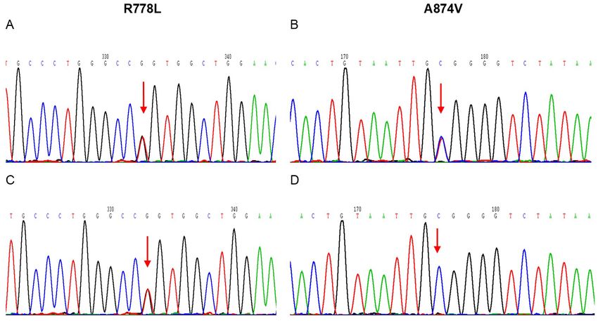
Hematuria of WD in twins

Figure 1. Liver ultrasound of the younger twin brother   Figure 2. T2WI-weighted image of the brain magnetic
shows the liver with a normal size, regular contour,     resonance imaging (MRI) of younger twin brother
and homogenous hyperechogenic parenchyma.                shows cord-like hyperintense foci in the left basal
                                                         nucleus (arrows) and small hyper-intense foci in the
                                                         right basal nucleus (triangular arrows).
urine and liver function tests every three mon-
ths thereafter. During 5-year follow-up period,
the laboratory tests were negative except for            4.25 μmol/L (normal range, 11-24.4 μmol/L),
mild to moderate microscopic hematuria exist-            and 24-h urine copper excretion of 1,583.06
ed. The patient developed proteinuria at age             μg/24 h (normal value,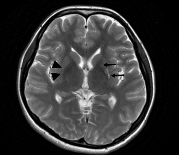
Hematuria of WD in twins
Though WD patients have cop-
per metabolism disorder from
birth, they generally have typi-
cal presentations only when
the accumulation or deposition
of copper reach the saturated
state. In our report, the young-
er twin brother had initial
symptom of mild-moderate
hematuria at the age of 5, oth-
erwise he was healthy. We
speculated that the copper
concentration did not reach
the saturation point until he
presented with typical WD
manifestations at the age of
11. The diagnosis of WD can
be challenging when non-typi-
cal abnormalities present prior
to typical symptoms such as
hepatic and neurological dam-
ages, which may result in de-
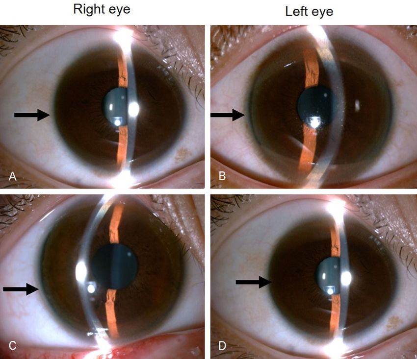
Figure 3. Slit-lamp fundoscopy shows Kayser-Fleischer (K-F) ring (arrow) in lay in diagnosis and treat-
the corneal periphery of both eyes of the younger twin brother (A, B) and the
older twin brother (C, D). ment. The case report indi-
cates that WD should be con-
sidered as one of differentials
The twin brothers were started on sodium when long-term renal abnormalities with un-
dimercaptopropane sulphonate (DMPS) treat- know etiology occur in pediatric population.
ment 20 mg/kg daily. Two weeks later, the urine
test for both of them were normal. And the Renal symptoms as initial signs of WD have
symptom of gait disturbance in the younger been reported as uncommon. Lai investigated
brother disappeared. After the two brothers the initial symptoms of WD in pediatric pati-
ents, 42% had liver injury, 34% showed neuro-
were discharged from our hospital, they re-
logical symptoms, and only 1% developed
ceived dimercaptosuccinate (DMSA) 1 g daily
renal abnormalities [11]. Renal damage can
and Gandou decoction (a Chinese traditional
occur in any stage of WD. Though the mecha-
medicine) therapy, and a low copper diet was nism is unknown, copper observed to be de-
also advised. The patients had been reported posited in the epithelial cell on the renal tu-
no renal, hepatic, or neurological symptoms bule may result in renal tubular dysfunction
during a 5.5-year follow-up period. [12]. Renal damage of WD is characterized by
the presence of hematuria, proteinuria, glycos-
This study was approved by the Ethics Com-
uria, and high phosphate and uric acid levels.
mittee of the First Affiliated Hospital, Anhui The younger twin brother was found to have
University of Chinese Medicine (2018AH-08). the first symptom of renal injury at the age of
Patients of this study have provided informed 5. It was consistent with the study by Wang et
consent for publication of the case. al. [13], in which the authors found that the
average age of the first symptom of renal injury
Discussion was 6.5 years old.
WD is a relatively rare autosomal recessive dis- Gait disturbance is a clinical presentation that
order with a clinically high variability in pheno- indicates the possibility of neurological dam-
type. This report described the case of 6-year- age [14]. Mild gait disturbance can be less
old identical twin brothers with initial symptom apparent in early stage of WD [15]. However,
of renal abnormalities who were misdiagnosed gait abnormalities show a tendency towards
due to a lack of typical signs of WD. progression with the progression of WD. The
2018 Int J Clin Exp Med 2021;14(6):2016-2021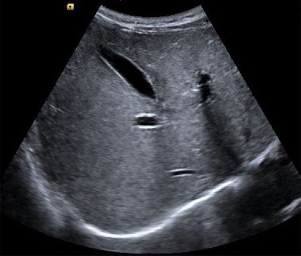
Hematuria of WD in twins

Figure 4. DNA sequencing shows compound mutations of R778L and A874V of ATP7B gene in the younger twin
brother (A, B) and the older twin brother (C, D).

Table 1. Clinical symptoms, laboratory tests, genetic analysis          was admitted to our hospital. Our
of the twin brothers upon admission                                     case suggests that complete neu-
                                              Younger    Older          rological examination is needed for
                                                twin     twin           early detection of WD.
                                              brother   brother
Clinical symptoms
                                                                     The genetic analysis confirmed
                                                                     R778L and A874V missense muta-
   Age at presence of hematuria           5-year-old 11-year-old
                                                                     tions in the twin brothers. These
   K-F ring                               Presence Absence
                                                                     mutations have been involved in a
   Neurological symptoms (gait disturbance)
                                          Presence Absence
                                                                     disorder of the copper-transporting
   Abnormalities on brain MRI             Presence Absence           ATPase, ATP7B [16-18]. R778L is
   Abnormalities on liver ultrasound      Presence Absence           the most common pathogenic gene
Urine test                                                           of WD in the Chinese population,
   Erythrocyte (µl)                         206.2      196.2         accounting for 30% of all WD
   24-h proteinuria (g/24 h)                 0.32       0.28         patients. It was reported that 74%
Copper metabolism                                                    of WD patients with R778L muta-
   Serum copper (µmol/L)                     4.25        3.3         tion had liver damage [19, 20].
   Ceruloplasmin (g/L)                      0.071      0.070         A874V is relatively rare in WD, only
   24-hour urine copper (µg/24 h)         1583.06     1121.41        accounting for 3.6% of cases [21].
Pathogenic gene analysis                   c.2333G>T; p.R778L        It’s impractical to clearly identify
                                           c.2621C>T; p.A874V        the correlation between gene phe-
 Leipzig scoring system                   11 points 10 points        notype and clinical phenotype
                                                                     since more than 700 ATP7B muta-
                                                                     tions were found in WD [21]. Of
younger twin brother was observed with mild              note, so far, no WD patients with both A874V
gait disturbance in physical examination upon            and R778L mutations have been reported. Ge-
admission, which provided critical diagnostic            netic analysis plays an important role in the
basis of WD. The patient might have developed            diagnosis of WD. The patient with mutations
gait disturbance before admission, but it was            on both alleles of ATP7B had four points on
not recognized due to the mild disorder until he         the Leipzig scoring system, and the diagnosis

2019                                                              Int J Clin Exp Med 2021;14(6):2016-2021
Hematuria of WD in twins
of WD should be made even with no clinical [2] Chang IJ and Hahn SH. The genetics of Wilson
manifestations presented. disease. Handb Clin Neurol 2017; 142: 19-34.
[3] Xie JJ and Wu ZY. Wilson’s disease in China.
In a study by Albert et al. [22], the authors Neurosci Bull 2017; 33: 323-330.
assessed the hepatic damages in 98 WD pa- [4] Ferenci P. Wilson disease. Semin Neurol 2007;
tients (26 children and 72 adults) using liver 27: 123-132.
biopsy. The study confirmed that pediatric pa- [5] Bandmann O, Weiss KH and Kaler SG. Wil-
son’s disease and other neurological copper
tients tended to develop hepatocyte steatosis
disorders. Lancet Neurol 2015; 14: 103-113.
(73% vs 46%) whereas the adults were more [6] Kumagi T, Horiike N, Michitaka K, Hasebe A,
likely to develop fibrosis (54% vs 27%). The Kawai K, Tokumoto Y, Nakanishi S, Furukawa
authors thus suggest that hepatocyte stea- S, Hiasa Y, Matsui H, Kurose K, Matsuura B
tosis is associated with early stage of WD. In and Onji M. Recent clinical features of Wilson’s
our report, the ultrasound examination of the disease with hepatic presentation. J Gastroen-
younger brother showed homogenous hyper- terol 2004; 39: 1165-1169.
echogenic parenchyma of the liver. This finding [7] Członkowska A, Litwin T and Chabik G. Wilson
implied the development of hepatocyte steato- disease: neurologic features. Handb Clin Neu-
sis though liver biopsy was not performed on rol 2017; 142: 101-119.
the patient. For the WD patients, liver ultra- [8] Dzieżyc K, Litwin T and Członkowska A. Other
organ involvement and clinical aspects of Wil-
sound should be annually used to evaluate the
son disease. Handb Clin Neurol 2017; 142:
liver damage, to adjust the treatment plan 157-169.
promptly in order to prevent the occurrence of [9] Poujois A and Woimant F. Challenges in the di-
liver fibrosis or cirrhosis [1]. agnosis of Wilson disease. Ann Transl Med
2019; 7 Suppl 2: S67.
In conclusion, WD is a rare hereditary disease. [10] Scheinberg IH. Wilson’s disease. J Rheumatol
Misdiagnosis can often occur due to the lack 1981; 8: 90-93.
of typical initial symptoms. The delay in diag- [11] Ferenci P, Czlonkowska A, Stremmel W, Houw-
nosis and treatment significantly affects the en R, Rosenberg W, Schilsky M, Jansen P, Mo-
prognosis of the disease. WD should be in- radpour D and Gitlin J. EASL clinical practice
cluded in the differential diagnoses in patients guidelines: Wilson’s disease. J Hepatol 2012;
with long-term unknown hematuria. Further- 56: 671-685.
more, liver ultrasound and brain MRI should [12] Zhuang XH, Mo Y, Jiang XY and Chen SM. Anal-
ysis of renal impairment in children with Wil-
be performed to assess organ damages, and
son’s disease. World J Pediatr 2008; 4: 102-
other family members should be genetically
105.
tested to early detect asymptomatic patients. [13] Wang H, Zhou Z, Hu JY and Han YZ. Renal im-
pairment in different phenotypes of Wilson dis-
Acknowledgements ease. Neurol Sci 2015; 36: 2111-2115.
[14] Machado A, Chien HF, Deguti MM and Canc E.
The authors thank the patients and their family Neurological manifestations in Wilson’s dis-
for their invaluable contribution to this study. ease: report of 119 cases. Mov Disord 2006;
21: 2192-2196.
Disclosure of conflict of interest [15] Dziezyc K, Litwin T, Chabik G and Członkowska
A. Frequencies of initial gait disturbances and
None. falls in 100 Wilson’s disease patients. Gait
Posture 2015; 42: 601-603.
Address correspondence to: Dr. Qiang Zhu, Depart- [16] Prashanth LK, Taly AB, Sinha S, Arunodaya GR
ment of Diagnostic Ultrasound, Beijing Tongren and Swamy HS. Wilson’s disease: diagnostic
Hospital, Capital Medical University, No. 1, Dong errors and clinical implications. J Neurol Neu-
Jiao Min Xiang Street, Dongcheng District, Beijing rosurg Psychiatry 2004; 75: 907-909.
100730, China. Tel: +86-10-58268134; Fax: +86- [17] Park S, Park JY, Kim GH, Choi JH, Kim KM, Kim
10-65131244; E-mail: qzhu_mail@126.com JB and Yoo HW. Identification of novel ATP7B
gene mutations and their functional roles in
References Korean patients with Wilson disease. Hum Mu-
tat 2007; 28: 1108-1113.
[1] Poujois A and Woimant F. Wilson’s disease: a [18] Lutsenko S. Modifying factors and phenotypic
2017 update. Clin Res Hepatol Gastroenterol diversity in Wilson’s disease. Ann N Y Acad Sci
2018; 42: 512-520. 2014; 1315: 56-63.
2020 Int J Clin Exp Med 2021;14(6):2016-2021Hematuria of WD in twins
[19] Wu ZY, Lin MT, Murong SX and Wang N. Mo- [22] Stättermayer A, Traussnigg S, Dienes H, Stau-
lecular diagnosis and prophylactic therapy for ber R, Lackner K, Hofer H, Stift J, Stadlmayr A,
presymptomatic Chinese patients with Wilson Datz C, Strasser M, Maieron A, Trauner M and
disease. Arch Neurol 2003; 60: 737-741. Ferenci P. Hepatic steatosis in Wilson disease
[20] Liu XQ, Zhang YF, Liu TT, Hsiao KJ, Zhang JM, - role of copper and PNPLA3 mutations. J Hep-
Gu XF, Bao KR, Yu LH and Wang MX. Correla- atol 2015; 63: 156-163.
tion of ATP7B genotype with phenotype in Chi-
nese patients with Wilson disease. World J
Gastroenterol 2004; 10: 590-593.
[21] Dong Y, Ni W, Chen WJ, Wan B, Zhao GX, Zhu Q,
Zhang Y, Wang N, Yu L, Xu JF and Wu ZY. Spec-
trum and classification of ATP7B variants in a
large cohort of chinese patients with Wilson’s
disease guides genetic diagnosis. Theranos-
tics 2016; 6: 638-649.
2021 Int J Clin Exp Med 2021;14(6):2016-2021You can also read