Whipple Disease Presenting as Cystic Brain Tumor: Case Report and Review of the Literature - Turkish ...
←
→
Page content transcription
If your browser does not render page correctly, please read the page content below
DOI: 10.5137/1019-5149.JTN.17111-16.2                                                Turk Neurosurg 28(3):495-499,2018
                Received: 08.02.2016 / Accepted: 05.05.2016
               Published Online: 08.08.2016

                       Case Report

Whipple Disease Presenting as Cystic Brain Tumor:
Case Report and Review of the Literature
Mohamed KILANI1, Leila NJIM2, Atef Ben NSIR1, Mohamed Nejib HATTAB1
1
    Fattouma Bourguiba University Hospital, Department of Neurosurgery, Monastir, Tunisia
2
    Fattouma Bourguiba University Hospital, Department of Pathology, Monastir, Tunisia

    ABSTRACT

     Although neurological features are commonly encountered in Whipple’s disease (WD), presentation with purely neurological patterns
     is uncommon. Exclusive confinement to the central nervous system (CNS) is extremely rare. In these cases, the development of an
     isolated cerebral mass is exceptional.
     In the present paper, the authors describe a case of a 68-year-old man who presented with partial seizures. The neurological
     examination was normal. The imaging showed a cystic lesion. This tumor-like lesion was removed by performing frontal
     craniotomy. A histopathological investigation revealed the presence of numerous perivascular foamy histiocytes infiltrating the
     brain parenchyma. The majority of these histiocytes showed Periodic acid–schiff (PAS)-positive intense staining, which is distinctive
     feature of cerebral WD. The diagnosis was confirmed by polymerase chain reaction (PCR) analysis of cerebrospinal fluid. There were
     no gastrointestinal symptoms and no PAS inclusions in intestinal mucosa. The patient received Ceftriaxone intravenously followed
     by oral trimethoprime-sulfamethoxazole (TMP-SMZ) for 12 months and recovered well. This case illustrates atypical WD, confined
     exclusively to the central nervous system.
     KEYWORDS: Whipple disease, Tropheryma whippelii, Encephalitis, Central nervous system

█      INTRODUCTION                                                        We present the first case of isolated cerebral Whipple’s
                                                                           disease presenting as a binocular cyst.

W
          hipple’s disease (WD) is a rare multisystem infectious
          disease caused by a slow growing soil-borne Gram-                █   CASE REPORT
          positive bacillus Tropheryma whippleii (T. whippleii),
related to the family of actinomyces. Humans are the only                  A 68-year-old right handed man was referred to our institution
known host for the infection (8). This chronic infection is                after suffering two simple partial epileptic seizures localized
characterized by predominant intestinal involvement. Weight                in the left side of his body. He had experienced problems of
loss, diarrhea, low-grade fever and arthralgia have all been               concentration and increasing irritability for a month without
recorded to be major symptoms of WD (11).                                  any headache. There was no history of fever, weight loss or
                                                                           recurring episodes of arthralgia.
The central nervous system (CNS) may be involved around
10-43% in patients with multisystem WD. The neurological                   On admission, the patient was afebrile and all vital signs were
manifestations are diverse and can mimic almost any neuro-                 normal. The results of a neurological examination were normal.
logical condition (9). These manifestations occur in three cir-            The digestive system and joints were normal.
cumstances: CNS relapse of previously treated classic WD,
                                                                           Magnetic resonance imaging (MRI) revealed a right frontal
neurological involvement in untreated classic WD, and isolat-
                                                                           binocular cyst in the subcortical white matter. The lesion was
ed neurological symptoms due to T. whippleii (3).

              Corresponding author: Mohamed KILANI
              E-mail: kilanineurochirurgien@gmail.com

                                                                                                                                       495
Kilani M. et al: Whipple Disease in the Brain
hypo-intense on T1-weighted images and hyper-intense on of perivascular foamy histiocytes. Periodic Acid-Schiff (PAS)
T2-weighted images. Transient contrast enhancement was preparation revealed intense staining in the macrophages and
noticed after injection of contrast agent (Figure 1A, B). There histiocytes which is a characteristic findings of cerebral WD
was a marked edema surrounding the lesion. The midline (Figure 2A, B). Special staining for acid-fast bacilli and fungi
structures were shifted to the left side. The lesion was thought were negative. There was no evidence of tumor.
to be a glioblastoma or a hydatid cyst.
The patient was placed on anti-epileptic medication on
The lesion was removed by a right frontal craniotomy. At admission. Upper intestinal endoscopy with duodenal and
surgery, the lesion appeared macroscopically like an old jejunal biopsies was performed. Histological examination of
hematoma. There was a fragile and thin membrane separating the specimens showed mostly normal tissue. Polymerase
two cysts. The tumor-like tissue could hardly be differentiated chain reaction (PCR) assay targeting the 16S rRNA gene of
from surrounding brain. The lesion vessels were vulnerable. T. whippleii showed a positive result in the cerebrospinal fluid
The removal was estimated to be complete macroscopically. (CSF).
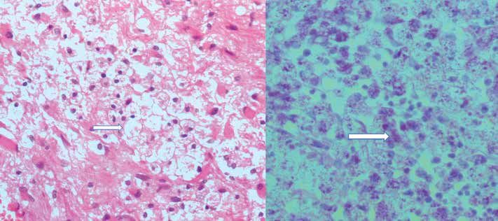
The neuropathological examination revealed reactive gliosis Antibiotic therapy with ceftriaxone (2g/day intravenously)
and infiltration of the brain parenchyma by a large number was given for 2 weeks followed by TMP-SMZ (160/800
Figure 1: Axial T1-weighted brain
MRI with gadolinium demonstrating
transient enhancing binocular
cyst in the right frontal lobe (A).
FLAIR sequence showing edema
A B surrounding the lesion and midline
structures shifted to the left side (B).
A B
Figure 2: Histological section shows an expansion of the brain tissue by foamy macrophages (arrow); H&E, original magnification×200
(A). PAS stain reveals brightly staining granular intracytoplasmic inclusions (arrow); original magnification×200 (B).
496 | Turk Neurosurg 28(3):495-499, 2018Kilani M. et al: Whipple Disease in the Brain
mg twice a day) for 12 months. His concentration trouble spread to the CNS and cause neurological symptoms without
gradually improved. At regular follow-up, the neurological systemic involvement.
examination remained stable. The patient had not developed
CNS Whipple’s disease presenting as a solitary mass is
any gastrointestinal symptoms. Follow-up MRI investigation
rather exceptional. Only 4 cases were reported (1,4,6,10).
performed 6 months after surgery showed no recurrence,
Among these, only 2 cases showed no systemic symptoms of
but a persisting pseudocyst defect (Figure 3). Currently, the
patient is seizure free. He continues to take TMP-SMZ on a Whipple’s disease (Table I).
regular basis. De Coen et al. (4) reported a case of a 49-year-old female who
suffered from dysarthria, right facial palsy and hemiparesis. MRI
█ DISCUSSION showed the lesion to be hypointense on T1-weighted images
with ring enhancement. It was thought to be a glioblastoma or
Whipple’s disease was first described in 1907 by George a metastasis. Microscopic examination showed the presence
Whipple at Johns Hopkins (16). At present, we recognize
of foamy macrophages with granules PAS positive. The patient
Tropheryma whippleii as the causative agent of WD. In fact,
was treated with trimethoprim. Follow-up MRI images showed
Relman et al.(13) identified the bacterium as a Gram-positive
complete resolution of the lesion.
actinomycete. Whipple’s disease typically involves multiple
organ systems (9,11). The symptoms of cerebral WD include Löhr et al. (10) report a case of a 40-year-old male suffering
oculomotor abnormalities, ataxia, seizures, psychiatric from bifrontal headache, speech disturbances and problems
disturbances, dementia, and aseptic meningitis (2). These of concentration. The lesion was hypo-intense on T1-weighted
neurological symptoms are varied and often complex but oculo- images with circular contrast enhancement. It was thought
masticatory myorhythmia (OMM) and oculo-facial-skeletal to be a low-grade glioma. The patient was operated. The
myorhythmia (OFSM) are considered to be pathognomonic of histopathological investigation showed pathological features
CNS WD (9). Cases of WD with isolated involvement of the of CNS WD. The patient received co-trimoxazole for one year
brain without any systemic affection appear to be rare. So far, post-operatively and remained symptom free.
only 21 cases have been reported in the literature (15).
As shown in these cases, brain MRI for diagnosis of CNS
The first question raised by the present report deals with the Whipple’s disease is paramount, but nonspecific as with most
hypothesis of isolated CNS WD. Pruss et al. (12) suggest that infectious processes affecting the CNS (14). Herein we report
the robust activity of duodenal lymphocytes may prevent bowel the first case of CNS WD, consisting of binocular cyst. When
disease. On the contrary, the concomitant impaired activity consisting of solitary mass, WD gives rise to many differential
of peripheral blood lymphocytes may lead to the bacterial diagnoses such as low-grade glioma, aggressive meningioma,
metastasis (1,4,10), glioblastoma and even a hydatid cyst.
Craniotomy and excision of the intracranial lesion leads to the
diagnosis, when CNS WD is presenting as a solitary lesion
(1,4,10). It is worth mentioning that, as in our case, when
the lesion seems to be macroscopically different from the
imaging diagnosis, WD should be suspected. In these four
cases, the diagnosis was established by a histopathological
analysis. It consists of an inflammatory reaction combining
gliosis and vasculitis replacing normal cortex and white
matter. Characteristic foamy macrophages are often seen,
and their presence should heighten the clinical suspicion of
WD. These macrophages stain very intensely with PAS stain
(5). PCR analysis performed to detect 16S ribosomal RNA of
T. whippleii is useful for both establishment of the diagnosis
and monitoring the treatment response (13).
Early treatment of WD leads to improvement of the lesion
(1,4,10) as shown in our case. Since CNS relapses carry a poor
prognosis, antibiotics should not be reduced or discounted.
They should be prescribed at least for one year to prevent
relapses (7).
█ CONCLUSION
Isolated cerebral Whipple’s disease often poses a great
diagnosis challenge since its symptoms and neuroimaging
Figure 3: Follow-up T1-weighted brain MRI obtained 6 months signs are not specific. Although this disease usually presents
after surgery revealed complete resolution of the lesion with a as diffuse lesions, this report demonstrates that it can
persisting pseudocyst defect. manifest as a solitary tumor–like lesion. This diagnosis should
Turk Neurosurg 28(3):495-499, 2018 | 497Table I: Review of the Literature of CNS Whipple’s Disease Presenting as an Intracranial Mass
Age/
Year/ Author Presentation Imaging Operation CSF Neural tissue Extraneural tissue
Gender
Let parietal lesion
FM
Dysartria, right facial T1: hypointense
1996/ (4) 49/F Tumor resection Normal PAS PG Duodenal : negative
palsy, right hemiparesis T2: hyperintense
Ring enhancement
Right frontoparietal solid mass
Diarrhea, headache, T1: isointense FM
2002/(1) 18/M partial epileptic seizures, T2: hypointense Tumor resection Normal PAS PG ND
left hemiparesis. Intense homogeneous enhancement LM : Positive
498 | Turk Neurosurg 28(3):495-499, 2018
Erosion of the tabula interna
Kilani M. et al: Whipple Disease in the Brain
Left frontal mass
Headache, speech Normal FM
T1: hypointense Duodenal/ jejunal:
2004/(10) 40/M disturbance, irritability, Tumor resection PCR: non PAS PG
T2: hyperintense negative
aggressiveness diagnostic
Ring enhancement
Right temporal lobe, hypothalamus and
History of Whipple
left temporomesial lesions FM
disease, headache, Temporal tumor
2009/ (6) 51/M T1 with Gado: intense contrast ND PAS PG ND
emesis, transient resection
enhancement
episodes of confusion.
T2: hyperintense
Right, frontal lesion
Partial epileptic FM
T1: hypointense Normal Duodenal/ jejunal:
Present case 68/M seizures, problems of Tumor resection PAS PG
T2: hyperintense PCR: positive negative
concentration, irritability
Transient contrast enhancement
ND: Not done, FM: foamy macrophages, PAS PG: PAS-positive granules, LM: light microscopy, CSF: cerebrospinal fluid.Kilani M. et al: Whipple Disease in the Brain
be suspected when no tumor tissue is found at surgery. The 8. Louis ED: Whipple’s disease. Curr Neurol Neurosci Rep 3:
excision or biopsy of the mass leads to the diagnosis. This 470-475, 2003
should be kept in mind since the CNS WD is treatable if 9. Louis ED, Lynch T, Kaufmann P, Fahn S, Odel J: Diagnostic
promptly diagnosed. guidelines in central nervous system Whipple’s disease. Ann
Neurol 40: 561-568, 1996
█ REFERENCES 10. Löhr M, Stenzel W, Plum G, Gross WP, Deckert M, Klug N:
Whipple disease confined to the central nervous system
1. Akar Z, Tanriover N, Tuzgen S, Canbaz B, Erman H, Oz B,
presenting as a solitary frontal tumor: Case report. J Neurosurg
Kuday C: Intracerebral Whipple disease: Unusual location and
101:336-339, 2004
bone destruction: Case report. J Neurosurg 97:988-991, 2002
11. Marth T, Schneider T: Whipple disease. Curr Opin Gastroenterol
2. Anderson M: Neurology of Whipple’s disease. J Neurol
24: 141-148, 2008
Neurosurg Psychiatry 68: 2-5, 2000
12. Pruss H, Katchanov J, Zschenderlein R, Loddenkemper C,
3. Compain C, Sacre K, Puéchal X, Klein I, Vital-Durand D,
Schneider T, Moos V: A patient with cerebral Whipple disease
Houeto JL, De Broucker T, Raoult D, Papo T: Central nervous
with gastric involvement but no gastrointestinal symptoms:
system involvement in Whipple disease: clinical study of 18
A consequence of local protective immunity? J Neurol
patients and long-term follow-up. Medicine (Baltimore) 92:
Neurosurg Psychiatry 78: 896-898, 2006
324-330, 2013
13. Relman DA, Schmidt TM, MacDermott RP, Falkow S:
4. De Coene B, Gilliard C, Indekeu P, Duprez T, Trigaux JP:
Identification of the uncultured bacillus of Whipple’s disease.
Whipple’s disease confined to the central nervous system.
N Engl J Med 327: 293-301, 1992
Neuroradiology 38: 325-327, 1996
14. Scholz KB, Henning S, Paulus W, Knauth M: MRI finding in
5. Durand DV, Lecomte C, Cathébras P, Rousset H, Godeau
isolated cerebral manifestation of Whipple’s disease: Case
P: Whipple disease: Clinical review of 52 cases. Medicine
report and review of the literature. Eur J Radiol 59: 1-5, 2006
(Baltimore) 76:170-184, 1997
15. Sung VW, Lyerly MJ, Fallon KB, Bashir K: Isolated CNS
6. Frazier JL, Quinones-Hinojosa A: Isolated Whipple disease of
Whipple disease with normal brain MRI and false-positive
the brain resembling a tumour. Acta Neurochir (Wien)151:173-
CSF 14-3-3 protein: A case report and review of the literature.
175, 2009
Brain Behav 2: 838-843, 2012
7. Gerard A, Sarrot-Reynauld F, Liozon E, Cathebras P, Besson
16. Whipple GH: A hiterto undescribed disease characterized
G, Robin C, Vighetto A, Mosnier JF, Durieu I, Vital Durand
anatomically by deposits of fat and fatty acids in the intestinal
D, Rousset H: Neurologic presentation of Whipple disease:
and mesenteric lymphatic tissues. John Hopkins Med Bull
Report of 12 cases and review of the literature. Medicine
231: 283-287, 1907
(Baltimore) 81: 443-457, 2002
Turk Neurosurg 28(3):495-499, 2018 | 499You can also read