Maxillofacial Prosthesis for Combined Intra and Extra-Oral Defect - A Case Report
←
→
Page content transcription
If your browser does not render page correctly, please read the page content below
Jemds.com Case Report
Maxillofacial Prosthesis for Combined Intra and Extra-Oral Defect
– A Case Report
Rajiv Dharampal Bhola1, Sweta Gajanan Kale Pisulkar2, Surekha Anil Dubey Godbole3,
Hetal Satish Purohit4, Anjali Bhoyar Borle5
1, 2, 3, 4, 5 Department
of Prosthodontics and Crown and Bridge, Sharad Pawar Dental College,
DMIMS (DU), Sawangi (Meghe), Wardha, Maharashtra, India.
INTRODUCTION
Combined intra and extra oral defects can be stated as those facial defects which have Corresponding Author:
an intraoral communicating route. Midfacial defects are aptly classified into 2 major Dr. Rajiv Bhola
categories by Marunick et al.1 as midline midfacial defects in which the nose and / or Senior Lecturer,
upper lip defects are included; and the second major group was lateral defects in Department of Prosthodontics and
Crown & Bridge, Sharad Pawar
which the cheek and orbital defects are categorized. However, defects which include
Dental College, DMIMS (DU),
combinations of the above-mentioned defects are in existence. Midfacial defects Sawangi (Meghe), Wardha,
which are acquired, present themselves often with severe disfigurement of structures Maharashtra, India.
and hence show impaired function. It is a meticulous task to rehabilitate the defects E-mail: rinkujii@gmail.com
which are caused as a result of cancerous lesion resection as they are huge. Such post
resection lesions frequently are rehabilitated by a facial prosthesis to maintain DOI: 10.14260/jemds/2021/119
function as well as the appearance in the normal form. In adjunction to the facial
prosthesis, an intraoral prosthesis which constitutes of an obturator is also required How to Cite This Article:
Bhola RD, Kale Pisulkar SGK, Godbole SAD,
to regain the natural speech and pattern of swallowing. Fabrication of such facial
et al. Maxillofacial prosthesis for combined
prosthesis not only requires the artistic capability but also excellent clinical decision intra and extra-oral defect - a case report. J
making of the prosthodontist. Mode of retention of the combined prosthesis should Evolution Med Dent Sci 2021;10(08):550-
also be kept in mind while fabricating as it is also a difficult task to retain them 554, DOI: 10.14260/jemds/2021/119
because of the size and weight of the same. Moreover the prosthesis should also be
secured in its place with these aids which can also prove as a challenge. This case Submission 25-10-2020,
report states rehabilitating a large surgically resected midfacial defect with the Peer Review 28-12-2020,
Acceptance 04-01-2021,
assistance of a “3-piece prosthesis” which constitutes a sectional intraoral obturator Published 22-02-2021.
along with maxillary and mandibular extraoral facial prosthesis.
Copyright © 2021 Rajiv Dharampal Bhola
et al. This is an open access article
P R E SE N T A T I O N O F C A S E distributed under Creative Commons
Attribution License [Attribution 4.0
International (CC BY 4.0)]
A woman aged 55 years with a history of surgical resection of a well differentiated
squamous cell carcinoma of left buccal mucosa with extra oral fungation, T4aN2aM0
before 6 months was brought up for definitive prosthetic rehabilitation of the defect.
Three reconstructive procedures were carried in a timeline of 3 months which were
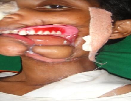
proven to be unsuccessful. The extra oral defect extends superoinferiorly from ala
tragus line to inferior border of mandible and antero-posteriorly from corner of
mouth to anterior border of ramus (Fig. 1). Intraorally, in maxilla missing teeth were
premolars and molars of left side and rest all teeth were present and in mandible all
anteriors and posterior teeth on left side were missing. The majority of the section of
the hard palate was surgically removed in the maxilla. The anatomical structures
which remained showed minimal area to provide support, along with retention, and
to provide stability of maxillary obturator. Hemimandibulectomy was performed for
the mandible and the remaining buccal mucosa was attached to the floor of the
mouth, so there was no available space for prosthetic rehabilitation in mandible
intraorally.
J Evolution Med Dent Sci / eISSN - 2278-4802, pISSN - 2278-4748 / Vol. 10 / Issue 08 / Feb. 22, 2021 Page 550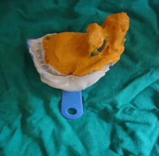
Jemds.com Case Report
D I SC U S SI O N O F M A N A G E M E N T
Owing to the large size of the facial defect, history of radiation
to the focus area, poor quality of the remaining mucosa, Figure 5B.
minimal bony supporting structures, and lack of natural Palatal View Extraorally
dentition, made the prognosis fair for prosthodontic
rehabilitation.
Figure 6.
Denture Prosthesis in Place
Figure 1.
Combined Intra–Extra Oral Defect
Figure 7.
Facial Moulage Cast Fabricated
in Type III Gypsum
Figure 2.
Maxillary Final Impression
Figure 8A
Clear Acrylic Facial Maxillary
Figure 8B Mandibular Stent
Figure 3. Extraorally.
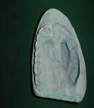
Maxillary Cast Showing Defect
Prosthetic rehabilitation commenced with the
construction of a partially edentulous maxillary obturator.
Impression of the maxillary arch was made (Fig. 2) and
working casts of maxillary arch with intraoral defect was
obtained (Fig. 3). Corner of mouth and occlusion at right side
guide for occlusion rim level of maxillary obturator. The
maxillary occlusion rim was contoured in correspond to upper
lip contour. The teeth were arranged and try-in done (Fig. 4)
and the intraoral prostheses (hollow maxillary obturator)
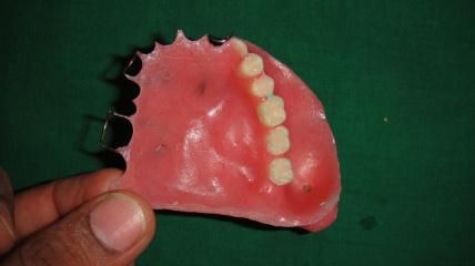
Figure 4.
Try-In of the Denture Intraorally fabricated (Fig. 5. a, b) and insertion done (Fig. 6).
Anatomical undercut and denture adhesive were considered
for the proper retention of the obturator. The patient was
advised to use gastric feeding tube throughout the prosthetic
rehabilitation procedure.
Intermediate Framework
Keeping the obturator in position impression of the face was
Figure 5A.
Prosthesis Occlusal made and facial moulage was obtained (Fig. 7). This facial
moulage helps to determine extend of extra oral defect,
J Evolution Med Dent Sci / eISSN - 2278-4802, pISSN - 2278-4748 / Vol. 10 / Issue 08 / Feb. 22, 2021 Page 551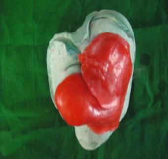
Jemds.com Case Report
planning and fabrication of extra oral maxillofacial prosthesis. Mandibular extraoral prosthesis was retained with the
For the fabrication of extra oral maxillofacial prosthesis first help of elastic. (Fig. 13). Pretreatment and post treatment view
clear acrylic maxillary and mandibular template is prepared of the patient (Fig. 14 a, b)
(Fig. 8. a, b, c, d) of the shape of maxilla and mandibular defect.
Figure 10A.
Wax Pattern Try in Over the
Acrylic Template
Figure 8C & D.
Evaluation of the Fit of the
Maxillary and Mandibular
Template on the Stone Model
Facial Prosthesis (Extra Oral Prosthesis)
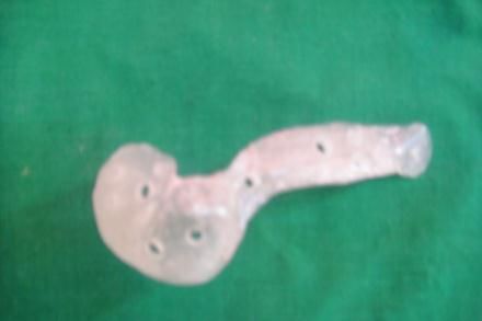
Retentive holes are made in the template for mechanical
retention of prosthesis (Fig. 9 a, b) Over the template wax
pattern of maxillary and mandibular extraoral prosthesis Figure 10B.
prepared and trying of wax pattern done (Fig. 10 a, b). Wax Pattern Try in over the
Acrylic Template
Wax pattern was used to make silicone prosthesis for
which MDX4-4210 base silicone was used. Later on the
silicone prosthesis was attached to the fabricated acrylic plate
with the assistance of adhesives and other mechanical
retentive aids. The processing of the prosthesis was carried
out at room temperature for about 48 hours and later on it was
deflasked, trimmed, cleaned, and bonded to a polyurethane
lining with medical adhesive type A (Factor II, Lakeside, Ariz.)
under vacuum as described by Lemon et al.2 Applying
polyurethane lining has added benefit of increasing the tear
Figure 11A.
resistance of the prosthesis at its margin. The prosthesis was Flasking of the Medical Grade
then trial fit and had extrinsically colored with Silicone
trichloroethane, medical adhesive type A, oil pigments, and
rayon flocking. (Factor II)
Figure 11B.
Prosthesis was Stained
Extrinsically
Figure 9A & B.
Retentive Holes are
Made in the Template
for Mechanical
Retention Figure 11C.
Extraoral Prosthesis
Adapted onto the Stone Model
Retention of magnets in the silicone prosthesis was carried
out by hose made up of nylon as stated by Lemon et al.3
Maxillary extra oral prosthesis is retained to maxilla with the
help of magnetic attachment, in magnet attached to acrylic
plate from inner side and magnetic keeper attached to
maxillary obturator buccal flange (Fig. 12).
J Evolution Med Dent Sci / eISSN - 2278-4802, pISSN - 2278-4748 / Vol. 10 / Issue 08 / Feb. 22, 2021 Page 552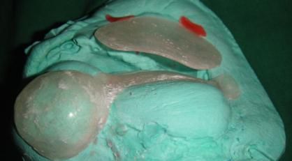
Jemds.com Case Report
eyeglasses,5,6 extensions from the denture7 that engage tissue
undercuts,6,8 magnets6,9 adhesives,6 combinations of the
above,6,8-10 and osseointegrated implants.6,8,11,12 Among all the
mentioned types of retention osseointegrated implants
provide the best mode of retention but it requires additional
surgeries and expenses. Also, patients with inadequate bone,
and a history of radiation to the focus area may prove as a
contraindication to the implant therapy.13,14
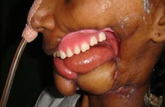
Figure 12A.
Pre-Treatment Photograph of
the Patient
CONCLUSIONS
In this case report the definitive prosthetic rehabilitation of a
55-year-old woman, with a history of cancer resection which
resulted in a combination of intraoral and extraoral defect is
illustrated. A “3-piece definitive prosthesis” which constituted
of a denture obturator along with a facial prosthesis (maxillary
Figure 12B. and mandibular), and an intermediate framework to support
Post-Treatment Photograph of the elements was fabricated. Magnet was incorporated into
the Patient with Prosthesis in maxillary facial prosthesis and elastics in the mandibular facial
Place
prosthesis, in adjunction with the intermediate framework
which assisted the retention of each element of the prosthesis.
Though it is a proven fact that osseointegrated implants not
Post treatment instructions were given, and maintenance only restore dentition but also retain extraoral prostheses, but
of silicone prosthesis explained to the patient. Patient is in this case radiation in the focus region ruled out their use in
recalled after 24 hours, then after one week and then 21 days this patient and hence another technique for retention was
and thereafter every 3 months.
mandatory. So, the intermediate framework, elastics and
magnets helped to achieve satisfactory retention for the
prostheses in this particular case. The patient was
D I SC U S SI O N recommended to remain on a gastric feeding tube for a period
of 8 weeks after the prosthodontic treatment was finished. The
Prosthetic treatment was planned to rehabilitate the defect stoppage of the use of feeding tube was aptly approved by
assisted by a “3-piece prosthesis” that included a sectional otolaryngologists along with speech and swallowing
intraoral obturator, maxillary and mandibular extra-oral therapists. It was duly checked that the patient is capable
maxillofacial prosthesis. First maxillary hollow obturator was enough to carry the whole weight with the supplemented soft
fabricated and then facial moulage was made by keeping diet along with the liquid nutritional ingredients ingested by
obturator in its intra oral position and then the maxillary and mouth. With the assistance of this prostheses, it was possible
mandibular template was prepared to provide retention, to achieve improvement in aesthetics as well as the capability
support and stability to extra oral prosthesis. Then wax to speak. The patient was not only able to swallow but also
pattern of the shape of maxilla and mandible prepared over
chew to a great extent which overall improved the quality of
template by using facial moulage as a guide and then try in
life of the patient.
done on patient and after satisfactory try in, wax pattern
A patient reporting for dental treatment also indirectly
converted into silicone prosthesis using MDX4-4210 base
gets treated for psychological issues in relation.15 Such defects
silicone. Maxillary facial prosthesis retained with magnets and
lead to social embarrassment and affect quality of life.16 The
mandibular with elastic. Insertion done and post treatment
definitive rehabilitation of a 55-year aged patient who
instruction given and patient kept on periodic recall.
presented with an intraoral as well as extra-oral defect is
Orofacial defects can be acquired or congenital. The
acquired defects are a result of either trauma or surgical introduced. “A 3-piece prosthesis” which constitutes denture
resection. These orofacial defects with bigger size lead to obturator, one maxillofacial prosthesis (maxillary and
severe functional impairment, disability to speak, leads to mandibular), with an intermediate framework was made.
improper mastication and disturbed swallowing patterns. Magnet was retained in the maxillary facial prosthesis and
Along with all these problems, the midfacial deformity also has elastics in the mandibular facial prosthesis, which is associated
a significant psychological impact due to its aesthetic with the intermediate framework with the purpose of
considerations. Also successful aesthetic results can still be retention of each element present in the combined prosthesis.
obtained but the retention of such prostheses poses a severe Though it is a proven fact that osseointegrated implants not
problem. With a proper knowledge and a comprehension of only restore dentition but also retain extra oral prostheses, but
the anatomic elements which are remaining, intraoral and in this case, radiation in the focus region ruled out their use in
extraoral prostheses combination that can retain each other this patient and hence another technique for retention was
with mutual effort is possible to fabricate. Hence for the aid mandatory. So, the intermediate framework, elastics and
other means of auxiliary retention for facial prostheses have magnets help to achieve satisfactory amount of retention for
been illustrated in the literature; they include eyepatches,4 the combined prostheses in this particular case.
J Evolution Med Dent Sci / eISSN - 2278-4802, pISSN - 2278-4748 / Vol. 10 / Issue 08 / Feb. 22, 2021 Page 553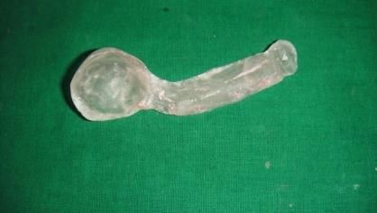
Jemds.com                                                                                                                   Case Report
Financial or other competing interests: None.                                [9] Dumbrigue HB, Fyler A. Minimizing prosthesis movement
Disclosure forms provided by the authors are available with the full              in a midfacial defect: a clinical report. J Prosthet Dent
text of this article at jemds.com.                                                1997;78(4):341-5.
                                                                             [10] Verdonck HW, Peters R, Vish LL. Retention and stability
                                                                                  problems in a patient with a large combined intra - and
                         REFERENCES                                               extraoral defect: a case report. J Facial Somato Prosthet
                                                                                  1998;4:123-8.
[1] Marunick MT, Harrison R, Beumer J. Prosthodontic                         [11] Menneking H, Klein M, Hell B, et al. Prosthetic restoration
    rehabilitation of midfacial defects. J Prosthet Dent                          of nasal defects: indications for two different
    1985;54(4):553-60.                                                            osseointegrated implant systems. J Facial Somato
[2] Lemon JC, Martin JW, King GE. Modified technique for                          Prosthet 1998;4:29-33.
    preparing a polyurethane lining for facial prostheses. J                 [12] Worthington       P,     Brånemark        PI.    Advanced
    Prosthet Dent 1992;67(2):228-9.                                               osseointegration     surgery:    applications     in    the
[3] Lemon JC, Martin JW, Chamber MS, et al. Technique for                         maxillofacial region. Carol stream ill. Quintessence
    magnet sreplacement in silicone facial prostheses. J                          1992:307-26.
    Prosthet Dent 1995;73(2):166-8.                                          [13] Arcuri M, LaVelle WE, Fyler E, et al. Prosthetic
[4] Tautin FS, Schoemann D. Retaining a large facial                              complications of extraoral implants. J Prosthet Dent
    prosthesis. J Prosthet Dent 1975;34(3):342-5.                                 1993;69(3):289-92.
[5] McClelland RC. Facial prosthesis following radical                       [14] Roumanas E, Nishumura R, Beumer J, et al.
                                                                                  Osseointegrated implants: six-year follow-up report on
    maxillofacial surgery. J Prosthet Dent 1977;38(3):327-30.
                                                                                  the success rates of craniofacial implants at UCLA.
[6] Thomas KF. Prosthetic rehabilitation. London:
                                                                                  International Journal of Oral and Maxillofacial Implants
    Quintessence Publishing 1994:93-103.
                                                                                  1994;9(5):579 -85.
[7] Fattore L, Edmonds DC. A technique for the obturation of
                                                                             [15] Pisulkar SK, Agrawal R, Belkhode V, et al. Perception of
    anterior maxillary defects with accompanying midfacial
                                                                                  buccal corridor space on smile aesthetics among specialty
    tissue loss. J Prosthet Dent 1987;58(2):203-5.
                                                                                  dentist and layperson. J Int Soc Prev Community Dent
[8] Beumer J, Curtis TA, Marunick MT. Maxillofacial                               2019;9(5):499-504.
    rehabilitation: prosthodontic and surgical considerations.               [16] Pisulkar SK, Purohit H, Mistry R, et al. Encasing the
    St Louis: Ishiyaku Euro America Inc 1996:408-16.                              encephalon: enhancing psychosocial rehabilitation. Int J
                                                                                  Cur Res Rev 2020;12(14).

J Evolution Med Dent Sci / eISSN - 2278-4802, pISSN - 2278-4748 / Vol. 10 / Issue 08 / Feb. 22, 2021                                Page 554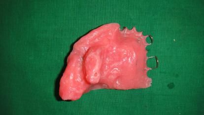
You can also read