Brain-Chip Characterization - Emulate
←
→
Page content transcription
If your browser does not render page correctly, please read the page content below
Technical Note

Brain-Chip
Characterization

Abstract
Despite the growing prevalence of neurodegenera-
tive disease globally, effective disease-modifying
                                                                 Key Highlights
therapies remain largely out of reach. One major
challenge has been the lack of robust preclinical             • Human-based model of the neurovascular
models that can be applied to model human brain                 unit—including neuronal compartment and
physiology and predict blood brain barrier (BBB) pen-           BBB
etration and efficacy of novel therapeutics. Here, we         • Tight BBB is formed with in vivo-like
present data characterizing the Emulate Brain-Chip              permeability that is improved by microglia.
as a comprehensive model of the neurovascular unit            • Exhibits high transcriptomic similarity to
(NVU), including a brain compartment recapitulating             adult human cortical tissue
morphological and functional characteristics of
                                                              • Enables neuroinflammatory disease
human brain tissue and a functional BBB with low
                                                                modeling, target validation, and BBB
permeability comparable to in vivo. Compared to                 penetration studies
traditional cell culture models, the transcriptomic
profile of the Brain-Chip more closely resembles
mature adult human cortical tissue and maintains           central nervous system (CNS).1 Due to the unique
expression stability for several days. Taken together,     and complex biology of the NVU, adequate models
the Brain-Chip is a physiologically relevant preclinical   are difficult to create.
model of the human NVU that can improve investiga-
tion of brain physiology, neurodegenerative disease,       Since the BBB prevents nearly all large molecule
drug efficacy, and drug transport across the BBB.          drugs and ~98% of small molecule therapeutics from
                                                           crossing the BBB, it can hinder the therapeutic effica-
Introduction                                               cy of drugs designed to target neurological diseases.3
                                                           In addition, despite the great need for more neu-
The neurovascular unit (NVU) is responsible for regu-      rotherapeutics, few drugs are being developed due to
lating cerebral blood in response to neural activity1,2    the low predictability of efficacy.3 Therefore, there is a
and is composed of brain endothelial cells, astro-         need to develop better models of the BBB that
cytes, mural cells such as pericytes, microglia, neu-      can accurately model drug delivery and disease
rons, as well as an extracellular matrix (ECM).2 Tight-    pathology.
ly bound microvascular endothelial cells make up the
blood-brain barrier (BBB),2 which separates the brain      Current in vitro NVU models range from traditional
microenvironment from the bloodstream and main-            cell monolayers to 3D organoid systems. Cellular
tains brain homeostasis by selectively regulating the      monolayers are widely utilized due to their ease of
transport of compounds from the bloodstream to the         use and high throughput capacity, but they lack

                                                                                                  © Emulate, Inc., 2021. All rights reserved.

                                              emulatebio.com
                                                                                                                Technical Note | June 2021
                                                                                   The technology herein may be covered by patents and/or
                                                                                        trademarks. Please contact Emulate for information.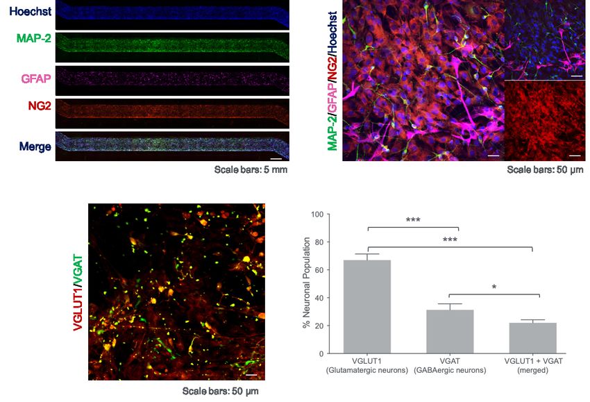
Technical Note
appropriate cytoarchitecture and a physiologically
relevant microenvironment.4 Many of these cell
cultures are limited in the number of cell types they can
support and subsequently lack cellular heterogeneity
required to form in vivo-like tight junctions and barrier
function.5 Organoid models have cell-cell interactions
that exhibit improved physiological characteristics over
cell monolayer models, but are not without their limita-
tions, including reproducibility due to variability,
absence of essential cell types (e.g., microglia), and
lack of vasculature and mechanical cues.6,7 While Figure 1. Schematic illustration of the Brain-Chip.
Top “brain” channel includes human induced pluripotent
animal models are often used to study the brain, stem cell (iPSC)-derived glutamatergic and GABAergic
species differences, low throughput, and ethical neurons, and primary human brain astrocytes,
pericytes, and microglia. The bottom “vascular” channel
concerns limit their successful translation to human
contains iPSC-derived brain endothelial cells.
response.8,9 More human-relevant models of the neu-
rovascular unit are needed to accelerate the develop-
ment of novel therapeutics and understand the patho- a.
genesis of neurological disorders.
MAP2 GFAP IBA1 αSMA DAPI
Goal
To develop and characterize an Organ-on-a-Chip
model of the NVU that emulates human brain and BBB
function by incorporating critical cell types in a dynam-
ic microenvironment. With morphological and function-
al characteristics of brain tissue, the Brain-Chip can be
Cortical Neurons in the Brain-Chip
applied to the study of drug transport, mechanisms of
b.
action, and disease pathology.
Materials a.
Hardware /
Cell Sources
Consumables
• Chip-S1® Emulate Brain Bio-Kit,
Stretchable Chip containing qualified human
iPSC-derived and primary cells: Glutamatergic neurons (VGLUT1)
• Zoë® Culture Module
GABAergic neurons (VGAT)
• Neurons (iPSC-derived)
• Orb® Hub Module Scale bar: 50 μm
• Microglia (primary)
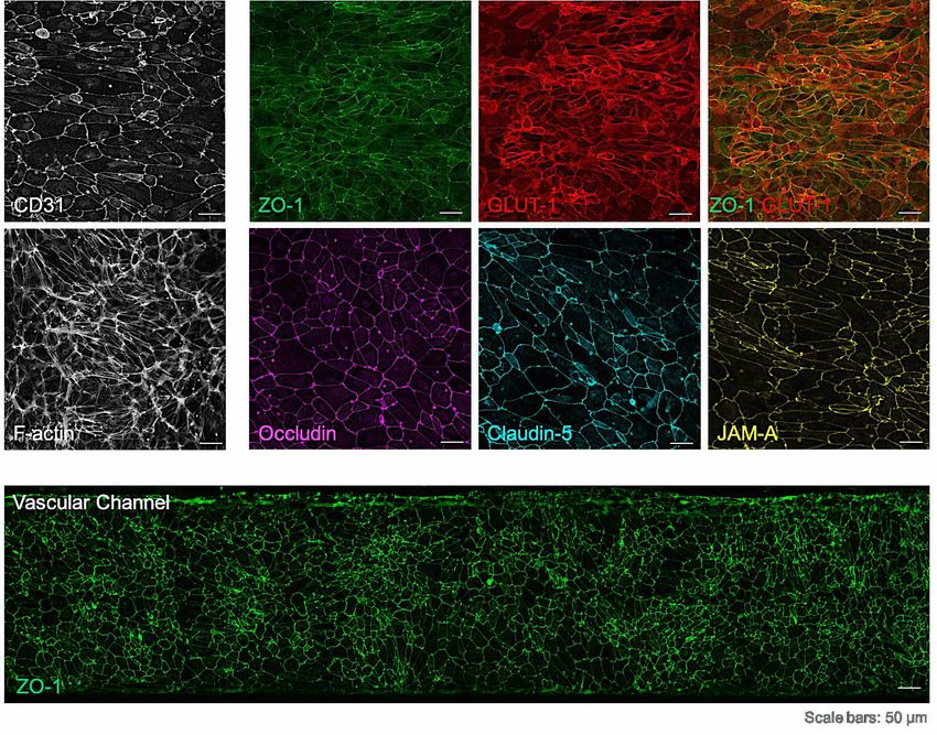
• Astrocytes (primary) Figure 2. Representative confocal images after 7
days of culture. Top channel stained for (A) neurons
• Pericytes (primary)
(MAP2, green), astrocytes (GFAP, magenta), and
• iPSCs (for differentiation into pericytes (NG2, red) (bar, 50 μm); and (B) co-stained
microvascular brain with for GABAergic (VGAT, green) and glutamatergic
endothelial cells) (VGLUT1, red) neurons (bar, 100 μm).
© Emulate, Inc., 2021. All rights reserved.
emulatebio.com
Technical Note | June 2021
The technology herein may be covered by patents and/or
trademarks. Please contact Emulate for information.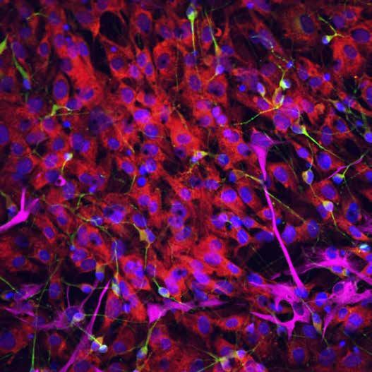
Technical Note

Results
                                                                                                                                   50

                                                                                                                  Glutamate (µM)
As shown in Figure 1, the Brain-Chip has a top and                                                                                 40
bottom fluidic channel separated by a thin, porous                       Neurotransmitter                                          30
                                                                             Release
polydimethylsiloxane (PDMS) membrane coated with                                                                                   20
                                                                            n = 6 Chips                                            10
a tissue-specific ECM that permits cellular communi-
cation. Excitatory and inhibitory cortical neurons,                                                                                 0
                                                                                                                                           5     6       7
microglia, astrocytes, and pericytes are co-cultured in                                                                                 Days in Culture (Time)
the top channel (also called the brain channel).            Figure 3. Secreted glutamate in the brain channel. Two
Cultured on the bottom channel (vascular channel),          technical replicates, 3 chips / condition. Data are expressed
are human induced pluripotent stem cell (iPSC)-de-          as mean ± S.E.M. ns, not significant.
rived brain microvascular endothelial cells (iBMECs).
The neurovascular unit is formed and maintained for
seven days in culture, including a BBB with in vivo-like          a.
apparent permeability (Papp).

After seven days of culture, the brain channel was
stained for neurons (microtubule-associated protein 2
[MAP2], green), astrocytes (Glial fibrillary acidic
protein [GFAP], magenta), microglia (ionized calci-
um-binding adaptor protein-1 [IBA-1], yellow) and
pericytes (alpha smooth muscle actin [αsma], red)
(Figure 2A) and for the two major neuronal subtypes,              b.
                                                                                                            Brain-Chip
GABAergic neurons (vesicular GABA transporter                             3 10-6                            Rat in vivo (Yuan et al., 2007)
                                                           Papp (cm/s)

                                                                                                            Rat in vivo (Shi et al., 2014)
[VGAT], green) and glutamatergic neurons (vesicular                       2 10-6
glutamate transporter [VGLUT1], red) (Figure 2B).
The confocal images verified an in vivo-like cell com-                    1 10-6
                                                                               0
position successfully incorporating both inhibitory and                                             3        4     10     20   40                      70
excitatory cortical neurons.                                      c.
                                                                                                                 Dextans (kDa)
                                                                                                  1 10-5
                                                                           Papp of 3kDa Dextran

To test glutamate transporter activity, glutamate levels                                          8 10-6
were established from effluent collected from the brain                                                                            ****
                                                                                   (cm/s)

                                                                                                  6 10-6
                                                                                                                                      ****
channel. As seen in Figure 3, glutamate levels are                                                4 10-6
stable across days five to seven, confirming appropri-                                            2 10-6                                     NS
ate and consistent synaptic activity.
                                                                                                        0
                                                                                                      glia   tes ons      el
                                                                                                  icro strocy neur ll mod
Barrier integrity was assessed by Papp levels with                                              m
                                                                                              no no a      no     fu
cascade-blue 3 kDa dextran on days five, six and
                                                           Figure 4. Assessment of apparent permeability (Papp)
seven (Figure 4A) and assessed with various sizes of
                                                           with fluorescent dextran. (A) Papp to 3KDa dextran using
dextran (Figure 4B). Results demonstrated that a           two iPSC endothelial donor lines. 4-6 chips / donor. Data
tight BBB was maintained for seven days with Papp          are expressed as mean ± S.E.M. (B) Papp to various sizes
values similar to those reported in vivo.10 In the         of fluorescent dextran and comparison to in vivo. (C) Papp to
                                                           3KDa fluorescent dextran with or without microglia,
absence of astrocytes or microglia, the Brain-Chip has
                                                           astrocytes, and neurons. 4-8 chips / condition. Data are
higher Papp values, confirming the positive effect of      expressed as mean ± S.E.M. **P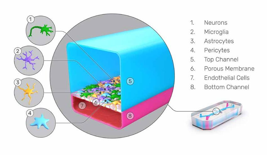
Technical Note

To confirm the establishment of a tight brain-specific
endothelial monolayer, the vascular channel was
stained for the tight junction protein marker (ZO-1,
green) and glucose transporter (GLUT1, red) after
seven days of culture (Figure 5). Results show the
endothelial cells did express tight junction proteins
and brain endothelium-specific GLUT-1 over the
entirety of the vascular channel.

One of the major routes for drug delivery of large
molecules across the BBB is receptor-mediated trans-          Figure 5. Vascular channel on day seven of culture.
                                                              Staining for tight junction protein marker (ZO-1, green)
cytosis, often through the transferrin receptor, which is
                                                              and glucose transporter-1 (GLUT1, red) (bar, 100 μm).
highly expressed in brain capillaries and is used for
therapeutic antibody transport.11 To assess the
suitability of the Brain-Chip for assessing active trans-
port of drug candidates, the transferrin receptor was
evaluated by quantifying levels of intracellular trans-
ferrin. Immunofluorescent staining (pHrodo Red
Transferrin Conjugate, red) revealed transferrin
that was internalized in the cytoplasm of iBMECs
(Figure 6)

Comparing differential gene expression (DGE) of the                         iPS-derived HBMECs
Brain-Chip to a Transwell model with the same                               Phalloidin, pHrodoRed Transferrin, Hoechst
constituent cells revealed that of the 57,500 genes                                                                Scale bar: 50 μm

annotated in the genome, 5,695 were significantly              Figure 6. Assessment of receptor-mediated
differentially expressed (DE), with 3,256 upregulated          transcytosis. Immunofluorescent staining for transferrin
                                                               (pHrodo Red Transferrin Conjugate, red) in the cytoplasm of
and 2,439 downregulated (Figure 7). Gene ontology              brain microvascular endothelial cells iBMECs (bar, 100 μm).
enrichment analysis performed on DE genes deter-
mined that the pathways enriched in the Brain-Chip
were associated with important biological processes
including ECM organization, cell adhesion, and tissue
development. Meanwhile, pathways enriched in Tran-
swells included axonogenesis, neurogenesis, and
neuron migration, demonstrating the enhanced matu-
                                                            -Log10 (padj)

ration of the Brain-Chip over Transwell models.                                                                                                          !

                                                                                                                                Not Significant

To compare the Brain-Chip and Transwell model to                                                                                Downregulated
                                                                                                                                Upregulated
adult human cortical tissue, Transcriptomic Signature                                                                        n-4 for each condition
                                                                                                                             adj.pvalue < 0.01
Distance (TSD) was calculated using the Human                                                                                |log2FoldChange| ≥1

Protein Atlas for human brain signature gene sets.12
TSD is a method that evaluates transcriptomic similar-                              Log2 Fold Change

ities of two tissues or groups of samples.13 Results          Figure 7. Differential gene expression. Brain-Chip and
revealed that the human Brain-Chip is genetically             Transwell cultures on day seven showing upregulated
                                                              (cyan) and downregulated genes (magenta).
more similar to adult human cortical brain tissue than
                                                                                                           © Emulate, Inc., 2021. All rights reserved.

                                               emulatebio.com
                                                                                                                         Technical Note | June 2021
                                                                                            The technology herein may be covered by patents and/or
                                                                                                 trademarks. Please contact Emulate for information.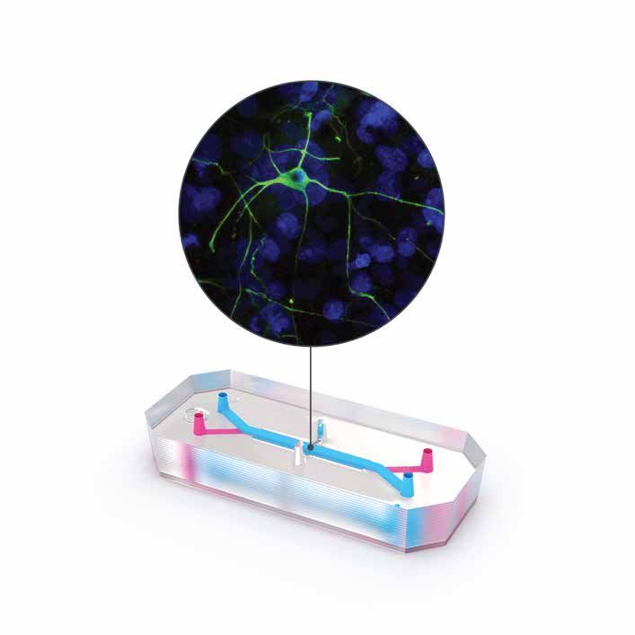
Technical Note

the Transwell model and maintains gene expression
over seven days (Figure 8).                                                            Distances from Human Brain (Cortex)
                                                                                                                    ***

Conclusion                                                                                            **

                                                            Transcriptomic Signature
                                                                                                                                                    ns

The human Brain-Chip emulates key functional char-

                                                                 Distance (TSD)
                                                                                                                          ns

acteristics of the neurovascular unit including a tight
blood-brain barrier with low Papp comparable to that
observed in the human brain. Over seven days of
culture, the Brain-Chip maintained in vivo-like cell
composition, barrier integrity and normal glutamate
levels, indicating synaptic activity. Gene expression
analysis supported that the Brain-Chip had enhanced                                     Human Brain    Transwells    Brain-Chips   Transwells   Brain-Chips

neural differentiation and maturation compared to                                                        (Day 5)       (Day 5)       (Day 7)      (day 7)

Transwell models. Further assessment using TSD                    Figure 8. Transcriptomic Signature Distance (TSD).
demonstrated that even with the same cellular compo-              Boxplots summarizing distributions of the corresponding
nents, the Brain-Chip more closely resembled adult                pairwise TSD. In each pair, one sample belongs to the
                                                                  reference tissue (adult human cortical tissue) and the other
human cortical tissue than did the Transwell model.
                                                                  either to the Brain-Chip or the Transwell. **P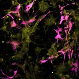
Technical Note

References

1.   McConnell, H. L., Kersch, C. N., Woltjer, R. L. & Neuwelt, E. A. The translational significance of the neurovascular
     unit. J. Biol. Chem. 292, 762–770 (2017).

2. Lochhead, J. J., Yang, J., Ronaldson, P. T. & Davis, T. P. Structure, Function, and Regulation of the Blood-Brain
   Barrier Tight Junction in Central Nervous System Disorders. Front. Physiol. 11, 914 (2020).

3. Pardridge, W. M. The blood-brain barrier: Bottleneck in brain drug development. NeuroRx 2, 3–14 (2005).

4. Helms, H. C. et al. In vitro models of the blood-brain barrier: An overview of commonly used brain endothelial cell
   culture models and guidelines for their use. J. Cereb. Blood Flow Metab. 36, 862–890 (2015).

5. Linville, R. M. et al. Human iPSC-derived blood-brain barrier microvessels: validation of barrier function and
   endothelial cell behavior. Biomaterials 190–191, 24–37 (2019).

6. Nikolakopoulou, P. et al. Recent progress in translational engineered in vitro models of the central nervous system.
   Brain 143, 3181–3213 (2021).

7.   Bhalerao, A. et al. In vitro modeling of the neurovascular unit: Advances in the field. Fluids Barriers CNS 17, 22
     (2020).

8. Song, H. W. et al. Transcriptomic comparison of human and mouse brain microvessels. Sci. Rep. 10, 12358 (2020).

9. Uchida, Y. et al. Quantitative targeted absolute proteomics of human blood-brain barrier transporters and receptors.
   J. Neurochem. 117, 333–345 (2011).

10. Shi, L., Zeng, M., Sun, Y. & Fu, B. M. Quantification of blood-brain barrier solute permeability and brain transport by
    multiphoton microscopy. J. Biomech. Eng. 136, (2014).

11. Sharma, B., Luhach, K. & Kulkarni, G. T. In vitro and in vivo models of BBB to evaluate brain targeting drug delivery.
    in Brain Targeted Drug Delivery System 53–101 (Elsevier, 2019). doi:10.1016/b978-0-12-814001-7.00004-4.

12. Uhlén, M. et al. Tissue-based map of the human proteome. Science (80-. ). 347, (2015).

13. Manatakis, D. V., Vandevender, A. & Manolakos, E. S. An information-theoretic approach for measuring the distance
    of organ tissue samples using their transcriptomic signatures. Bioinformatics 36, 5194–5204 (2020).

                                                                                                          © Emulate, Inc., 2021. All rights reserved.

                                                  emulatebio.com
                                                                                                                        Technical Note | June 2021
                                                                                           The technology herein may be covered by patents and/or
                                                                                                trademarks. Please contact Emulate for information.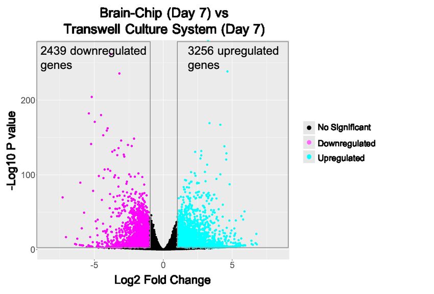
You can also read