International Journal of Veterinary Science
←
→
Page content transcription
If your browser does not render page correctly, please read the page content below
P-ISSN: 2304-3075; E-ISSN: 2305-4360

                       International Journal of Veterinary Science
                                                www.ijvets.com; editor@ijvets.com

Research Article
Flea Species Isolated from the Human and Animals: Prevalence and
Ultrastructural Studies
Marwa M Attia1* Nagla MK Salaeh2 and Sohila M El-Gameel1
1
 Parasitology Department, Faculty of Veterinary Medicine, Cairo University, 12211, Giza, Egypt; 2Zoology department,
Faculty of Science, Aswan University. Aswan, Egypt
*Corresponding author: marwaattia.vetpara@yahoo.com; marwaattia.vetpara@cu.edu.eg

    Article History: 19-662    Received: September 05, 2019 Revised: December 08, 2019 Accepted: December 16, 2019

    AB S T RA C T
    Fleas are one of the insects that belong to the order Siphonaptera. They are a wingless, compressed laterally, and non-
    host specific insect parasitizing a wide range of hosts, so they able to transmit diseases from animals to human. This
    study aimed to identify the fleas in and around human and animals with their prevalence and ultrastructure study. Flea
    samples were collected from four governorates (Cairo; Giza; Fayoum and South Sinai); Egypt during the period from
    January 2017 to May 2018.One-hundred and twenty fleas were collected from five animals (donkeys, sheep, goats,
    dogs, and cats) surrounding humans. All the collected fleas were identified as Ctenocephalides felis felis (C. f. felis).
    All the animals were infested with C. f. felis with flea's allergic dermatitis recorded in dogs, human, and donkeys, while
    goats and sheep showed anemic mucous membrane. The highest infestation was recorded in spring and summer. The
    morphological characters of 50 specimens were recorded and measured using the stereoscopic and light microscope as
    well as scanning electron microscope (SEM). The ultramorphological characters of C.f. felis head, thorax, and abdomen
    with the genital organs of male and female were fully described. The antennae have three segments with the third one
    had several adhesive circular disks and numerous sensory hairs in antennal grooves. The maxillary palps were well
    developed with four segments which supported with sensory small hairs. The first genal comb is half of the second one
    or nearly equal to its length. These results about identification and prevalence studies were used to update the knowledge
    about the flea species present in investigated localities in Egypt; to detect the appropriate control measures which could
    be applied in flea's infestation.
    Key words: Ctenocephalides felis felis, Flea allergic dermatitis, Siphonaptera, Prevalence, Ultrastructure

                      INTRODUCTION                                          The cat flea is the most dominant flea type in
                                                                       domesticated and even wild animals in different countries
     Fleas are blood-sucking ectoparasites of human,                   such as China, where all the fleas collected was C.f. felis
animals, and birds. They are non-host specific wingless                (Slapeta et al., 2011). In comparison, in Australia, C. f. felis
insects, parasitizing a wide range of hosts. So, they can              is the predominant flea on animals in veterinary clinics in
transmit diseases from animals to humans and between                   which no Ctenocephalides canis cases were recorded
animals (Kettle, 1995).                                                (Slapeta et al., 2011 and Lawrence et al., 2015). The life
     Ctenocephalides felis felis (C.f. felis), the cat flea is a       cycle of C. f. felis is defined as complete metamorphosis. It
non-host-specific type of fleas that a wide range of hosts all         lasts for about 14 days at 32ᵒC and m ay be as long as 140
over the world. It acts as an intermediate host for                    days at low temperature, at 13ᵒC and humidity above 50%.
Dipylidium caninum, some filarial nematodes, and                       All stages of fleas are present on dusty corners, feces and
different pathogens as Rickettsia felis and Bartonella spp.            housing bedding (Kettle, 1995 and Soulsby, 1986). The
(Kettle, 1995 and Šlapeta et al., 2018). It infests a wide             geographical distribution of the fleas is dependent upon the
range of mammals including sheep, goats, cattle, buffaloes,            presence of its hosts; while C. f. felis has multiple hosts so,
horses, and donkeys which reared in the barn with straw                it also has a worldwide distribution. In Egypt; a recent
bedding in which the life cycle can develop well. Death was            arthropod survey on donkeys which reveals 58.33%
reported in young unusual hosts (lamb and calves) which                infestation with C. f. felis by Attia et al. 2018. So, the aim
were severely infested with C. f. felis (Yeruham et al., 1989).        of the current studies is to update the knowledge about the

Cite This Article as: Attia MM, NMK Salaeh and SM El-Gameel, 2020. Flea Species Isolated from the Human and
Animals: Prevalence and Ultrastructural Studies. Int J Vet Sci, x(x): xxxx. www.ijvets.com (©2020 IJVS. All rights
reserved)

                                                                   1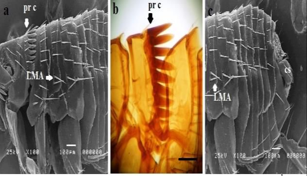
Int J Vet Sci, 2020, x(x): xxx.
Fleas present in different localities in Egypt in different RESULTS
animals which surround the human with special references
to their ultramorphological structure. The collected fleas
One-hundred and twenty fleas were collected from the
MATERIALS AND METHODS four governorates. The fleas were identified as C. f. felis.
The fleas were collected from different animals (donkeys;
Collection of fleas goats; sheep; dogs; cats) around the humans. All the
The fleas samples were collected from four examined animals were infested with C. f. felis with
governorates (Cairo, Giza, Fayoum, and South Sinai) different infestation degrees which were the highest in
during the period from January 2017 to May 2018; the spring and summer (the highest percentage occurring on
Cairo governorate is located at E ″07ʹ14031° and N goats) in comparison to autumn and winter. All of the
″30ʹ0230°, and the other governorates are far from Cairo by animals had different clinical manifestation as, fleas
7, 70 and 244 km, respectively. The fleas were collected allergic dermatitis recorded in dogs, human and donkeys,
from different animals, such as donkeys; goats; sheep; anemic mucous membrane recorded in goats. Prevalence
dogs; cats, animals which come in contact with human studies of C. f. felis on different hosts which come in
(Tables 1). The fleas which were collected from human as
contact to humans recorded in Table 2.
(human keeping the sheep herd and human rearing a pet
All the fleas which were collected from human were
animals) the number of human 30 collected in spring and
identified as C. felis felis in spring and summer (100%).
autumn in the highest percentage of infestation in all
animals. All Institutional and National Guidelines for the
Adult fleas
care and use of animals were followed.
Under stereoscopic loupe; fleas were dark brown and
wingless insects with a laterally compressed body of about
Preservation of fleas
1-3.5 mm mean length. The whole length of female body
The collected fleas were divided into two tubes; one
ranged from 2-3.5 mm ± 0.3 and whole length in male 1-
tube had 70% ethanol and the other tube had 2.5%
2.5 mm ± 0.5. They possess six long and strong legs.
glutaraldehyde [diluted in PBS (Phosphate- Buffered
Ultramorphological structure of C.f. felis.
Saline; pH: 7.4)]. Then all samples were sent to the
The body composed of head; 3 segmented thorax and
parasitology department, Faculty of Veterinary Medicine,
large abdomen. The head has an acutely angled frons which
Cairo University, Egypt for further studies.
specific for C. f.felis and pointed clypeus with the head
Identification of the collected fleas length 350 -500 μm ± 0.6. (Fig. 1). The antennae have three
The identification of the collected fleas was carried out segments with the third one being well developed with
according to the morphological characters reported by several adhesive circular disks with its length 140 - 180 μm
(Kettle, 1995 and Soulsby, 1986). The morphological ± 0.7 and numerous sensory hairs in antennal grooves
characters from 50 specimens were measured under the which are present around the edge of the groove (Fig. 2).
light microscope (Labomed × 40 and × 100). The maxillary palps were well developed with four
segments which supported with sensory small hairs at their
Preparation of permanent specimens posterior end (Fig. 3), with the presence of two maxillae at
For detailed morphological structures, all fleas were the posterior end of the head. There were two simple eyes
fixed immediately in 70% ethanol to stop their movement. at the two sides called ocelli, the genal combs which are
After the fleas’ relaxation, they were mounted according to present posteriorly to the ocelli; were eight in numbers and
(Attia, 2018). Briefly; all the fleas were placed in 5% present on each side of the head; the first genal comb is half
sodium hydroxide (NaOH) for 1 hour then washed with of the length in comparison to the second one ranging from
distilled water and dehydrated in ethanol series for 1 hour 60–80 μm which is important criteria to distinguish this
each. The specimens were cleared in clove oil then put in fleas with other Siphonaptera (Fig. 1 and 3). The number
Xylene. Finally, all specimens were mounted using Canada of postocciptal hairs were from 5-10 in mumber. The
balsam and incubated at 40°C overnight in order to dry thorax is composed of three segments; the first thoracic
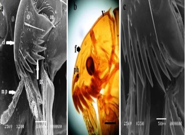
(Attia and Salaeh, 2019). segment (prothorax) is supported with eight pronotal
combs; with total dimension of prothorax were 100 – 150
Preparation of specimens for scanning electron
μm ± 1.3, and the pronotal length was 140-180 μm ±
microscope (SEM)
1.1.The lateral metanotal area (LMA) has two lateral setae
The ultrastructure of the fleas was identified using
(Fig. 4 and 5). The legs are supported with two long claws
SEM, according to (Attia and Salaeh, 2019). Summarized
as, the fleas were washed using buffered saline (PH 7.2) which are radiated in shape and two triangular pullvilli. The
then fixed in 2.5% glutaraldehyde at 4°C for 24 hours as mesothorax dimension was 150 – 200 μm ± 2; and in
described by (Hilali et al., 2015). All fleas were dehydrated metathorax 150- 300 μm ± 1.6. The posterior end of the
in ascending ethanol series (10% -30%-50% -70% -90% abdomen has several sensory sensilium which contains
and 100%) and desiccated on CO2 critical point drier several sensory pits with sensory hairs which are opposite
(Autosamdri-815, Germany). Flea specimens were glued with the antisensilial seta and posterior to it and there can be
and coated on gold; photographed with SEM (JSM 5200, seen several stylets called anal stylet. The posterior end of
Electron prob, Microanalyzer, Jeol, Japan) at Faculty of the male has a clasper with numbers of bristles on its outer
Agriculture, Cairo University, Egypt. Finally, fleas were surface which is supported by manubrium in abdomen
identified according to (Beaucournu and Ménier, 1998). and at the surface there is an apical arm of 9 th sternite.
2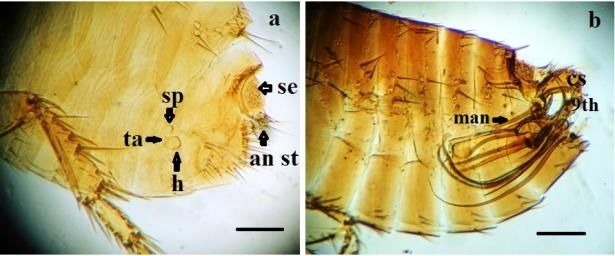
Int J Vet Sci, 2020, x(x): xxx.

Table 1: collection of fleas in different governorates in Egypt.
 Locality                                    Number of the collected fleas           Number of the collected male and female fleas
 Cairo                                                      20                                  12♀ and 8♂ C. f. felis
 Giza                                                       30                                  25♀ and 5♂ C.f. felis
 Fayoum                                                     50                                  42♀ and 8♂ C.f. felis
 South Sinai                                                20                                  15♀and 5 ♀ C. f. felis
 Total number of collected fleas                           120

Table 2: Prevalence studies of C. f. felis on different hosts which come in contact to humans.
Animals          Donkeys                       Sheep                   Goats                   Dogs                      Cats
Seasons No. Ex. No. Pos. % No. Ex. No. Pos. % No. Ex. No. Pos. % No. Ex. No. Pos. % No. Ex. No. Pos.                                 %
Spring       36       20     55.5       30        20     66.6     30     25     83.33     20    15      75      25        20         80
Summer       36       30     83.3       25        20      80      30     30      100      25    18      72      30        25        83.3
Autumn       36       15     41.6       25        8       32      30     15       50      30    15      50      30        20        66.6
Winter       36        5     13.8       25        5       20      30      8      26.6     30     6      20      30        10        33.3
Total       144       70     48.6 105             53     50.5 120        78       65      105   54     51.4 115           75        65.2
No. Ex.: number of examined animals; No. Pos.: number of positive animals for C. f. felis; %: percentage of infestation.

                                                                         Fig. 3: Scanning electron micrograph (SEM) of C. f. felis head
                                                                         showing in a and b; the maxillary palps having 4 segments which
                                                                         are supported with sensory small hairs at their posterior end
                                                                         (showed by arrows).

Fig. 1: Scanning electron micrograph (SEM) of C. f. felis head;
A: showing genal comb (gc) with the first spine equal to the
second one; maxillary palp (mp) and simple eye (Ocelli) (o) and
clypeus (cl) with antenna (an); b: permanent mount of cat flea
head showing pharynx (ph) frons(f); vertex (V) and also the genal
comb well developed and the maxillary palp; C: large
magnification of SEM showing the shape of genal comb.

                                                                         Fig. 4: Scanning electron micrograph (SEM) of C. f. felis thorax;
                                                                         a: The thorax composed from three segments; the first thoracic
                                                                         segment (prothorax) supported with 8pronotal combs (pr c). The
                                                                         metanotal area (LMA) of C. f. felis bearing 2 setae. b: light
                                                                         microscopic photograph of C. f felis thorax showing 8 pronotal
                                                                         combs (pr c). c: abdomen of male C. f. felis showing; the lateral
                                                                         metanotal area (LMA) which bears two setae and claspers at the
                                                                         posteror end (cs); in photo (b): Scale bar: 100μm.

                                                                                                 DISCUSSION
Fig. 2: Scanning electron micrograph (SEM) of C. f. felis head; a;
b; c: showing antenna (an) which has 3 segments, the third one
                                                                              The cat flea (C. felis felis) is world-widely distributed
being well developed with a number of adhesive circular discs and        and it has a wide range of hosts because it lacks the host
many sensory hairs (s) in antennal grooves which were present            specificity (Yeruham and Koren, 2003) and is important to
around the edge of the groove; O: ocelli.                                livestock as well as pet animals (Rust, 2017). Several flea
                                                                         important transmitted diseases were explanated with
The abdomen of female with its posterior end showing                     Blagburn and Dryden (2009) with fleas allergic dermatitis
several sensory sensilium and posterior to it, a number of               and many diseases transmitted. In this study, with severe
stylets called anal stylets; and had female genital organ                infestation of the examined animals and human with the
which is the spermatheca; tall and head. (Fig. 6 and 7).                 world-wide species of the fleas, we mention that it is

                                                                     3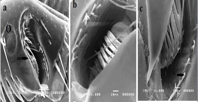
Int J Vet Sci, 2020, x(x): xxx.

                                                                                   Different survey studies were done in only one or two
                                                                             animals species as in Israel by Yeruham and Koren; 2003
                                                                             on donkeys and by Attia et al. 2018 on donkeys in Egypt.
                                                                             Survey occurred in water buffalo in India by Singh et al.
                                                                             2011. Different morphological studies were done on C. f.
                                                                             felis but none gave the full description of the different parts
                                                                             of fleas with scanning electron microscope as (Kettle,
                                                                             1995, Lawrence et al., 2015 and Marrugal et al., 2013).
                                                                             Recent study on morphological and molecular studies on
                                                                             Ctenocephalides spp by Azarm et al. 2016, this study
Fig. 5: Scanning electron micrograph (SEM) of C. f. felis legs: a;b          occureed on one animal species which is the dogs while our
showing the legs which are supported with two long claws (cl)                study occurred in different animals species and human. The
which are radiated and also have two triangular Pullvilli (pu).              previous study gives light microscopic explanation of the
                                                                             two species C. canis and C. felis which based on
                                                                             description of the shape of the head and legs setae (Azarm
                                                                             et al. 2016). So, our results give the exact morphological
                                                                             identification on the widely spread fleas in Egypt, C. f. felis.

                                                                             Conclusions
                                                                                   The high prevalence and infestation of C. felis felis in
                                                                             different domestic animals could be due to the direct
                                                                             contact of cats with goats, sheep, and donkeys as well as
                                                                             the presence of dogs which collect these fleas from these
                                                                             animals. These animals surround humans so the fleas infest
                                                                             the human of the same species as animalswhichare C. felis
Fig. 6: Scanning electron micrograph (SEM) of male C.f. felis                felis. There must urgently be found the way to control these
abdomen: a; b: The posterior end has several sensory sensilium               fleas by using different insecticides, a topic which had been
(se) which contain several sensory pits with sensory hairs (pointed          severely studied before.
by arrows in (c)); the sensilium is opposite with the antisensilial
seta (ans). The posterior end of the male has a clasper (cs) with                                   REFERENCES
numerous bristles on its outer surface in a; b.
                                                                             Attia MM, Khalifa MM and Atwa MT, 2018. The prevalence and
                                                                                  intensity of external and internal parasites in working
                                                                                  donkeys (Equus asinus) in Egypt. Vet World, 11: 1298-1306.
                                                                             Attia MM and Salaeh NMK, 2019. Ultrastructure of adult
                                                                                  Gasterophilus intestinalis (Diptera: Gasterophilidae)
                                                                                  and its puparium. Int J Trop Insect Sci, DOI:
                                                                                  10.1007/s42690-019-00084-9.
                                                                             Azarm A, Dalimi A, Mohebali M, et al., 2016. Morphological and
                                                                                  molecular characterization of Ctenocephalides spp isolated
                                                                                  from dogs in north of Iran. J Entomol Zool Stud, 4: 713-717.
                                                                             Baker KP and Elharam S, 1992. The biology of Ctenocephalides
Fig. 7: Light microscopic phpotograph of C. f. felis abdomen: a:                  canis in Ireland. Vet Parasitol, 45: 141-6.
abdomen of female with its posterior end showing several sensory             Blagburn BL and Dryden MW, 2009. Biology, treatment, and
sensilium (se) and posterior to it, a number of stylets called anal               control of flea and tick infestations. Vet Clin Small Anim,
stylets (an st); the sp: spermatheca; ta: tall; h: head. The posterior            39: 1173–1200.
end of the male has a clasper (cs) which is supported by                     Dryden MW and Rust MK, 1994. The cat flea: biology, ecology
manubrium in abdomen and at the surface there is an apical arm                    and control. Vet Parasitol, 52: 1-19.
of 9 th sternite. Scale bar: 100μ m.                                         Hilali M, Mahdy OA and Attia MM, 2015. Monthly variations of
                                                                                  Rhinoestrus spp. (Diptera: Oestridae) larvae infesting
important to find a possible way to control such disease                          donkeys in Egypt: Morphological and molecular
                                                                                  identification of third stage larvae. J Adv Res, 6: 1015-1021.
infestations. Studies on successful control strategies of                    Kettle DS, 1995. Medical and Veterinary Entomology. CAB
fleas were based on the identification of the parasites, their                    International.
life cycle, and bionomics of their different stages. The life                Lawrence AL, Hii SF, Jirsova D, et al., 2015. Integrated
cycle of C. f. felis and C. canis had been studied several                        morphological and molecular identification of cat fleas
times (Baker and Elharam,1992). The adult C. f. felis                             (Ctenocephalides felis) and dog fleas (Ctenocephalides
begins feeding within a few minutes from emergence. Fleas                         canis) vectoring Rickettsia felis in central Europe. Vet
mate after the blood meal and the female starts the egg-                          Parasitol, 210: 215–23.
laying (Dryden and Rust, 1994). Eggs are laid on the host                    Marrugal A, Callejón R, de Rojas M, et al., 2013. Morphological,
nest, bedding wool, and carpet (Dryden and Rust, 1994).                           biometrical      and      molecular      characterization   of
                                                                                  Ctenocephalides felis and Ctenocephalides canis isolated
The hatched C. f. felis larvae are free-living, they feed on                      from dogs from different geographical regions. Parasitol
organic matter and the adult flea feces. The larvae become                        Res, 112: 2289–2298.
a cocoon in which they pupate (Shanks et al., 2000). The                     Rust MK, 2017. The Biology and ecology of cat fleas and
explanation of the life cycle was to determine the exact                          advancements in their pest management: A Review. Insects,
time for fleas' control.                                                          8: 118.

                                                                         4
Int J Vet Sci, 2020, x(x): xxx.
Shanks DJ, Rowan TG, Jones R, et al., 2000. Efficacy of selamectin Singh NK, Haque M, Rath SS, et al., 2011. First report of
in the treatment and prevention of flea (Ctenocephalides felis Ctenocephalides felis felis infestations of buffalo calves in
felis) infestations on dogs and cats housed in simulated home Punjab, India J Parasitol Dis, 35: 235–236.
environments. Vet Parasitol, 91: 213–222. Yeruham I and Koren O, 2003. Severe infestation of a she-ass
Slapeta J, King J, McDonell D, et al., 2011. The cat flea with the cat flea Ctenocephalides felis felis (Bouche, 1835).
(Ctenocephalides f. felis) is the dominant flea on domestic Vet Parasitol, 115: 365–367.
dogs and cats in Australian veterinary practices. Vet Yeruham I, Rosen S and Hadani A, 1989. Mortality in calves,
Parasitol, 180: 383–388. lambs and kids caused by severe infestation with the cat flea
ŠlapetaJ, Lawrence A and Reichel MP, 2018. Cat fleas Ctenocephalides felis felis (Bouche, 1835) in Israel. Vet
(Ctenocephalides felis) carrying Rickettsia felis and Parasitol, 30: 351–356.
Bartonella species in Hong Kong. ParasitolInt, 67: 209-212. Yeruham I, Rosen S and Perl S 1997. An apparent flea-allergy
Soulsby EJL,1986. Helminthes, arthropods and protozoa of dermatitis in kids and lambs. J Vet Med, 44: 391–397.
domesticated animals. (7th ed) Bailliere Tindall London,
pp:371-372.
5You can also read